Abstract
Abstract 2725
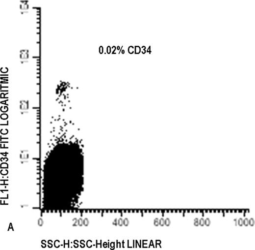
Already for decades, controversy exists as to whether CD34 expression predicts for poor prognosis (Kanda et al, Cancer 88:2529, 2000). One of the complicating factors between studies was the use of different cut-off values, ranging from 5% to 25% CD34 (% of WBC). Such cut-offs usually result in arbitrary boundaries between positive (+) and negative (-) cells. Earlier, we have identified a particular subtype of AML with a small (≤1%) CD34+ population present, which was homogeneous in the flowcytometry properties forward- and sideward scatter and CD34 expression (Figure A) and was of non-malignant origin (van der Pol et al, Haematol. 88: 983, 2003). We considered this type of AML as truly negative for CD34+ AML cells (called: CD34- AML). With this definition, patients with >1% CD34+ cells thus have CD34+ AML. In a retrospective analysis of 98 patients with long-term follow up (median= 14.8 months; range 0.4–169), CD34- patients (n=24) had a much better prognosis compared to CD34+ AML patients (n=74): median OS not reached (>160 months) versus 13 months (p<0.005; figure B). For RFS this was >160 months versus 10 months (p=0.009). Importantly, when applying the historical cut-offs of 5%, 10%, 15% and 20%, no significant prognostic value was found, with only trends in some cases. When excluding all CD34- patients, no prognostic impact al all was left for the 4 cut-offs of 5%-20%. This proves that, when applying the cut-off strategy (5%-20%) in the whole patient group (including the CD34- patients) it is the CD34- sub-group herein that is responsible for the better prognosis of patients below these (5-20%) other cut-off levels. The difference in prognosis implies differences in therapy resistance. P-glycoprotein activity is one of the functional factors that best predict therapy resistance and prognosis in AML. Almost all CD34- patients (22/24) had P-glycoprotein activity lower than the median of normal bone marrow (nBM) CD34+ cells. Above 1% CD34 level, increasing CD34 percentages were accompanied by increasing P-glycoprotein activity (38/74 higher than nBM CD34+), as is known from literature too. The prognostic value of CD34-negativity was confirmed in a retrospective setting in a large (n= 394; 85 CD34- and 309 CD34+) group of uniformly treated patients (<60 yrs). The results confirmed the retrospective results: Figure C shows that median OS is not reached (> 41 months) for CD34- patients and 19 months for CD34+ patients (p=0.006). For RFS (n=225) median RFS was not reached but differences were significant ((p=0.003). However, in contrast to the retrospective study, now also the cut-offs of 5–20% had significant, be it low, prognostic impact both for OS and RFS (not shown). This could be explained by the impact of CD34- patients within the patient groups below the cut-offs: when applying the cut-offs of 5%-20% after exclusion of the CD34- group, no prognostic impact at all was left. Altogether these results show that finding a prognostic impact for CD34 percentage using the historical cut-offs strongly depends on the size of the patient group studied and likely also on the size of the CD34- group herein. These phenomena fully explain the discrepancies in literature up to date. NPM1 mutated leukemias are reported as CD34 negative, although without using our strict definition of CD34 negativity. With our definition, 46/68 evaluable CD34- cases were NPM1 mutated, 31/46 with single mutation (group 1) and 15/46 with an additional FLT3-ITD (group 2); 22 patients had neither NPM1 mutations nor FLT3-ITD (group 3) [only 1 patient with cytogenetic good risk aberrancy t(8;21)]. All these 3 CD34 negative subgroups showed similar good prognosis (70% survival after 30–40 months follow up). For comparison, in the CD34+ group, NPM1 single mutation (n=19/260) had good prognosis; NPM1 mutation+FLT3-ITD (n=17/260), similar to FLT3-ITD alone (n=22/260), had a very poor prognosis. In conclusion, we show that a particular group of AML patients, who completely lack malignant CD34 positive cells, have CD34 percentages ≤1 %, with a specific profile on flowcytometer, have a good prognosis compared to CD34+ patients. Cells from CD34- patients show therapy sensitive characteristics and contain both NPM1 wild type and NPM1 mutated cases. The global way in which prognostic impact of CD34 percentage has been applied for decades, thus has prevented the clear statement that CD34 has indeed strong prognostic impact in AML.
No relevant conflicts of interest to declare.
Author notes
Asterisk with author names denotes non-ASH members.


This feature is available to Subscribers Only
Sign In or Create an Account Close Modal