Abstract
Rho kinases belong to a family of serine/threonine kinases whose role in recruitment and migration of inflammatory cells is poorly understood. We show that deficiency of ROCK1 results in increased recruitment and migration of macrophages and neutrophils in vitro and in vivo. Enhanced migration resulting from ROCK1 deficiency is observed despite normal expression of ROCK2 and a significant reduction in overall ROCK activity. ROCK1 directly binds PTEN in response to receptor activation and is essential for PTEN phosphorylation and stability. In the absence of ROCK1, PTEN phosphorylation, stability, and its activity are significantly impaired. Consequently, increased activation of downstream targets of PTEN, including PIP3, AKT, GSK-3β, and cyclin D1, is observed. Our results reveal ROCK1 as a physiologic regulator of PTEN whose function is to repress excessive recruitment of macrophages and neutrophils during acute inflammation.
Introduction
Neutrophils and macrophages are a major cellular component of the innate immune response that are rapidly recruited in large numbers to sites of infection.1-3 In response to inflammation, neutrophils and macrophages migrate from blood to infected sites in various tissues and protect the host by destroying invading bacterial and fungal pathogens. This process is thought to involve chemokines, cytokines, extracellular matrix proteins, and members of the β1 integrin family, including α4β1and α5β1. Importantly, interfering with the function of β1 integrins impairs the ability of macrophages to be recruited to the sites of inflammation.4,5 Although it is clear that cytokines such as macrophage colony stimulating factor (M-CSF), chemokines such as monocyte chemotactic protein-1 (MCP-1), and β1 integrins play a significant role in regulating adhesion and migration of macrophages and neutrophils, the signaling pathways responsible for coordinating these processes downstream from these molecules are poorly defined.
Recent studies have implicated phosphatidylinositol 3,4,5-trisphosphate (PIP3) in regulating several aspects of cytoskeleton-based functions, including adhesion and migration in response to activation of a variety of cell surface receptors.6,7 In macrophages and neutrophils, PIP3 regulates adhesion, migration, and polarization as a consequence of activation of the enzyme phosphatidyl-inositol-3 kinase (PI3K). End products of PI3K are partly regulated by phosphatase and tensin homolog deleted on chromosome 10 (PTEN). PTEN is a tumor suppressor gene that is often mutated in several tumors.8 PTEN inactivates PI3K by dephosphorylating PIP3 to PIP2.9 The structure of PTEN involves multiple domains, including the phosphatase domain (C2), PDZ binding domain, as well as various phosphorylation sites implicated in regulating the activity/stability of PTEN. Although it is widely accepted that deletion or mutation of PTEN can contribute to tumor formation, recent studies suggest that modulation in the levels of PTEN expression may also contribute to tumorigenesis.10 To this end, studies have shown that PTEN protein expression is reduced in a significant number of breast cancers.11 Although the precise reason behind reduced PTEN protein levels in these cancers is poorly understood, alteration in the transcription of PTEN as well as changes in the activity and stability of PTEN protein have been proposed.
In neutrophils, the intracellular localization and activity of PTEN are regulated by chemoattractants.12,13 In these cells, during chemotaxis, PI3K activity is localized at the leading edge (front) of the cells, whereas PTEN is localized at the back of the cell.14 This process of differential localization of PTEN and PI3K activity in the same cell contributes to restricted PIP3 levels in specific compartments (ie, leading edge during migration) of the cell, thereby regulating migration/recruitment.12,15 Whereas a direct link exists between PTEN and PI3K in regulating the recruitment of inflammatory cells during acute inflammation, the mechanism(s) by which PTEN activity is regulated by cytokines/chemokines and/or integrins in primary neutrophils and macrophages and the functional consequence(s) of deregulated PTEN activity during acute inflammation is(are) not known.
Rho GTPase family members are important regulators of cell migration, proliferation, and apoptosis.16,17 Rho stimulates contractility and adhesion by inducing the formation of actin stress fibers and focal adhesions in fibroblasts and aggregation of platelets and lymphocytes by regulating the avidity of surface integrins.18 Rho cycles between guanosine diphosphate–bound inactive and guanosine triphosphate (GTP)–bound active forms, and the GTP-bound form binds to specific targets to exert its biologic functions.19,20 Two closely related Rho kinases, ROCK1 and ROCK2, have been identified as key downstream effectors of Rho GTPases and contribute to multiple cytoskeleton functions in nonhematopoietic cells.21,22 Whereas ROCK1 and ROCK2 share significant sequence homology in the kinase domain (> 90%), the regulatory domains at the C terminus show significant divergence.23,24 Although the functional differences between ROCK1 and ROCK2 are poorly understood, pharmacologic studies as well as studies conducted with activated and dominant active versions of Rho kinases in nonhematopoietic cells suggest that Rho kinases contribute to increased actin-myosin II–mediated contractility by directly phosphorylating myosin light chain and negatively regulating myosin light chain phosphatase by phosphorylating myosin binding subunit of myosin light chain phosphatase.23-27 Rho kinases also activate LIM kinase, phosphorylate cofilin, and inhibit its actin-depolymerizing activity, leading to stabilization of actin stress fibers in fibroblasts.28-30
Although a large body of evidence has demonstrated that Rho kinases are intimately associated with different phases of cellular functions primarily based on pharmacologic studies, the physiologic role of ROCK1 or ROCK2 in blood cells is poorly understood. To date, most of the conclusions with regard to ROCK function have been drawn using pharmacologic inhibitors as well as dominant negative approaches. Although useful, these studies do not distinguish between the physiologic function of ROCK1 and ROCK2 in primary cells. Here, we investigated the role of ROCK1 mediated signaling in regulating macrophage and neutrophil function(s) in vivo.
Methods
Mice
Six- to 8-week-old wild-type (WT) and ROCK1−/− mice (FVB background) were used to isolate macrophage and neutrophil progenitor cells from bone marrow (BM).31 Mice used in the present study were fully backcrossed and maintained in the FVB genetic background and are viable. All mouse studies were approved by the Indiana University Animal Care and Use Committee.
Macrophage and neutrophil migration in vivo
WT and ROCK1−/− mice were challenged with 4% thioglycollate (Sigma-Aldrich) by intraperitoneal injection (2 mL) as described previously.32 For macrophage migration assay in vivo, mice were killed by cervical dislocation after 4 days of injections, and peritoneal cells were harvested by injecting 6 mL of Dulbecco phosphate-buffered saline through the peritoneal wall. Peritoneal lavage was collected, and red blood cells (RBCs) present in the lavage fluid were removed by RBC lysis buffer. Cells were centrifuged and resuspended in 1 mL of RPMI medium containing 10% fetal bovine serum. The number of cells in the lavage was determined using a hemocytometer. Cells were stained with phycoerythrin-conjugated anti- F4/80 antibody and analyzed by flow cytometry (BD Biosciences PharMingen). Macrophage content was evaluated by cytospin preparations, and cells were stained with Giemsa stain (Dade Behring). For neutrophil migration assay in vivo, mice were challenged with 4% thioglycollate (Sigma-Aldrich) by intraperitoneal injection (2 mL) as described previously.33 Peritoneal lavage was performed 4 hours after the challenge as described.33 Cells were counted by hemocytometer and then costained with phycoerythrin (PE)–conjugated anti–Gr-1 (Ly-6G, clone RB 6-8C 5), FITC–Mac-1 (CD11b, clone M1/70) antibodies and analyzed by FACSCalibur flow cytometer (BD Biosciences PharMingen).
Isolation of bone marrow–derived macrophage and neutrophil progenitors
Murine bone marrow–derived macrophages (BMMs) and neutrophils (BMNs) were generated from 6- to 8-week-old WT and ROCK1−/− femurs, tibias, and iliac crest. Briefly, bone marrow cells were flushed into 50-mL Falcon tubes using syringe needle and Iscove modified Dulbecco medium (IMDM; Invitrogen). Cells were collected by centrifugation at 800g for 5 minutes (Beckman Coulter) at room temperature, and RBCs were lysed with RBC lysis buffer containing 155mM ammonium chloride, 10mM potassium bicarbonate, and 0.1mM ethylenediaminetetraacetic acid for 5 minutes at room temperature. The cells were centrifuged and resuspended in IMDM. Low-density bone marrow (LDBM) cells were isolated by density gradient centrifugation using histopaque 1083 (Sigma-Aldrich). For BMMs, LDBM cells were cultured in complete media consisting of IMDM, 20% fetal bovine serum supplemented with 1% penicillin/streptomycin, and in the presence of 100 ng/mL M-CSF (PeproTech). BMMs were passaged and, on reaching confluence, were used between 3 and 6 passages.34 For BMNs, LDBM cells were cultured in IMDM, 10% fetal bovine serum supplemented with 1% penicillin/streptomycin, and in the presence of 1 ng/mL granulocyte colony-stimulating factor and 5 ng/mL interleukin-3 (PeproTech) as described.33,35
Macrophage migration assay in vitro
Haptotactic cell migration assay was performed as previously described.36 Briefly, the bottom of transwell filters (8μM pore filter; Costar) was coated with 20 μg/mL fibronectin CH296 peptide (Takara) for 2 hours at 37°C and rinsed twice with phosphate-buffered saline (PBS) containing 2% bovine serum albumin. The fibronectin peptide-coated filters were placed in the lower chamber containing 500 μL of complete medium with or without M-CSF (100 ng/mL) or MCP-1 (PeproTech). BMMs (2.5 × 10⁵ for M-CSF and 0.5 × 10⁶ for MCP-1) were resuspended in 100 μL of IMDM and allowed to migrate toward the bottom of the top chamber. After 20 hours (for M-CSF) and 8 hours (for MCP-1) of incubation at 37°C, nonmigrated cells in the upper chamber were removed with a cotton swab, and the migrated cells attached to the bottom surface of the membrane were stained with 0.1% crystal violet dissolved in 0.1M borate, pH 9.0, and 2% ethanol for 5 minutes at room temperature. The number of migrated cells per membrane was counted in 10 random fields with an inverted microscope using a 20× objective lens.
Neutrophil migration assay in vitro
BMN migration was performed using a 48-well micro-chemotaxis chamber (3μM membrane; Neuro Probe) as described previously.37
Wound-healing assay
Wound-healing assay was performed as described previously.36
Flow cytometric analysis
Flow cytometry was performed as described.34 F-actin staining of macrophages was performed according to the manufacturer's instructions (Invitrogen).
Adhesion assay on fibronectin
Adhesion assay was performed as described earlier.38
Confocal microscopy
Intracellular staining was performed using a Millipore kit according to the manufacturer's instructions (Actin Cytoskeleton/Focal Adhesion Staining Kit).
Western blot analysis
Western blot analysis was performed using anti–phospho-Akt (serine 473), anti–phospho PTEN antibody (S380/Thr382/Thr383), and anti–phospho GSK-β (serine 9) antibody from Cell Signaling Technology. Anti-PTEN (FL-403), anticyclinD1 (72-13G), anti-ROCK1 (H-85), and anti-ROCK2 (C-20) antibodies were obtained from Santa Cruz Biotechnology. Supersignal West Dura extended duration detection system (Pierce Chemical) was used according to the manufacturer's instructions to visualize protein signal.
ROCK kinase activity assay
ROCK kinase activity was assessed by the phosphorylation of exogenously added ROCK substrate, MYPT1, as described previously.39
Phosphatase activity assay
PTEN activity was evaluated by assessing the rate of phosphate release using malachite green assay (Upstate Biotechnology). Briefly, cell lysates were lysed in a phosphatase extraction buffer containing 20mM imidazole-HCl, pH 7.0, 2mM dithiothreitol, 2mM ethyleneglycoltetraacetic acid, 1mM phenylmethylsulfonyl fluoride, 10 μg/mL aprotinin, and 10 μg/mL leupeptin. To initiate enzyme reaction, an equal amount of lysate was incubated with 50μM synthetic diC8-PI 3,4,5 P3 (Echelon) and 50μM 1,2-dioleoyl-sn-glycero-3-phospho-L-serine sodium salt for 10 minutes at 37°C. The reaction was stopped by adding 100 μL of malachite green reagent. The amount of phosphate released was measured by reading the absorbance at 650 nm.
Phospholipid analysis
Phospholipid extraction and thin-layer chromatography analysis was performed as described previously.40
Pulse-chase labeling
WT and ROCK1−/− BMMs were replated on tissue-culture plates and on reaching 70% confluence were washed with PBS and incubated with methionine-free medium for 2 hours. Cells were then pulse-labeled with radioactive [35S]-methionine (PerkinElmer) containing medium (150 μCi/mL) for 1 hour, after which cells were washed and incubated in chase medium containing 10 mL of regular Dulbecco modified Eagle medium, 5mM methionine, and 5mM cysteine for the indicated time period. BMMs were collected and then lysed. PTEN was immunoprecipitated using an anti-PTEN antibody from total lysates. The immune complex was resolved by sodium dodecyl sulfate–polyacrylamide gel electrophoresis and visualized by autoradiography.
Results
Increased recruitment of ROCK1−/− macrophages and neutrophils in an in vivo model of acute peritonitis
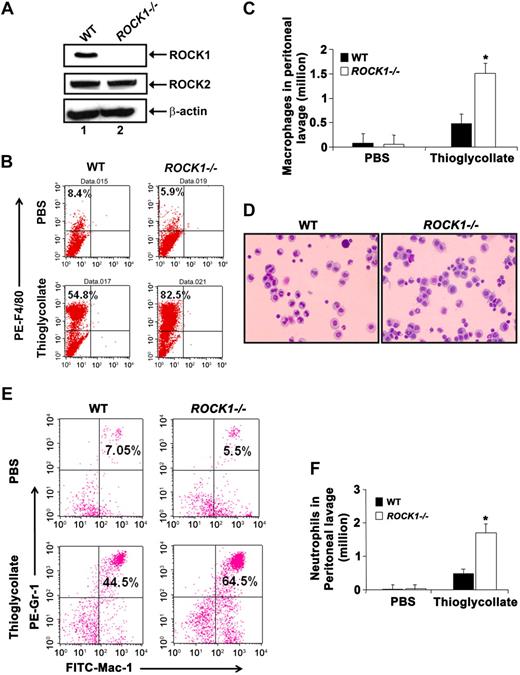
We reasoned that investigation of the role of ROCK1 in a gene-targeted mouse model would more precisely elucidate the physiologic role of individual Rho kinases in regulating inflammation in vivo instead of pharmacologic inhibitors, which are known to inhibit the activation of both ROCK1 and ROCK2 equally and demonstrate nonspecific inhibition of protein kinase C as well as protein kinase A at higher concentrations.41 To this end, we first examined the expression of ROCK1 and ROCK2 in BMMs from WT and ROCK1−/− mice. We observed no detectable ROCK1 expression in ROCK1−/− BMMs (Figure 1A). The expression of ROCK2 in ROCK1−/− BMMs was unaltered and similar to that observed in WT BMMs, suggesting that deficiency of ROCK1 in BMMs does not induce compensatory changes in the expression of ROCK2 (Figure 1A). We next assessed the role of ROCK1 in regulating the recruitment of macrophages and neutrophils to the peritoneal cavity in vivo. For these studies, we used a well-characterized in vivo model of aseptic peritonitis. Briefly, thioglycollate (4%) was injected intraperitoneally into WT and ROCK1−/− mice, and the recruitment of macrophages and neutrophils in the peritoneal cavity after 4 days (for macrophages) or 4 hours (for neutrophils) was assessed. As seen in Figure 1B through D, a significant increase in the recruitment of ROCK1−/− macrophages was observed in the peritoneal cavity compared with WT controls (P < .01, Figure 1C). The increase in the recruitment of ROCK1−/− macrophages in the peritoneal cavity was not the result of an enhanced number of circulating monocytes in ROCK1-deficient mice (data not shown). Similar to the increased recruitment of ROCK1-deficient macrophages in response to thioglycollate treatment, deficiency of ROCK1 also resulted in an increased recruitment of neutrophils in the peritoneal cavity as early as 4 hours after thioglycollate challenge (Figure 1E-F). The increase in neutrophil recruitment was not the result of increased cycling of ROCK1−/− Gr-1/Mac-1 cells in the bone marrow (supplemental Figure 1, available on the Blood website; see the Supplemental Materials link at the top of the online article). These results suggest that deficiency of ROCK1 results in increased recruitment of both macrophages and neutrophils in an in vivo model of acute peritonitis.
Increased recruitment of macrophages and neutrophils into the inflamed peritoneum of ROCK1−/− mice. (A) Western blot analysis demonstrating the expression of ROCK1 and ROCK2 in BMMs from WT and ROCK1−/− mice. Equal amounts of cell lysate were probed with antibodies specific for the coiled-coil region of ROCK1 or ROCK2, respectively. The same blot was reprobed for β-actin to show protein loading in each lane. Lanes 1 and 2 represent lysates derived from WT and ROCK1−/− BMMs, respectively. The top arrow indicates expression of ROCK1 in WT BMMs but not in ROCK1−/− BMMs; the middle arrow indicates the expression of ROCK2 in both WT and ROCK1−/− BMMs; the bottom arrow indicates β-actin expression in WT and ROCK1−/− BMMs; n = 3. (B) WT and ROCK1−/− mice were given intraperitoneal injections of 4% thioglycollate. Peritoneal lavage was collected 4 days after injection, and mature macrophage-specific marker F4/80 expression was examined by flow cytometry. Top left quadrant of each dot blot represents the percentage of PE-conjugated F4/80-positive cells in WT and ROCK1−/− peritoneum after PBS or thioglycollate injections. Dot blots shown are representative of macrophages recruited into the peritoneal cavity of WT and ROCK1−/− mice, analyzed after PBS or thioglycollate injection. Similar results were obtained in 7 other mice from each genotype examined. (C) A representative bar graph shown here is the total number of macrophages recruited into the peritoneal cavity. Results shown are from one independent experiment containing 3 pairs of mice from each genotype after PBS or thioglycollate injection. WT vs ROCK1−/− mice, *P < .01. Similar results were obtained in 2 other independent experiments using 5 mice of each genotype (n = 3, 8 pairs of WT and ROCK1−/− mice). (D) Representative micrograph shown is a cytospin preparation of macrophages elicited by thioglycollate treatment from the peritoneal cavity of WT and ROCK1−/− mice. Peritoneal macrophages were stained with Wright-Giemsa. Images were acquired through a Zeiss Axioskop 2 Plus microscope equipped with a Plan-Neofluar 20×/0.5 objective lens, and were captured with an Axiocam MRC-5 camera and Axiovision 4 software (all from Carl Zeiss). (E) Increased recruitment of neutrophils to the inflamed peritoneum of ROCK1−/− mice. WT and ROCK1−/− mice were given intraperitoneal injections of 4% thioglycollate. Peritoneal lavage was collected after 4 hours of injection, and Gr-1/Mac-1 expression was examined by flow cytometry. Top right quadrant of each dot blot represents percentage of Gr-1 and Mac-1 double-positive cells in the peritoneum after PBS or thioglycollate injections. Dot blots shown are representative of neutrophils migrated into the peritoneum of WT and ROCK1−/− mice, analyzed after PBS or thioglycollate injection. Similar results were obtained in 4 other mice from each genotype examined. (F) A representative bar graph shows the total number of neutrophils recruited into the peritoneal cavity. Results are from one independent experiment containing 3 pairs of mice of each genotype after PBS or thioglycollate injection. WT vs ROCK1−/− mice, *P < .05. Similar results were obtained from one other independent experiment containing 2 mice of each genotype (n = 2, 5 pairs WT and ROCK1−/− mice).
Increased recruitment of macrophages and neutrophils into the inflamed peritoneum of ROCK1−/− mice. (A) Western blot analysis demonstrating the expression of ROCK1 and ROCK2 in BMMs from WT and ROCK1−/− mice. Equal amounts of cell lysate were probed with antibodies specific for the coiled-coil region of ROCK1 or ROCK2, respectively. The same blot was reprobed for β-actin to show protein loading in each lane. Lanes 1 and 2 represent lysates derived from WT and ROCK1−/− BMMs, respectively. The top arrow indicates expression of ROCK1 in WT BMMs but not in ROCK1−/− BMMs; the middle arrow indicates the expression of ROCK2 in both WT and ROCK1−/− BMMs; the bottom arrow indicates β-actin expression in WT and ROCK1−/− BMMs; n = 3. (B) WT and ROCK1−/− mice were given intraperitoneal injections of 4% thioglycollate. Peritoneal lavage was collected 4 days after injection, and mature macrophage-specific marker F4/80 expression was examined by flow cytometry. Top left quadrant of each dot blot represents the percentage of PE-conjugated F4/80-positive cells in WT and ROCK1−/− peritoneum after PBS or thioglycollate injections. Dot blots shown are representative of macrophages recruited into the peritoneal cavity of WT and ROCK1−/− mice, analyzed after PBS or thioglycollate injection. Similar results were obtained in 7 other mice from each genotype examined. (C) A representative bar graph shown here is the total number of macrophages recruited into the peritoneal cavity. Results shown are from one independent experiment containing 3 pairs of mice from each genotype after PBS or thioglycollate injection. WT vs ROCK1−/− mice, *P < .01. Similar results were obtained in 2 other independent experiments using 5 mice of each genotype (n = 3, 8 pairs of WT and ROCK1−/− mice). (D) Representative micrograph shown is a cytospin preparation of macrophages elicited by thioglycollate treatment from the peritoneal cavity of WT and ROCK1−/− mice. Peritoneal macrophages were stained with Wright-Giemsa. Images were acquired through a Zeiss Axioskop 2 Plus microscope equipped with a Plan-Neofluar 20×/0.5 objective lens, and were captured with an Axiocam MRC-5 camera and Axiovision 4 software (all from Carl Zeiss). (E) Increased recruitment of neutrophils to the inflamed peritoneum of ROCK1−/− mice. WT and ROCK1−/− mice were given intraperitoneal injections of 4% thioglycollate. Peritoneal lavage was collected after 4 hours of injection, and Gr-1/Mac-1 expression was examined by flow cytometry. Top right quadrant of each dot blot represents percentage of Gr-1 and Mac-1 double-positive cells in the peritoneum after PBS or thioglycollate injections. Dot blots shown are representative of neutrophils migrated into the peritoneum of WT and ROCK1−/− mice, analyzed after PBS or thioglycollate injection. Similar results were obtained in 4 other mice from each genotype examined. (F) A representative bar graph shows the total number of neutrophils recruited into the peritoneal cavity. Results are from one independent experiment containing 3 pairs of mice of each genotype after PBS or thioglycollate injection. WT vs ROCK1−/− mice, *P < .05. Similar results were obtained from one other independent experiment containing 2 mice of each genotype (n = 2, 5 pairs WT and ROCK1−/− mice).
Deficiency of ROCK1 results in increased macrophage and neutrophil migration in vitro
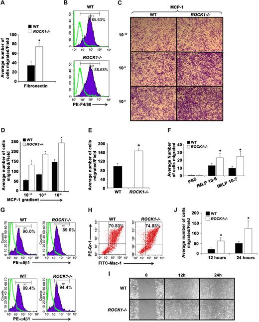
In an effort to understand the mechanism behind the enhanced recruitment of ROCK1-deficient macrophages and neutrophils in vivo in response to thioglycollate challenge, we conducted in vitro experiments to assess the migratory capacity of ROCK1-deficient BMMs and neutrophils to multiple migration-inducing stimuli, including cytokines, chemokines, and integrins. As seen in Figure 2A, loss of ROCK1 results in a significant increase in the migration of BMMs on fibronectin compared with WT controls. Importantly, the increase in migration of ROCK1−/− BMMs was not the result of impaired differentiation of ROCK1-deficient cells. Figure 2B demonstrates similar expression of macrophage specific marker F4/80 on WT and ROCK1−/− BMMs, demonstrating similar maturation in the 2 genotypes. Consistent with the enhanced migratory capacity of ROCK1-deficient BMMs, peritoneal cavity-derived ROCK1−/− macrophages also demonstrated enhanced migration in response to a gradient of MCP-1 (Figure 2C-D) as well as via β1 integrins (Figure 2E). Likewise, ROCK1−/− BMNs also demonstrated enhanced migration in response to a gradient of formyl-methionyl-leucyl-phenylalanine (fMLP; Figure 2F). This increase was not the result of differences in the maturation of these cells as the expression of β1 integrin as well as that of Gr-1 and Mac-1 on WT and ROCK1−/− BMNs was comparable (Figure 2G-H). Taken together, these data demonstrate that the increased recruitment of macrophages and neutrophils to the inflamed peritoneum in an acute model of peritonitis is probably the result of loss of ROCK1 in these cells.
Deficiency of ROCK1 in BMMs, peritoneal macrophages, and neutrophils results in increased migration and chemotaxis in vitro. (A) WT and ROCK1−/− BMMs (2.5 × 10⁵) were subjected to a migration assay on fibronectin fragment CH-296, which contains the binding sites for both integrins α4β1 and α5β1. Shown is the quantitative assessment of the number of WT and ROCK1−/− BMMs migrated on fibronectin-coated wells. The bar graph represents the average number of WT and ROCK1−/− BMMs migrated per field in an independent experiment performed in replicates of 3. WT versus ROCK1−/−, *P < .05. Similar results were obtained in 3 other independent experiments. (B) Expression of F4/80 on WT and ROCK1−/− BMMs. BMMs were stained with PE-conjugated anti-F4/80 antibody and subjected to flow cytometric analysis. Solid histograms represent the level of F4/80 expression on the surface of WT and ROCK1−/− BMMs, whereas open histograms represent the level of expression using an isotype control antibody. Percentage of F4/80 expression on WT and ROCK1−/− BMMs is shown in the top right corner of each histogram (n = 4, 6 pairs of WT and ROCK1−/− mice). (C-D) BMM migration using 8-μm pore size transwell filters coated with fibronectin. Representative images shown here are from WT and ROCK1−/− BMMs migrated on fibronectin in response to the MCP-1 gradient. The bar graph represents the average number of WT and ROCK1−/− BMMs migrated in response to MCP-1 gradient per field in an independent experiment. Similar results were obtained using 2 other mice of each genotype (total 3 pairs of WT and ROCK1−/− mice, *P < .05). (E) WT and ROCK1−/− peritoneal macrophages (2.5 × 10⁵) were subjected to a migration assay on fibronectin. The bar graph represents average number of migrated peritoneal macrophages derived from WT and ROCK1−/− mice from an independent experiment. WT versus ROCK1−/−, *P < .05. Similar results were obtained using 2 other mice of each genotype (total n = 3). (F) BMN migration assay was performed using the Boyden chamber. Migration was evaluated without fMLP, or in a gradient of 1μM fMLP. The bar graph represents the number of migrated BMNs per field (mean ± SD). Shown is a representative experiment performed in replicates of 3. WT versus ROCK1−/−, *P < .05. Similar results were obtained in 2 additional independent experiments (total n = 3). (G) Expression of integrins on WT and ROCK1−/− BMNs. BMNs were stained with PE-conjugated anti-α4β1 or anti-α5β1 antibody and subjected to flow cytometric analysis. Solid histograms indicate the level of α4β1 and α5β1 expression on the surface of WT and ROCK1−/− BMNs, whereas open histograms in both panels demonstrate the level of expression using an isotype control antibody. Percentage of α4β1 or α5β1 integrin expression on BMNs is shown in the top right quadrant of each histogram. Histograms shown are from a representative experiment. Similar results were obtained in 3 independent experiments (total n = 4). (H) WT and ROCK1−/− BMNs were harvested after 1 week and stained with antibodies that recognize Gr-1 and Mac-1. Stained cells were analyzed by flow cytometry to determine the level of Gr-1 and Mac-1 expression. Percentage of Gr-1 and Mac-1 double-positive cells in either genotype are indicated in the top right quadrant of each dot blot. (I) Increased migration of ROCK1−/− BMMs in a wound-healing assay. WT and ROCK1−/− BMMs were cultured for 8 days in 24-well plates in the presence of 100 ng/mL M-CSF. An artificial wound was created in the macrophage monolayer using a pipette tip. Images were taken immediately and again at indicated time periods after creating the wound. Top panels represent the migration of WT cells; bottom panels represent migration of ROCK1−/− cells. (J) Bar graph shows quantitative analysis of the number of migrated cells in the wounded area from 1 independent experiment. WT versus ROCK1−/−, *P < .05. Similar results were obtained in 2 other independent experiments (total n = 3). (C, I) Images were acquired through a Zeiss Axioskop 2 Plus microscope equipped with a Plan-Neofluar 20×/0.5 objective lens, and were captured with an Axiocam MRC-5 camera and Axiovision 4 software (all from Zeiss).
Deficiency of ROCK1 in BMMs, peritoneal macrophages, and neutrophils results in increased migration and chemotaxis in vitro. (A) WT and ROCK1−/− BMMs (2.5 × 10⁵) were subjected to a migration assay on fibronectin fragment CH-296, which contains the binding sites for both integrins α4β1 and α5β1. Shown is the quantitative assessment of the number of WT and ROCK1−/− BMMs migrated on fibronectin-coated wells. The bar graph represents the average number of WT and ROCK1−/− BMMs migrated per field in an independent experiment performed in replicates of 3. WT versus ROCK1−/−, *P < .05. Similar results were obtained in 3 other independent experiments. (B) Expression of F4/80 on WT and ROCK1−/− BMMs. BMMs were stained with PE-conjugated anti-F4/80 antibody and subjected to flow cytometric analysis. Solid histograms represent the level of F4/80 expression on the surface of WT and ROCK1−/− BMMs, whereas open histograms represent the level of expression using an isotype control antibody. Percentage of F4/80 expression on WT and ROCK1−/− BMMs is shown in the top right corner of each histogram (n = 4, 6 pairs of WT and ROCK1−/− mice). (C-D) BMM migration using 8-μm pore size transwell filters coated with fibronectin. Representative images shown here are from WT and ROCK1−/− BMMs migrated on fibronectin in response to the MCP-1 gradient. The bar graph represents the average number of WT and ROCK1−/− BMMs migrated in response to MCP-1 gradient per field in an independent experiment. Similar results were obtained using 2 other mice of each genotype (total 3 pairs of WT and ROCK1−/− mice, *P < .05). (E) WT and ROCK1−/− peritoneal macrophages (2.5 × 10⁵) were subjected to a migration assay on fibronectin. The bar graph represents average number of migrated peritoneal macrophages derived from WT and ROCK1−/− mice from an independent experiment. WT versus ROCK1−/−, *P < .05. Similar results were obtained using 2 other mice of each genotype (total n = 3). (F) BMN migration assay was performed using the Boyden chamber. Migration was evaluated without fMLP, or in a gradient of 1μM fMLP. The bar graph represents the number of migrated BMNs per field (mean ± SD). Shown is a representative experiment performed in replicates of 3. WT versus ROCK1−/−, *P < .05. Similar results were obtained in 2 additional independent experiments (total n = 3). (G) Expression of integrins on WT and ROCK1−/− BMNs. BMNs were stained with PE-conjugated anti-α4β1 or anti-α5β1 antibody and subjected to flow cytometric analysis. Solid histograms indicate the level of α4β1 and α5β1 expression on the surface of WT and ROCK1−/− BMNs, whereas open histograms in both panels demonstrate the level of expression using an isotype control antibody. Percentage of α4β1 or α5β1 integrin expression on BMNs is shown in the top right quadrant of each histogram. Histograms shown are from a representative experiment. Similar results were obtained in 3 independent experiments (total n = 4). (H) WT and ROCK1−/− BMNs were harvested after 1 week and stained with antibodies that recognize Gr-1 and Mac-1. Stained cells were analyzed by flow cytometry to determine the level of Gr-1 and Mac-1 expression. Percentage of Gr-1 and Mac-1 double-positive cells in either genotype are indicated in the top right quadrant of each dot blot. (I) Increased migration of ROCK1−/− BMMs in a wound-healing assay. WT and ROCK1−/− BMMs were cultured for 8 days in 24-well plates in the presence of 100 ng/mL M-CSF. An artificial wound was created in the macrophage monolayer using a pipette tip. Images were taken immediately and again at indicated time periods after creating the wound. Top panels represent the migration of WT cells; bottom panels represent migration of ROCK1−/− cells. (J) Bar graph shows quantitative analysis of the number of migrated cells in the wounded area from 1 independent experiment. WT versus ROCK1−/−, *P < .05. Similar results were obtained in 2 other independent experiments (total n = 3). (C, I) Images were acquired through a Zeiss Axioskop 2 Plus microscope equipped with a Plan-Neofluar 20×/0.5 objective lens, and were captured with an Axiocam MRC-5 camera and Axiovision 4 software (all from Zeiss).
Increased migration of ROCK1-deficient macrophages in a wound-healing assay
Macrophages migrate into numerous tissues during inflammation and play a significant role in regulating wound healing. Previous studies have suggested a role for fibronectin and VCAM-1 in the migration of macrophages to sites of inflammation. Studies using macrophage cell lines, as well as pharmacologic inhibitors of Rho kinase and siRNA knockdown approaches, have suggested a role for Rho kinases in wound healing.39,42 However, no genetic studies have been conducted to demonstrate a direct role for ROCK1 and/or ROCK2 in regulating this process. To examine the role of ROCK1 in wound repair, WT and ROCK1−/− BMMs were subjected to a wound healing assay in vitro. As seen in Figure 2I-J, a significant increase in the number of migrating ROCK1−/− BMMs was observed in the wounded area compared with WT controls. This increase was noted at all time points examined, including at the end of both 12 and 24 hours after wound creation (n = 3, P < .05).
Deficiency of ROCK1 in BMMs results in increased adhesion on fibronectin in vitro
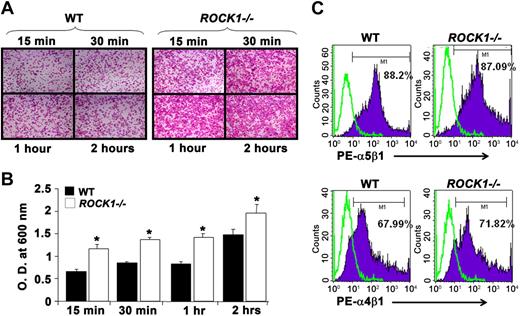
Cellular attachment is mediated by the direct ligation of integrin extracellular domains to defined sequences within the extracellular matrix. To understand the physiologic role of ROCK1 in integrin-mediated adhesion, we performed adhesion assay on fibronectin using BMMs derived from WT and ROCK1−/− mice. Loss of ROCK1 resulted in a significant increase in the adhesion of BMMs on fibronectin fragment CH296 (consisting of both α4β1 and α5β1 binding sites) compared with WT BMMs at all time points examined (Figure 3A-B). To determine whether the increased adhesion of ROCK1−/− BMMs to fibronectin was a result of enhanced integrin expression, we performed flow cytometric analysis on WT and ROCK1−/− cells using antibodies that recognize integrins α4β1 and α5β1. Our results revealed a similar level of expression of α4β1 and α5β1 integrins on WT and ROCK1−/− BMMs (WT; 67.99% and 88.2% vs ROCK1−/−; 71.82% and 87.09%, respectively, n = 4, Figure 3C), suggesting that deletion of ROCK1 does not affect the expression of β1 integrins on BMMs.
Deficiency of ROCK1 results in increased adhesion on fibronectin. (A) WT and ROCK1−/− BMMs (5 × 10⁵) were subjected to an in vitro adhesion assay on fibronectin fragment CH-296. Representative images shown here are of adherent WT (left panel) and ROCK1−/− BMMs (right panel) on fibronectin at different time points. Results shown here are from 1 independent experiment. Similar results were obtained in 2 other independent experiments. Images were acquired through a Zeiss Axioskop 2 Plus microscope equipped with a Plan-Neofluar 20×/0.5 objective lens, and were captured with an Axiocam MRC-5 camera and Axiovision 4 software (all from Zeiss). (B) Quantitative adhesion was assessed by measuring absorbance at indicated times. Bar graph represents the optical density of adherent cells at 600 nm from 1 independent experiment. WT versus ROCK1−/−, *P < .05. Similar results were obtained in 3 other independent experiments (total n = 4). (C) Expression of β1 integrins on WT and ROCK1−/− BMMs. BMMs were stained with PE-conjugated anti-α4β1 or anti-α5β1 antibody and subjected to flow cytometric analysis. Top panel (solid histograms) represents the level of α5β1 expression on the surface of WT and ROCK1−/− BMMs; bottom panel (solid histograms) represents the level of α4β1 expression on the surface of WT and ROCK1−/− BMMs, whereas open histograms demonstrate the level of expression using an isotype control antibody in both panels. Percentage of α4β1 or α5β1 integrin expression on BMMs is shown in the top right quadrant of each histogram (total n = 4).
Deficiency of ROCK1 results in increased adhesion on fibronectin. (A) WT and ROCK1−/− BMMs (5 × 10⁵) were subjected to an in vitro adhesion assay on fibronectin fragment CH-296. Representative images shown here are of adherent WT (left panel) and ROCK1−/− BMMs (right panel) on fibronectin at different time points. Results shown here are from 1 independent experiment. Similar results were obtained in 2 other independent experiments. Images were acquired through a Zeiss Axioskop 2 Plus microscope equipped with a Plan-Neofluar 20×/0.5 objective lens, and were captured with an Axiocam MRC-5 camera and Axiovision 4 software (all from Zeiss). (B) Quantitative adhesion was assessed by measuring absorbance at indicated times. Bar graph represents the optical density of adherent cells at 600 nm from 1 independent experiment. WT versus ROCK1−/−, *P < .05. Similar results were obtained in 3 other independent experiments (total n = 4). (C) Expression of β1 integrins on WT and ROCK1−/− BMMs. BMMs were stained with PE-conjugated anti-α4β1 or anti-α5β1 antibody and subjected to flow cytometric analysis. Top panel (solid histograms) represents the level of α5β1 expression on the surface of WT and ROCK1−/− BMMs; bottom panel (solid histograms) represents the level of α4β1 expression on the surface of WT and ROCK1−/− BMMs, whereas open histograms demonstrate the level of expression using an isotype control antibody in both panels. Percentage of α4β1 or α5β1 integrin expression on BMMs is shown in the top right quadrant of each histogram (total n = 4).
Increased F-actin and podosomes in ROCK1−/− BMMs
Changes in actin cytoskeleton and podosome formation are hallmarks of migrating cells. To determine whether deletion of ROCK1 alters cytoskeleton rearrangement, F-actin reorganization was examined in BMMs derived from WT and ROCK1−/− mice. WT BMMs, on stimulation, induced the formation of lamellipodia at the leading edge accompanied by a more punctuated pattern of F-actin located behind the cells leading edge, whereas ROCK1-deficient cells displayed increased F-actin-rich extensions around the cell (Figure 4A). A subset of ROCK1-deficient cells demonstrated multiple “heads” or leading edges (Figure 4A). Finally, the punctuate organization of F-actin was absent in a significant number of ROCK1-deficient cells, and F-actin was arranged on the lamellipodia of the cell (Figure 4A). Consistent with the confocal observations, flow cytometric analysis also revealed that ROCK1-deficient BMMs have more F-actin than WT controls (Figure 4B-D, P < .05).
Increased F-actin in ROCK1−/− BMM. (A) BMMs were seeded on a glass slide overnight. The next day, cells were fixed and stained with Alexa 488–phalloidin to detect F-actin shown in green. Representative confocal microscopy images demonstrate the expression of F-actin on WT, ROCK1−/− unstimulated, and stimulated BMMs. Top panel represents F-actin content in green on WT and ROCK1−/− BMMs unstimulated condition; bottom panel represents F-actin content in green on WT and ROCK1−/− BMMs on stimulation with M-CSF. Images were acquired through a Zeiss Axioskop 2 Plus microscope equipped with a Plan-Neofluar 20×/0.5 objective lens, and were captured with an Axiocam MRC-5 camera and Axiovision 4 software (all from Zeiss). (B) Images were captured by a confocal laser-scanning microscope (Zeiss LSM 510), and F-actin content was quantified using ImageJ software. Quantification data are represented as F-actin content expressed as arbitrary units. Bar graph represents compiled data from 3 independent experiments in which 50 BMMs were randomly quantified from 10 different fields (n = 3, *P < .05). (C) BMMs were stained with Alexa 488–phalloidin to determine F-actin content and subjected to flow cytometric analysis. Bottom right quadrant represents the percentage of F-actin–positive WT (left panel) and ROCK1−/− (right panel) BMMs. Data shown are from a representative experiment. Similar results were obtained in 3 other independent experiments. (D) Quantification of F-actin content in WT and ROCK1−/− BMMs is represented as percentage of F-actin content from 3 experiments, shown as bar graph (total n = 4, *P < .05).
Increased F-actin in ROCK1−/− BMM. (A) BMMs were seeded on a glass slide overnight. The next day, cells were fixed and stained with Alexa 488–phalloidin to detect F-actin shown in green. Representative confocal microscopy images demonstrate the expression of F-actin on WT, ROCK1−/− unstimulated, and stimulated BMMs. Top panel represents F-actin content in green on WT and ROCK1−/− BMMs unstimulated condition; bottom panel represents F-actin content in green on WT and ROCK1−/− BMMs on stimulation with M-CSF. Images were acquired through a Zeiss Axioskop 2 Plus microscope equipped with a Plan-Neofluar 20×/0.5 objective lens, and were captured with an Axiocam MRC-5 camera and Axiovision 4 software (all from Zeiss). (B) Images were captured by a confocal laser-scanning microscope (Zeiss LSM 510), and F-actin content was quantified using ImageJ software. Quantification data are represented as F-actin content expressed as arbitrary units. Bar graph represents compiled data from 3 independent experiments in which 50 BMMs were randomly quantified from 10 different fields (n = 3, *P < .05). (C) BMMs were stained with Alexa 488–phalloidin to determine F-actin content and subjected to flow cytometric analysis. Bottom right quadrant represents the percentage of F-actin–positive WT (left panel) and ROCK1−/− (right panel) BMMs. Data shown are from a representative experiment. Similar results were obtained in 3 other independent experiments. (D) Quantification of F-actin content in WT and ROCK1−/− BMMs is represented as percentage of F-actin content from 3 experiments, shown as bar graph (total n = 4, *P < .05).
Because the phagocytic activity of macrophages is critical for bacterial clearance at sites of inflammation and tissue injury, we assessed phagocytosis in WT and ROCK1−/− BMMs. We examined the ability of WT and ROCK1−/− BMMs to engulf opsonized sheep red blood cells (sRBCs). After incubation at 37°C for 15 minutes, the percentage of phagocytosing cells was similar in WT and ROCK1−/−BMMs (data not shown). Furthermore, the phagocytic index was also similar between the 2 genotypes. No difference in the expression of CD16/32 between WT and ROCK1−/− BMMs was observed (data not shown).
Enhanced phosphorylation of AKT and GSK-3β in ROCK1-deficient BMMs
We next assessed the biochemical basis for the enhanced migration and recruitment of ROCK1-deficient macrophages. To this end, we first measured ROCK activity in ROCK1−/− BMMs. As seen in Figure 5A, approximately 60% reduction in ROCK activity was observed in ROCK1-deficient BMMs in response to M-CSF stimulation compared with controls. Paradoxically, despite a 60% reduction in overall ROCK activity in ROCK1-deficient BMMs, these cells continued to demonstrate enhanced migration, which was associated with enhanced PIP3 levels (Figure 5B). The enhanced migration was PI3K dependent because it was inhibited in the presence of LY294002 (supplemental Figures 2-3). Interestingly, neutrophils deficient in PTEN also demonstrate enhanced migration and recruitment using similar functional readouts as the ones described in this study.43 Given the similarities between the observed phenotypes, we hypothesized that perhaps ROCK1 is involved in regulating the expression, phosphorylation, and/or activity of PTEN in response to cytokine/chemokine stimulation in BMMs. To test this, we first assessed PTEN protein levels in WT and ROCK1−/− BMMs and found significantly enhanced levels of PTEN protein degradation in ROCK1−/− BMMs compared with controls (Figure 6A). Previous studies have shown that PTEN protein stability is largely regulated by the phosphorylation of serine and threonine residues in the carboxy tail, including serine 380 and threonine 382 and 383.44 To test whether deficiency of ROCK1 might contribute to impaired phosphorylation of serine 380, threonine 382, and threonine 383 of PTEN, we performed Western blot analysis on control and M-CSF–stimulated cell lysates derived from WT and ROCK1−/− BMMs using a phospho-specific antibody against all 3 residues. As seen in Figure 6B, deficiency of ROCK1 in BMMs resulted in a significant reduction in the phosphorylation of all 3 residues in PTEN′s carboxy terminal region compared with controls. Consistent with enhanced PTEN degradation and reduced phosphorylation, overall PTEN activity and stability in ROCK1−/− BMMs were also significantly impaired compared with WT controls (Figure 6C-D).
Reduced phosphorylation of MYPT1 in ROCK1−/− macrophages. (A) WT BMMs were starved overnight and stimulated with 100 ng/mL M-CSF for 5 minutes. Equal amounts of lysates were subjected to immunoprecipitation with an anti-ROCK1 antibody. Rho kinase activity was initiated by adding MYPT1 as a substrate for Western blot analysis using an anti-phospho MYPT1 antibody. Lanes 1 and 2 consist of lysates derived from unstimulated and stimulated WT BMMs, respectively; lanes 3 and 4 consist of lysates derived from unstimulated and stimulated ROCK1−/− BMMs, respectively. The top arrow indicates phospho MYPT1 levels in WT and ROCK1−/− BMMs on stimulation with M-CSF; the bottom arrow indicates β-actin levels in WT and ROCK1−/− BMMs in unstimulated and stimulated conditions. The bar graph on top indicates the density of each band as analyzed by NIH Image software. The bar graph is presented as intensity of pMYPT1 in arbitrary units in WT and ROCK1−/− BMMs (n = 3). (B) Enhanced PIP3 levels in ROCK1−/− BMMs. WT and ROCK1−/− BMMs were starved in serum-free medium for 24 hours and then labeled with [32P] orthophosphate (0.5 mCi/mL) for 2 hours. Cells were then stimulated with M-CSF (100 ng/mL) for 5 minutes before harvesting. Phospholipids were extracted and analyzed on a thin layer chromatography plate. Arrow indicates radiolabeled PIP3 in WT and ROCK1−/− BMMs. Lanes 1 and 2 indicate WT and ROCK1−/− BMMs, respectively, stimulated with M-CSF. Lanes 3 and 4 indicate WT and ROCK1−/− BMMs, respectively, unstimulated with M-CSF. The bar graph on top shows quantification of PIP3 levels in WT and ROCK1−/− BMMs. Amount of radioactive labeled 32P incorporated in PIP3 was quantified with ImageJ software, and PIP3 levels are shown as arbitrary units.
Reduced phosphorylation of MYPT1 in ROCK1−/− macrophages. (A) WT BMMs were starved overnight and stimulated with 100 ng/mL M-CSF for 5 minutes. Equal amounts of lysates were subjected to immunoprecipitation with an anti-ROCK1 antibody. Rho kinase activity was initiated by adding MYPT1 as a substrate for Western blot analysis using an anti-phospho MYPT1 antibody. Lanes 1 and 2 consist of lysates derived from unstimulated and stimulated WT BMMs, respectively; lanes 3 and 4 consist of lysates derived from unstimulated and stimulated ROCK1−/− BMMs, respectively. The top arrow indicates phospho MYPT1 levels in WT and ROCK1−/− BMMs on stimulation with M-CSF; the bottom arrow indicates β-actin levels in WT and ROCK1−/− BMMs in unstimulated and stimulated conditions. The bar graph on top indicates the density of each band as analyzed by NIH Image software. The bar graph is presented as intensity of pMYPT1 in arbitrary units in WT and ROCK1−/− BMMs (n = 3). (B) Enhanced PIP3 levels in ROCK1−/− BMMs. WT and ROCK1−/− BMMs were starved in serum-free medium for 24 hours and then labeled with [32P] orthophosphate (0.5 mCi/mL) for 2 hours. Cells were then stimulated with M-CSF (100 ng/mL) for 5 minutes before harvesting. Phospholipids were extracted and analyzed on a thin layer chromatography plate. Arrow indicates radiolabeled PIP3 in WT and ROCK1−/− BMMs. Lanes 1 and 2 indicate WT and ROCK1−/− BMMs, respectively, stimulated with M-CSF. Lanes 3 and 4 indicate WT and ROCK1−/− BMMs, respectively, unstimulated with M-CSF. The bar graph on top shows quantification of PIP3 levels in WT and ROCK1−/− BMMs. Amount of radioactive labeled 32P incorporated in PIP3 was quantified with ImageJ software, and PIP3 levels are shown as arbitrary units.
Increased PTEN cleavage, reduced phosphorylation, stability, and activity of PTEN in ROCK1−/− BMMs. (A) Equal amounts of lysates from WT and ROCK1−/− BMMs were subjected to Western blot analysis using an anti-PTEN antibody. Lanes 1 and 2 consist of lysates derived from unstimulated and stimulated WT BMMs, respectively; lanes 3 and 4 consist of lysates derived from unstimulated and stimulated ROCK1−/− BMMs, respectively. Top arrow represents total PTEN protein, whereas the middle arrow and stars indicate cleaved PTEN products in WT and ROCK1−/− BMMs (n = 3). The same blot was reprobed for β-actin to show protein loading in each lane (bottom panel). (B) Equal amounts of lysates were subjected to Western blot analysis using an anti-phospho PTEN antibody. Lanes 1 and 2 consist of lysates derived from unstimulated and stimulated WT BMMs, respectively; lanes 3 and 4 consist of lysates derived from unstimulated and stimulated ROCK1−/− BMMs, respectively. The top arrow (on left) indicates the level of phospho PTEN levels in WT and ROCK1−/− BMMs; bottom arrow indicates total β-actin levels in each lane. Western blot shown is representative of at least 3 independent experiments. The bar graph indicates the band intensity of phospho PTEN (n = 4). (C) PTEN activity assay. WT and ROCK1−/− BMMs cultured for 10 days in the presence of M-CSF were starved overnight and stimulated with 100 ng/mL M-CSF for 5 minutes. Equal amount of lysates were subjected to PTEN activity assay using the malachite green assay kit. PTEN activity is expressed as the amount of phosphate released. Data shown are the mean value of phosphate release in 3 independent experiments (n = 3, *P < .05). (D) Pulse-chase analysis of PTEN stability. WT and ROCK1−/− BMMs were metabolically labeled with [35S]-methionine. Medium was replaced with methionine-containing chase medium at time zero and chased at the indicated time points. PTEN was immunoprecipitated from labeled cell extracts, separated by gel electrophoresis, and detected by autoradiography. Lanes 1 to 3 represent PTEN expression in WT BMMs chased at different time points; lanes 4 to 6 indicate expression of PTEN in ROCK1−/− BMMs chased for the same amount of time as WT cells (n = 2).
Increased PTEN cleavage, reduced phosphorylation, stability, and activity of PTEN in ROCK1−/− BMMs. (A) Equal amounts of lysates from WT and ROCK1−/− BMMs were subjected to Western blot analysis using an anti-PTEN antibody. Lanes 1 and 2 consist of lysates derived from unstimulated and stimulated WT BMMs, respectively; lanes 3 and 4 consist of lysates derived from unstimulated and stimulated ROCK1−/− BMMs, respectively. Top arrow represents total PTEN protein, whereas the middle arrow and stars indicate cleaved PTEN products in WT and ROCK1−/− BMMs (n = 3). The same blot was reprobed for β-actin to show protein loading in each lane (bottom panel). (B) Equal amounts of lysates were subjected to Western blot analysis using an anti-phospho PTEN antibody. Lanes 1 and 2 consist of lysates derived from unstimulated and stimulated WT BMMs, respectively; lanes 3 and 4 consist of lysates derived from unstimulated and stimulated ROCK1−/− BMMs, respectively. The top arrow (on left) indicates the level of phospho PTEN levels in WT and ROCK1−/− BMMs; bottom arrow indicates total β-actin levels in each lane. Western blot shown is representative of at least 3 independent experiments. The bar graph indicates the band intensity of phospho PTEN (n = 4). (C) PTEN activity assay. WT and ROCK1−/− BMMs cultured for 10 days in the presence of M-CSF were starved overnight and stimulated with 100 ng/mL M-CSF for 5 minutes. Equal amount of lysates were subjected to PTEN activity assay using the malachite green assay kit. PTEN activity is expressed as the amount of phosphate released. Data shown are the mean value of phosphate release in 3 independent experiments (n = 3, *P < .05). (D) Pulse-chase analysis of PTEN stability. WT and ROCK1−/− BMMs were metabolically labeled with [35S]-methionine. Medium was replaced with methionine-containing chase medium at time zero and chased at the indicated time points. PTEN was immunoprecipitated from labeled cell extracts, separated by gel electrophoresis, and detected by autoradiography. Lanes 1 to 3 represent PTEN expression in WT BMMs chased at different time points; lanes 4 to 6 indicate expression of PTEN in ROCK1−/− BMMs chased for the same amount of time as WT cells (n = 2).
To further elucidate the mechanism of PTEN regulation by ROCK1, we examined the binding of PTEN and ROCK1 in response to M-CSF stimulation in BMMs. We first performed immunoprecipitation experiments using lysates derived from WT BMMs. As seen in Figure 7A, immunoprecipitation of lysates using an anti-PTEN antibody followed by Western blotting using an anti-ROCK1 antibody revealed a significant increase in the association between PTEN and ROCK1. Importantly, this association did not occur in BMMs deficient in the expression of ROCK1. Interestingly, the functional and biochemical defects in ROCK1 BMMs were observed despite continued expression of ROCK2 in ROCK1-deficient BMMs (Figure 1A). Consistent with reduced PTEN activation and accumulation of PIP3 in ROCK1−/− BMMs, loss of ROCK1 in response to M-CSF also resulted in enhanced activation of downstream PTEN targets, including AKT, GSK-3β, and cyclin D1 (Figure 7B-D). Taken together, these results demonstrate that ROCK1 plays a significant role in regulating the activation of PTEN in response to M-CSF stimulation in macrophages, which probably contributes to the enhanced recruitment of macrophages in vivo and migration in vitro.
ROCK1 interacts with PTEN. (A) WT and ROCK1−/− BMMs cultured for 10 days in the presence of M-CSF were starved overnight and stimulated with 100 ng/mL M-CSF for 5 minutes. Equal amount of lysates were subjected to immunoprecipitation with an anti-PTEN antibody, and Western blot analysis was performed using an anti-ROCK1 antibody. Lanes 1 and 2 consist of lysates derived from unstimulated and stimulated ROCK1−/− BMMs, respectively; lanes 3 and 4 consist of lysates derived from unstimulated and stimulated WT BMMs, respectively. The arrow indicates the interaction of ROCK1 with PTEN in WT BMMs stimulated with M-CSF. (B-C) Enhanced phosphorylation of AKT and GSK-3β in ROCK1−/−-deficient BMMs. WT and ROCK1−/− BMMs were starved for 24 hours and stimulated with 100 ng/mL M-CSF for 5 minutes. Equal amounts of lysates were subjected to Western blot analysis using an anti-phospho AKT or phospho GSK3-β antibody. Lanes 1 and 2 consist of lysates derived from unstimulated and stimulated WT BMMs, respectively; lanes 3 and 4 consist of lysates derived from unstimulated and stimulated ROCK1−/− BMMs, respectively (n = 3). (D) Increased expression of cyclin D1 in ROCK1−/− BMMs. WT and ROCK1−/− BMMs were harvested and lysed in lysis buffer. Equal amounts of protein lysates were subjected to Western blot analysis using an antibody against cyclin D1. The top arrow indicates the expression of cyclin D1 in WT and ROCK1−/− BMMs. The bottom arrow indicates β-actin expression in each lane (n = 3).
ROCK1 interacts with PTEN. (A) WT and ROCK1−/− BMMs cultured for 10 days in the presence of M-CSF were starved overnight and stimulated with 100 ng/mL M-CSF for 5 minutes. Equal amount of lysates were subjected to immunoprecipitation with an anti-PTEN antibody, and Western blot analysis was performed using an anti-ROCK1 antibody. Lanes 1 and 2 consist of lysates derived from unstimulated and stimulated ROCK1−/− BMMs, respectively; lanes 3 and 4 consist of lysates derived from unstimulated and stimulated WT BMMs, respectively. The arrow indicates the interaction of ROCK1 with PTEN in WT BMMs stimulated with M-CSF. (B-C) Enhanced phosphorylation of AKT and GSK-3β in ROCK1−/−-deficient BMMs. WT and ROCK1−/− BMMs were starved for 24 hours and stimulated with 100 ng/mL M-CSF for 5 minutes. Equal amounts of lysates were subjected to Western blot analysis using an anti-phospho AKT or phospho GSK3-β antibody. Lanes 1 and 2 consist of lysates derived from unstimulated and stimulated WT BMMs, respectively; lanes 3 and 4 consist of lysates derived from unstimulated and stimulated ROCK1−/− BMMs, respectively (n = 3). (D) Increased expression of cyclin D1 in ROCK1−/− BMMs. WT and ROCK1−/− BMMs were harvested and lysed in lysis buffer. Equal amounts of protein lysates were subjected to Western blot analysis using an antibody against cyclin D1. The top arrow indicates the expression of cyclin D1 in WT and ROCK1−/− BMMs. The bottom arrow indicates β-actin expression in each lane (n = 3).
Discussion
Rho and ROCK participate in a variety of important physiologic functions, including proliferation, adhesion, migration, and many aspects of inflammatory responses.16,45-47 Contribution of the Rho and ROCK pathway has been largely demonstrated using pharmacologic inhibitors of ROCK kinases. Although informative, pharmacologic inhibitors of ROCK inhibit the function of ROCK1 and ROCK2 as well as other related kinases, thereby underscoring the importance of ROCK1 and ROCK2 in specifically regulating these functions.41 Whereas ROCK1 and ROCK2 share many common substrates, specificity with respect to their substrates has also been described. To this end, ROCK1 can be activated during apoptosis by cleavage via caspases, whereas granzyme B-induced proteolytic cleavage has been shown to induce ROCK2 activation.48,49 This type of selectivity occurs as a result of a lack of consensus sequence for caspase 3 and granzyme B cleavage in ROCK2 and ROCK1, respectively. In addition, basal ROCK2 activity has been reported to be higher than ROCK1 activity in lung endothelial cells.50 In contrast, in fibroblasts, ROCK1 activity has been reported to be greater than ROCK2 activity, and ROCK1 is required for stress fiber formation and myosin light chain phosphorylation.51,52 In addition, RhoE uniquely binds ROCK1 but not ROCK2 and inhibits ROCK1 activation. ROCK1 phosphorylation of RhoE increases RhoE stability.53 In contrast, ROCK2 shows stronger binding to PIP3 than ROCK1.51 Thus, activity of ROCK1 and ROCK2 may be restricted at specific sites in a cell, giving rise to distinct functional outcomes, highlighting the need to examine the role(s) of ROCK isoforms in a cell type- and stimulus-specific manner.
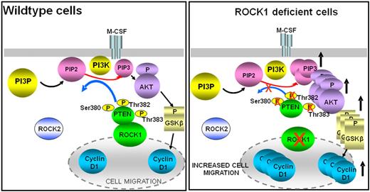
Our findings demonstrate an unexpected role for ROCK1 in negatively regulating the recruitment and migration of macrophages and neutrophils (Figure 8). Our results show enhanced migration of these 2 cell types in response to multiple stimuli, including M-CSF, fibronectin, MCP-1, and fmlp. The defects we observe prevail in BMMs, peritoneal cavity-derived macrophages, and PMNs. Importantly, the observed defects are not the result of changes in the maturation, expression of β1 integrins, or changes in the expression of ROCK2 in ROCK1−/− BMMs. At the biochemical level, our results show a critical and unique role for ROCK1 in regulating the intracellular levels of PIP3/AKT in response to receptor activation by regulating the phosphorylation, degradation, stability, and activity of PTEN (Figure 8). Specifically, our results show that the serine/threonine kinase ROCK1 binds to PTEN in response to M-CSF and that this binding is enhanced when PTEN is phosphorylated. We also show that M-CSF-induced ROCK1 activity induces the phosphorylation of at least serine 380, threonine 382, and threonine 383 in PTEN′s carboxy tail (Figure 8). In the absence of ROCK1, no physical association between PTEN and ROCK1 is observed. As a consequence, carboxy-terminal serine 380, threonine 382, and threonine 383 show significant impairment in phosphorylation, which results in enhanced degradation, reduced stability, and reduced overall PTEN activity in these cells. Importantly, the phenotypic changes in ROCK1−/− BMMs occur despite the presence of ROCK2, suggesting that ROCK1 functions with specificity in regulating the phosphorylation, stability, and activity of PTEN and in repressing the recruitment of inflammatory cells during infection.
Proposed model of ROCK1-mediated PTEN regulation. ROCK1 plays a critical role in regulating the intracellular levels of PIP3/AKT in response to receptor activation by regulating the phosphorylation, degradation, stability, and activity of PTEN.
Proposed model of ROCK1-mediated PTEN regulation. ROCK1 plays a critical role in regulating the intracellular levels of PIP3/AKT in response to receptor activation by regulating the phosphorylation, degradation, stability, and activity of PTEN.
Level and localization of PIP3 within cells have been shown to be essential for regulating migration/chemotaxis and recruitment of both neutrophils and macrophages in response to activation of multiple distinct receptors.6,7 BMMs deficient in the expression of p85α subunit of class IA PI3K demonstrate significant reduction in migration on fibronectin in the presence and/or absence of M-CSF.34 Likewise, BMMs deficient in the expression of p110δ catalytic subunit of class IA PI3K also demonstrate reduced migration in response to M-CSF.54 Furthermore, BMMs lacking the expression of Src family kinases show reduced migration via β1 integrins resulting from impaired localized PIP3 activity.32 Importantly, neutrophils deficient in the expression of PTEN demonstrate enhanced AKT phosphorylation and F-actin polymerization, which is associated with enhanced chemoattractant-induced transwell migration and cell speed in vitro.43 In vivo, deficiency of PTEN in PMNs results in enhanced recruitment to the peritoneal cavity during acute peritonitis.43 Similar defects have been reported in PTEN-deficient B cells in response to SDF-1 as well as in PTEN-deficient fibroblasts in response to β1 integrins.55,56 Furthermore, enhanced F-actin polymerization has also been reported in PTEN-depleted Dictyostelium and Jurkat T cells.57,58 Our findings in ROCK1-deficient macrophages and PMNs are consistent with those observed in PTEN-deficient neutrophils, fibroblasts, and B cells. We show that deficiency of ROCK1 in macrophages results in elevated PIP3/AKT levels as a result of impaired PTEN activation, which results in enhanced migration of these cells, increased F-actin content, and increased activation of AKT, GSK-3β, and cyclin D1, all downstream targets of PTEN (Figure 8). Consistent with our observations in ROCK1−/− BMMs, fibroblasts as well as macrophages deficient in the expression of either GSK-3β or cyclin D1 also show enhanced and reduced migration, respectively.59,60 Taken together, our findings using primary macrophages define a physiologically relevant biochemical pathway involving ROCK1 and PTEN, which appears to be important for suppressing the recruitment of inflammatory cells during acute inflammation.
Our findings in BMMs suggest that M-CSF is responsible for inducing the phosphorylation of PTEN on several serine and threonine residues in vivo. We further show that phosphorylation of PTEN is dependent on ROCK1 and is probably mediated by direct physical association between ROCK1 and PTEN as little to no physical association occurs between PTEN and ROCK1 in the absence of M-CSF stimulation. Remarkably, despite significant sequence similarity between ROCK1 and ROCK2, ROCK2 is unable to compensate for the loss of ROCK1 in these cells. Our findings in BMMs with respect to ROCK1 and PTEN interaction are in agreement with those of Li et al using an in vitro overexpression system.14 Li et al14 demonstrated a critical role for ROCK in regulating PTEN localization using a pharmacologic inhibitor of ROCK. They found that PTEN colocalizes with active RhoA at the posterior of the cell. Our results provide further evidence to suggest that, in primary macrophages, ROCK1 is the principal regulator of PTEN phosphorylation on serine and threonine residues in response to M-CSF and plays an indispensable role in regulating the stability and activity of PTEN. Importantly, deregulation of this pathway in ROCK1-deficient cells results in enhanced recruitment of macrophages in vivo.
Our findings demonstrating enhanced degradation and reduced stability of PTEN resulting from reduced phosphorylation of PTEN on serine and threonine residues in ROCK1-deficient macrophages in response to M-CSF stimulation are consistent with previous reports implicating these 3 residues in regulating PTEN stability and degradation.44 Using serine or threonine to alanine mutants of PTEN and overexpression systems, Vazquez et al showed that point mutations in either serine or threonine residues rendered PTEN less stable.44 Our data in primary macrophages show that enhanced degradation and reduced half-life of PTEN in ROCK1-deficient cells result in reduced overall PTEN activity. Although the precise mechanism by which the PTEN protein in ROCK1-deficient cells is degraded is unclear, PTEN tail contains 2 putative PEST sequences, which have been implicated in targeting proteins for proteolytic degradation. One of the putative PEST sequence includes the 3 residues that show defective phosphorylation of PTEN in ROCK1-deficient cells (ie, Ser380, Thr382, and Thr383), raising the possibility that PTEN might be targeted for proteosomal or caspase-3 mediated degradation in ROCK1-deficient macrophages in both starved and stimulated conditions.
Our in vitro and in vivo findings using mice completely deficient in the expression of ROCK1 are in contrast to the in vivo findings described using ROCK1 heterozygous mice. Noma et al using ROCK1 haplo-insufficient mice (ie, ROCK1+/−) demonstrated reduced recruitment of leukocytes in the areas of vascular injury.61 Several possibilities may contribute to these differences: (1) the types of signals generated in cells that are completely devoid of ROCK1 versus those consisting of 50% ROCK1 are probably distinct; (2) our results are obtained using ROCK1−/− mice generated in the FVB background,31 whereas Noma et al61 used ROCK1 heterozygous mice generated in the C57/BL6 background. Our mice are viable as adults with no obvious hematologic or other abnormalities, whereas homozygous deletion of ROCK1 in C57/BL6 strain results in embryonic and perinatal lethality.61 Therefore, it is conceivable that the genetic differences may contribute to differences in the recruitment of leukocytes; studies by Noma et al characterized macrophages using a monoclonal antibody MOMA-2.61 This antibody only recognizes a subset of macrophages.62 In the present study, macrophages were identified using the F4/80 antibody, which identifies all mature macrophages. Prior studies have shown that, on thioglycollate injection, one observes very few MOMA-2+ cells 4 to 8 hours after injection, respectively. In contrast, 17% and 73% F4/80 positive cells can be seen at this same time point.62 Furthermore, BMMs cultured in vitro for 7 days result in only 30% MOMA-2+ cells. In contrast, 90% F4/80-positive cells can be seen under identical culture conditions.62 Therefore, it is conceivable that only a subset of macrophages were being investigated in the Noma et al study.61
In conclusion, our studies identify a critical role for ROCK1 in negatively regulating macrophage and neutrophil function by binding to PTEN and regulating its activation. We think that, as a physiologic regulator of PTEN, ROCK1 should be a promising therapeutic target for modulating neutrophil and macrophage functions in various infectious and inflammatory diseases. In addition, given the fact that approximately 20% of known tumor-associated PTEN mutations are confined to the carboxy tail of PTEN, which result in rapid degradation of PTEN, our results might provide novel mechanistic insights into the function of PTEN as a tumor suppressor. Because the level of PTEN expression plays a profound role in influencing tumor susceptibility, a better understanding of the pathways involved in regulating PTEN levels is probably of therapeutic benefit.
The online version of this article contains a data supplement.
The publication costs of this article were defrayed in part by page charge payment. Therefore, and solely to indicate this fact, this article is hereby marked “advertisement” in accordance with 18 USC section 1734.
Acknowledgments
The authors thank Marilyn Wales for her administrative support.
This work was supported by the National Institutes of Health (grants HL077177 and HL07816, R.K.; grant HL085098, L.W.) and the Showalter Trust Award (89829, J.S.).
National Institutes of Health
Authorship
Contribution: S.V. designed and performed research and wrote the manuscript; J.S. performed research and provided reagent; P.H. performed research; L.W. provided reagent; and R.K. designed research and wrote the manuscript.
Conflict-of-interest disclosure: The authors declare no competing financial interests.
Correspondence: Reuben Kapur, Herman B Wells Center for Pediatric Research, Indiana University School of Medicine, Cancer Research Institute, 1044 W. Walnut St, Rm 408, Indianapolis, IN 46202; e-mail: rkapur@iupui.edu; or Sasidhar Vemula, Herman B Wells Center for Pediatric Research, Indiana University School of Medicine, Cancer Research Institute, 1044 W. Walnut St, Rm 408, Indianapolis, IN 46202; e-mail: svemula@iupui.edu.


![Figure 5. Reduced phosphorylation of MYPT1 in ROCK1−/− macrophages. (A) WT BMMs were starved overnight and stimulated with 100 ng/mL M-CSF for 5 minutes. Equal amounts of lysates were subjected to immunoprecipitation with an anti-ROCK1 antibody. Rho kinase activity was initiated by adding MYPT1 as a substrate for Western blot analysis using an anti-phospho MYPT1 antibody. Lanes 1 and 2 consist of lysates derived from unstimulated and stimulated WT BMMs, respectively; lanes 3 and 4 consist of lysates derived from unstimulated and stimulated ROCK1−/− BMMs, respectively. The top arrow indicates phospho MYPT1 levels in WT and ROCK1−/− BMMs on stimulation with M-CSF; the bottom arrow indicates β-actin levels in WT and ROCK1−/− BMMs in unstimulated and stimulated conditions. The bar graph on top indicates the density of each band as analyzed by NIH Image software. The bar graph is presented as intensity of pMYPT1 in arbitrary units in WT and ROCK1−/− BMMs (n = 3). (B) Enhanced PIP3 levels in ROCK1−/− BMMs. WT and ROCK1−/− BMMs were starved in serum-free medium for 24 hours and then labeled with [32P] orthophosphate (0.5 mCi/mL) for 2 hours. Cells were then stimulated with M-CSF (100 ng/mL) for 5 minutes before harvesting. Phospholipids were extracted and analyzed on a thin layer chromatography plate. Arrow indicates radiolabeled PIP3 in WT and ROCK1−/− BMMs. Lanes 1 and 2 indicate WT and ROCK1−/− BMMs, respectively, stimulated with M-CSF. Lanes 3 and 4 indicate WT and ROCK1−/− BMMs, respectively, unstimulated with M-CSF. The bar graph on top shows quantification of PIP3 levels in WT and ROCK1−/− BMMs. Amount of radioactive labeled 32P incorporated in PIP3 was quantified with ImageJ software, and PIP3 levels are shown as arbitrary units.](https://ash.silverchair-cdn.com/ash/content_public/journal/blood/115/9/10.1182_blood-2009-08-237222/4/m_zh89990948520005.jpeg?Expires=1769867778&Signature=cNbAt98AM2O7DsbC4iMQXLQSVB2uLT84-5Wvdg0PuHnUMQrFlGBpWV54L-xxk16yHRt2Jf814cL4InvPiNZ3yOsAMNNKk9joMkNXdzsD5IYoR4KFpdqshsqdQl~WF1oPxNya-okhFEuPaW-LLlkBelE-SCpaq3Ou~iqQghr78ewJCb-~9nyEnlGliIYiTDSsMIrOndUUtTUbc7~Af~aY~jlJdY2mZrSIFPdJ-g2Nf---u-Wm7SwtRiiShwdnZsyeMEr1Q00D4b~u49SlgDNT1hvHKgTKESONWl7co0R9aIPXBY6qcdBwtkg4~5cUUkvCgE0gVejG2yu34mcW~54h8w__&Key-Pair-Id=APKAIE5G5CRDK6RD3PGA)
![Figure 6. Increased PTEN cleavage, reduced phosphorylation, stability, and activity of PTEN in ROCK1−/− BMMs. (A) Equal amounts of lysates from WT and ROCK1−/− BMMs were subjected to Western blot analysis using an anti-PTEN antibody. Lanes 1 and 2 consist of lysates derived from unstimulated and stimulated WT BMMs, respectively; lanes 3 and 4 consist of lysates derived from unstimulated and stimulated ROCK1−/− BMMs, respectively. Top arrow represents total PTEN protein, whereas the middle arrow and stars indicate cleaved PTEN products in WT and ROCK1−/− BMMs (n = 3). The same blot was reprobed for β-actin to show protein loading in each lane (bottom panel). (B) Equal amounts of lysates were subjected to Western blot analysis using an anti-phospho PTEN antibody. Lanes 1 and 2 consist of lysates derived from unstimulated and stimulated WT BMMs, respectively; lanes 3 and 4 consist of lysates derived from unstimulated and stimulated ROCK1−/− BMMs, respectively. The top arrow (on left) indicates the level of phospho PTEN levels in WT and ROCK1−/− BMMs; bottom arrow indicates total β-actin levels in each lane. Western blot shown is representative of at least 3 independent experiments. The bar graph indicates the band intensity of phospho PTEN (n = 4). (C) PTEN activity assay. WT and ROCK1−/− BMMs cultured for 10 days in the presence of M-CSF were starved overnight and stimulated with 100 ng/mL M-CSF for 5 minutes. Equal amount of lysates were subjected to PTEN activity assay using the malachite green assay kit. PTEN activity is expressed as the amount of phosphate released. Data shown are the mean value of phosphate release in 3 independent experiments (n = 3, *P < .05). (D) Pulse-chase analysis of PTEN stability. WT and ROCK1−/− BMMs were metabolically labeled with [35S]-methionine. Medium was replaced with methionine-containing chase medium at time zero and chased at the indicated time points. PTEN was immunoprecipitated from labeled cell extracts, separated by gel electrophoresis, and detected by autoradiography. Lanes 1 to 3 represent PTEN expression in WT BMMs chased at different time points; lanes 4 to 6 indicate expression of PTEN in ROCK1−/− BMMs chased for the same amount of time as WT cells (n = 2).](https://ash.silverchair-cdn.com/ash/content_public/journal/blood/115/9/10.1182_blood-2009-08-237222/4/m_zh89990948520006.jpeg?Expires=1769867778&Signature=nzZlL5ysErb~D4l5Xu7uA3csn6cb0kG6Mh-EtLn-qDEXm2a7UvBrfEz5WdinlUM8io1kGd0WjY8sc1mQCHlSIkF92bqoSfnJTAfa1DQwCvxIBTqbQZAbF3bwEvBCgOrztLFnawUSxQTDvuj9hy~qMA3iSsudA6ckHjt~QgzqEvVeSvwyjYZnDVvy0YXBynUxTekBdAWwHraF2ucQlQB8bGzcRay7Ct-bom~viXhy3Jepp4v1yVNHnulrU7N~gbOUo5G0EflMCDVUalz1aIbsC4vKPYJTiX1P1Xh22C~2LMo3MQG40IPyrdLPgnk5gv~yASkdvMBRaExglSt~Gji~fA__&Key-Pair-Id=APKAIE5G5CRDK6RD3PGA)


This feature is available to Subscribers Only
Sign In or Create an Account Close Modal