Abstract
T cells contribute to host-tumor interactions in patients with monoclonal gammopathies. Expansions of CD8+CD57+ T-cell receptor Vβ–positive (TCRVβ+)–restricted cytotoxic T-cell (CTL) clones are found in 48% of patients with multiple myeloma and confer a favorable prognosis. We now report that CTL clones with varying TCRVβ repertoire are present in 70% of patients with Waldenström macroglobulinemia (WM; n = 20). Previous nucleoside analog (NA) therapy, associated with increased incidence of transformation to aggressive lymphoma, significantly influenced the presence of TCRVβ expansions (χ2 = 11.6; P < .001), as 83% of patients without (n = 6) and only 7% with (n = 14) TCRVβ expansions had received NA. Clonality of CD3+CD8+CD57+TCRVβ+-restricted CTLs was confirmed by TCRVβ CDR3 size analysis and direct sequencing. The differential expression of CD3+CD8+CD57+TCRVβ+ cells was profiled using DNA microarrays and validated at mRNA and protein level. By gene set enrichment analysis, CTL clones expressed not only genes from cytotoxic pathways (GZMB, PRF1, FGFBP2) but also genes that suppress apoptosis, inhibit proliferation, arrest cell-cycle G1/S transition, and activate T cells (RAS, CSK, and TOB pathways). Proliferation tracking after stimulation confirmed their anergic state. Our studies demonstrate the incidence, NA sensitivity, and nature of clonal CTLs in WM and highlight mechanisms that cause anergy in these cells.
Introduction
T-cell expansions unassociated with viral infections have been reported to be present in the peripheral blood of patients with a variety of non–T-cell hematologic malignancies including chronic myeloid leukemia (CML),1-3 myelodysplastic syndromes4,5 and monoclonal gammopathies.6-15 We previously reported that 48% of 120 patients with multiple myeloma have peripheral blood T-cell expansions that show the phenotype of CD3+CD8+ T-cell receptor Vβ–positive (TCRVβ+) CD57+, and we confirmed the clonal nature of these expanded T cells by determining TCRVβ CDR3 size distribution and direct sequencing.10,11,16 Although the specificity of these T-cell clones has not yet been determined, several patient cohorts have shown that they persist for long periods and are associated with a good prognosis, suggesting that they may arise after chronic stimulation by a tumor-associated antigen.5,9,11 Thus, these cells are likely to be tumor-specific cytotoxic lymphocytes with tumor-induced dysfunction. In contrast to patients with multiple myeloma, aged-matched control samples rarely have T-cell expansions and if present are almost exclusively CD4+ T cells.6-9
Waldenström macroglobulinemia (WM) is a B-cell malignancy with an increased serum monoclonal immunoglobulin M (IgM). Whether patients with WM also develop clonal T expansions has not been reported. Unlike multiple myeloma, many WM patients receive nucleoside analog therapy that causes lymphopenia and thus may inhibit the proliferation of clonal T cells and alter host-tumor interactions. As nucleoside analog therapy in WM results in a significantly increased incidence of transformation to a more aggressive lymphoma, there needs to be consideration of balancing the benefits with the risks associated with nucleoside analogs in these patients.17,18
In this study, we have searched for T-cell clones in the blood of patients with WM and related their presence with nucleoside analog therapy and transformation. We have also sorted CD3+CD4−TCRVβ+CD57+ from CD3+CD4−TCRVβ+CD57− cells and confirmed clonality by both TCRVβ CDR3 size determination and sequencing and have used carboxy-fluorescein diacetate succinmidyl ester (CFSE) tracking to monitor the proliferation of CD3+CD4−TCRVβ+ clones. Finally, we have performed microarray analysis to search for significantly differentially expressed genes and pathways in the clonally expanded CD3+CD8+TCRVβ+CD57+ cells, not only to confirm the cytotoxic pathways but also to identify other changes in these cells including the mechanisms that led to anergy. Expression of a selection of the most differentially expressed genes detected by microarray was validated at both mRNA and protein level.
Methods
Patient and control samples
Details of the 20 patients with WM included in this study are outlined in Table 1. Peripheral blood (ethylenediaminetetraacetic acid) was obtained from 42 age-matched controls and 20 patients with WM attending the clinic at Royal Prince Alfred Hospital. Six patients had previously received nucleoside analog therapy. Written informed consent was obtained from each patient and approval was given by the institutional ethics review committee at Royal Prince Alfred Hospital.
Details of patients with WM in this study
| Patient . | Sex/age, y . | Paraprotein isotype . | IgM, g/L . | TCRVβ clone expanded . | TCRVβ as % of CD3 cells . | Previous nucleoside analogue therapy . | Transformed to NHL . |
|---|---|---|---|---|---|---|---|
| 1 | F/77 | IgM L | 14.6 | 22 | 9.2 | N | N |
| 2 | M/85 | IgM K | 37.1 | 17 | 11.7 | N | N |
| 3 | F/89 | IgM K | 20.1 | 2 | 17.8 | Y | N |
| 3 | 14.1 | ||||||
| 5.1 | 9.3 | ||||||
| 4 | F/88 | IgM K | 22.5 | 20 | 21 | N | N |
| 5 | F/82 | IgM K | 32.1 | 21.3 | 8 | N | N |
| 6 | M/83 | IgM L | 17.4 | 3 | 9.7 | N | N |
| 13.6 | 6.3 | ||||||
| 23 | 4.4 | ||||||
| 7 | M/76 | IgM K | 16.8 | 3 | 27.4 | N | N |
| 8 | M/86 | IgM K | 21.4 | 5.1 | 9.9 | N | N |
| 9 | M/86 | IgM L | 13.8 | 22 | 4.2 | N | N |
| 10 | M/76 | IgM K | 20.9 | 17 | 44 | N | N |
| 11 | F/79 | IgM L | 24.4 | 5.1 | 8.0 | N | N |
| 23 | 4.0 | ||||||
| 12 | F/71 | IgM K | 42.2 | 9 | 18.9 | N | N |
| 13.1 | 20.4 | ||||||
| 13 | M/58 | IgM K | 22.9 | 7 | 5.1 | N | N |
| 14 | M/75 | IgM K | 40.8 | 5.1 | 8.5 | N | N |
| 15 | M/86 | IgM L | 2.7 | None | NA | Y | Y |
| 16 | M/68 | IgM K | 2.0 | None | NA | Y | N |
| 17 | M/71 | IgM K | 27.0 | None | NA | Y | N |
| 18 | F/64 | IgM K | 0.3 | None | NA | Y | N |
| 19 | M/75 | IgM L | 26.2 | None | NA | Y | N |
| 20 | M/69 | IgM L | 11.6 | None | NA | N | N |
WM indicates Waldenström macroglobulinemia; TCRVβ, T-cell receptor Vβ; NHL, non-Hodgkin lymphoma; None, no TCRVβ clone; NA, not applicable; Y, yes; and N, no.
Flow cytometry and cell sorting
TCRVβ repertoire analysis and the procedure for isolation of CD3+CD4−TCRVβ+CD57+ and CD3+CD4−TCRVβ+CD57− cells have been previously described.8 Briefly, peripheral blood mononuclear cells (PBMCs) were isolated by Ficoll gradient centrifugation and assayed to determine TCRVβ repertoire using an IOTest Beta Mark TCRVβ Repertoire Kit (Beckman Coulter). Residual PBMCs were frozen and stored in liquid nitrogen for additional analysis and sorting. After thawing, the cells were incubated on ice for 1 hour with a cocktail of anti-CD3–allophycocyanin(APC) anti-CD4–phycoerythrin (PE)–cyanin 5 (cy5), anti-CD57–fluorescein isothiocyanate (FITC) or –PE (all from BD Biosciences), and a specific anti–TCRVβ-PE for Vβ1, 2, 5.1, 5.2, 5.3, 7.2, 8, 9, 11, 12, 14, 17, 18, 20, 22, 23 or anti–TCRVβ-FITC for Vβ3, 13.1, 13.6, 16, 21.3 (Beckman Coulter). These stained cells, after washing with phosphate-buffered saline (PBS) were analyzed by FACSAria II flow cytometry (BD Biosciences) and sorted to CD3+CD4−TCRVβ+CD57+ and CD3+CD4−TCRVβ+CD57− cells. Cells were reanalyzed after sorting to check purities, which were typically more than 95%. The sorted T cells were immediately extracted for total RNA.
RNA sample processing
Total RNA was extracted using an RNeasy Micro Kit (QIAGEN) from freshly purified CD3+CD4−TCRVβ+CD57+ and CD3+CD4−TCRVβ+CD57− T cells separately. To obtain a sufficient amount of RNA for microarray hybridization, 2 rounds of RNA amplification were conducted and then labeled using the MessageAmp II aRNA Kit and the MessageAmp II Biotin Enhanced Kit (Ambion). After fragmenting, the amplified RNA (aRNA) samples were stored at −80°C until hybridization for microarray analysis. The qualities of total RNA, aRNA amplification, and aRNA fragmentation were checked by RNA6000 Pico LabChip kit and 2100 Bioanalyzer (Agilent Technologies). The quantities were determined by NanoDrop ND-1000 spectrophotometer (Thermo Scientific).
Analysis of clonality
The clonality of the T-cell expansions was verified by analysis of the size of the CDR3 region of the TCRVβ chain and by direct sequencing.19 In brief, total RNA of CD3+CD4−TCRVβ+CD57+ and CD3+CD4−TCRVβ+CD57− T cells was reverse transcribed to cDNA using a SuperScript VILO cDNA Synthesis Kit (Invitrogen) according to the manufacturer's instructions. Aliquots of cDNA were amplified using a TCRVβ-specific primer (Vβ20, 5′-AGCTCTGAGGTGCCCCAGAATCTC-3′; Vβ21.3, 5′-GAGTGTGGCTTTTTGGTGCAA-3′; Vβ23, 5′-GACAGCTGATCAAAGAAAAGA-3′, and a fluorescent-Cβ primer (5′-TTCTGATGGCTCAAACAC-3′, labeled at 5′ end with 6-FAM; Sigma-Aldrich).20,21 Polymerase chain reaction (PCR) mixture (25 μL) contained the following components at the final concentrations: 500nM each primer with 2 μL of cDNA template, 0.2mM deoxynucleotide triphosphate, 1× PCR buffer, 1.5mM MgCl2, and 0.625 U of Ampli Taq Gold DNA polymerase (Promega). The amplification was performed on a DNA Engine thermal cycler PTC-200 (MJ Research). After 10-minute denaturation at 95°C, 40 cycles of 94°C for 30 seconds, annealing at 55°C for 30 seconds, and extension at 72°C for 30 seconds, with a single final extension at 72°C for 10 minutes, 6-FAM–labeled PCR products were analyzed by SUPAMAC (University of Sydney). Briefly, 2.0-μL aliquots of FAM-labeled PCR products were mixed with 8.0 μL of Hi-Di Formamide (Applied Biosystems) and 0.1 μL of GeneScan 500 LIZ Size Standard (Applied Biosystems) before loading onto a 3730xl Capillary DNA Analyzer (Applied Biosystems). The data were analyzed with GeneMarker software (SoftGenetics LLC). Samples with 1 reproducible dominant peak were regarded as clonal. DNA sequencing of PCR products was used to confirm clonality. In brief, unlabeled PCR products were prepared using Vβ-specific primers and unlabeled Cβ primer in the same PCR conditions, then purified from 1.5% agarose gel by the QIAquick Gel Extraction Kit (QIAGEN). The purified products were directly sequenced on a 3730xl Capillary DNA Analyzer (Applied Biosystems). The data were analyzed by Sequence Scanner Software Version 1.0 (Applied Biosystems), and then aligned with the T-cell receptor sequences based on IMGT/JunctionAnalysis V-D-J region (http://www.imgt.cines.fr/IMGT_vquest/vquest?livret=0&Option=humanTcR, The International Immunogenetics Information System), and the CDR3 region was identified.19,22
Affymetrix microarray and data analysis
Fragmented biotin-RNA samples (20 μg) were processed at the Ramaciotti Center (University of New South Wales) on Affymetrix GeneChip Human Genome U133 plus 2.0 Arrays, following the manufacturer's protocol. CEL files were normalized using RMA, and present/marginal/absent calls were generated using the affy library in R/Bioconductor.23-25 We deemed a probe set to be detected if it had at least 1 present call. Differential expression because of CD57 positivity or negativity for the noncontrol probe set (determined in at least 1 sample) was assessed. We performed gene set enrichment analysis (GSEA) with GSEA software (MIT) to assess functional significance at the level of sets of genes.26 We ranked these gene symbols by decreasing moderated t statistic, and created a preranked list, and used this as input for running GSEA in preranked mode with 1000 permutations. We compared the moderated t statistic to the “c2_all” collection of curated gene sets from the Molecular Signatures Database MsigDB 1.0 (http://www.broad.mit.edu/gsea/msigdb/index.jsp, May 4, 2009), consisting of 1892 gene sets corresponding to biologic pathways and published microarrays studies. The minimum size of tested gene sets was 15 genes and the maximum was 500. Gene sets were compared using the normalized enrichment score (NES) and deemed significant at 0.05 with a false discovery rate (FDR) less than 0.25. Heat maps were generated using the TIGR multiexperiment viewer (MeV; http://www.tm4.org/mev.html).27 All analyses were performed using R 2.8.0 and Bioconductor 2.3. The entire dataset for all microarray experiments has been deposited with Gene Expression Omnibus under accession number GSE18313.28
Validation of differential expression using real-time quantitative RT-PCR
To confirm the differentially expressed genes identified by microarray analysis, real-time PCR was performed for 6 selected genes with significant biologic function that were in the top or bottom of a ranked gene list. Total RNA of samples was used for quantification. Briefly, after reverse transcription, each sample of cDNA was subjected to real-time PCR with the ABI 7500 (Applied Biosystems). PCR mixture (20 μL) contained 1× EXPRESS SYBR GreenER Universal SuperMix Universal with 50nM ROX reference dye (Invitrogen), 200nM each primer, and 1 μL of cDNA template. Thermal cycling conditions were 50°C for 2 minutes, 95°C for 2 minutes followed by 40 cycles of 95°C for 15 seconds, and 60°C for 1 minute. Melting curves recorded at the end of reaction were used to verify the specificity of the PCRs. Primers of these selected genes and reference genes are shown in format of 5′-3′ as follows:29-32 GZMH, CCATTCCTCCTCCTGTTGG and ACTAGGATGCCGCCACAC; GZMB, GGTGGCTTCCTGATACAAGACG and GGTCGGCTCCTGTTCTTTGAT; EBI2, GAATCGGAGATGCCTTGTGT and GCCTCCTGCTTTGACATAGG; CD28, TTTCCCGGACCTTCTAAGC and CGGGGAGTCATGTTCATGTAG; PRF1, GTGGAGTGCCGCTTCTACAGTT and TGCCGTAGTTGGAGATAAGCCT; FGFBP2, CTTCCGAGGGTGACAGGTGA and TCCAGTGTGAGAACGTTGGATTG; GAPDH, TGCCAAATATGATGACATCAAGAA and GGAGTGGGTGTCGCTGTTG; and β-ACTIN, TTGTTACAGGAAGTCCCTTGCC and ATGCTATCACCTCCCCTGTGTG. Quantification of the results was performed on the relative standard curve method. Relative expression levels of target genes were calculated from the results of at least triplicate samples, normalized to the average of 2 reference genes, GAPDH and β-ACTIN.
Validation at protein level
For immunophenotyping of TCRVβ+ expanded cells, expression was determined on the FACSAria II flow cytometer (BD Biosciences) using anti–CD3-PE-cy7, anti–CD8-APC or anti–CD4-peridinin-chlorophyll-protein complex (PerCP)–cy5.5, specific anti–TCRVβ-PE or -FITC, anti–CD57-PE or -FITC, with either anti–CD28-PerCP-cy5.5 or anti–granzyme-B–APC (all from BD Biosciences). Intracellular perforin expression was stained using anti–CD3-APC, anti–CD4-PerCP-cy5.5, specific anti–TCRVβ-PE, CD57 biotin, and streptavidin-PE-cy7 (eBioscience) and then anti–perforin-FITC after fixation and permeabilization with BD cytofix/cytoperm and BD GolgiPlug (BD Biosciences). 4,6-Diamidino-2-phenylindole–negative gating was used to ensure analysis of viable cells.
In vitro proliferation assays
PBMCs (5 × 10⁵ per well) from patients were labeled with 2 μL of 5mM CFSE using CellTrace CFSE Cell Proliferation Kit (Invitrogen) and stimulated with MACSiBead particles (Miltenyi Biotec) at a bead-to-cell ratio of 1:10 and 1:20 for 4 days at 37°C in a humidified CO2 incubator. Cells were stained with anti–CD3-PE-cy7, anti–CD8-APC (BD Biosciences), and specific anti–TCRVβ-PE antibodies (Beckman Coulter). Analysis involved a determination of percentage of cells that had progressed through at least 1 cell cycle as determined by CFSE staining on CD3+CD8+TCRVβ+ and CD3+CD8+TCRVβ− T cells.
Statistics
GraphPad Prism software (GraphPad) was used for statistical analysis of data other than microarray. The association between NA therapy and the presence of TCRVβ clones was examined using the χ2 test, and linear correlations were calculated using standard Pearson correlation coefficient. P values were 2 tailed and less than .05 was deemed statistically significant. Microarray data were analyzed to determine different expression of individual genes using a paired moderated t test implemented in limma followed by multiple testing corrections using FDR, which assigns each probe set a q value.33-35 The significance level of individual genes for FDR values was set at 0.05.
Results
Incidence of TCRVβ expansions in WM and clinical correlations
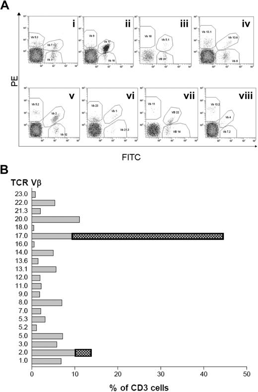
We analyzed the TCRVβ repertoire of 20 patients with WM and 42 age-matched controls using monoclonal antibodies to 24 TCRVβ families. A representative sample (patient no. 10) is illustrated with flow cytometric scatterplots in Figure 1A, and this patient's TCRVβ profile is compared with the mean of 42 age-matched controls in Figure 1B. The size of the TCRVβ expansions for all patients is reported in Table 1. A TCRVβ expansion was considered to be present when the percentage of cells in a particular TCRVβ family was more than mean plus 3 SDs of the healthy age-matched control. There was evidence of at least one such TCRVβ expansion in 70% (14 of 20) of the patients. We have previously reported an incidence of 48% in patients with multiple myeloma (n = 120) before autologous transplantation using the same criteria.16 Five of the 6 patients without TCRVβ expansions had previously received nucleoside analog therapy, and 1 later transformed to non-Hodgkin lymphoma, whereas only 1 of the 14 patients with significant T-cell expansions had received nucleoside analog therapy (Table 1). Thus, previous nucleoside analog therapy had a significant influence on the presence of TCRVβ expansions (χ2 = 11.6; P < .001). Table 1 shows that a broad spread of different TCRVβ families was used and that the expansions were up to 44% of the total T cells. There was no correlation between the IgM concentration and the number of expanded T cells.
TCRVβ repertoire analysis by flow cytometry. (A) Expression of TCRVβ in PBMCs of a representative WM patient (patient no. 10) after cells were stained with a panel of TCRVβ antibodies from the Beta Mark kit and anti–CD3-ECD. (B) Percentage of CD3+ T cells expressing the TCRVβ of patient no. 10 compared with the mean ± 3 SD of the age-matched normal range.
TCRVβ repertoire analysis by flow cytometry. (A) Expression of TCRVβ in PBMCs of a representative WM patient (patient no. 10) after cells were stained with a panel of TCRVβ antibodies from the Beta Mark kit and anti–CD3-ECD. (B) Percentage of CD3+ T cells expressing the TCRVβ of patient no. 10 compared with the mean ± 3 SD of the age-matched normal range.
CD3+CD4−TCRVβ+CD57+ of expanded cytotoxic T cells are monoclonal
CD3+CD4−TCRVβ+CD57+ and CD3+CD4−TCRVβ+CD57− T cells were sorted from WM PBMCs by fluorescence-activated cell sorting (FACS) as shown in Figure 2A. Clonality of these sorted cells could be readily identified by PCR using fluorescently labeled Cβ and TCRVβ-specific PCR primers, followed by measuring the length of those products by DNA fragment analysis. A single-peak profile suggests the sizes of CDR3 region of expanded T cells are identical, which normally results from a monoclonal expansion. In comparison, multipeaks of CDR3 profiles suggest polyclonal expansion. We tested 3 WM patients (patient nos. 4-6) who had significant T-cell expansions, and all samples showed a single peak in sorted CD3+CD4−TCRVβ+CD57+ T cells but not CD3+CD4−TCRVβ+CD57− T cells as shown in Figure 2B. To further confirm that the single-peak PCR products are monoclonal, we performed direct DNA sequencing of the PCR products. As expected, the sequences of PCR products from CD3+CD4−TCRVβ+CD57+ T cells could be determined clearly and also matched the expected TCRVβ subfamily sequence and length (Table 2). In contrast, the direct sequencing of CD3+CD4−TCRVβ+CD57− T cells yielded a highly mixed sequence electrophoresis pattern, failing to obtain a clear sequence of the CDR3 region (Figure 2C). These results demonstrate that the CD3+CD4−TCRVβ+CD57+ T cells from WM patients are monoclonally expanded T cells, whereas the CD3+CD4−TCRVβ+CD57− T cells are polyclonal.
The nature of WM T-cell clonality from 1 representative WM patient (patient no. 4). (A) Flow cytometric cell sorting of CD3+CD4−TCRVβ+CD57+ and CD3+CD4−TCRVβ+CD57− cells from PBMCs of WM patient no. 4. (B) CDR3 length profile of CD3+CD4−TCRVβ+CD57+ and CD3+CD4−TCRVβ+CD57− cells using 6-FAM-Cβ and specific-Vβ20 primer for WM patient no. 4. (C) DNA sequence analysis in sorted cells from WM patient no. 4. The sequences are shown below the trace pattern.
The nature of WM T-cell clonality from 1 representative WM patient (patient no. 4). (A) Flow cytometric cell sorting of CD3+CD4−TCRVβ+CD57+ and CD3+CD4−TCRVβ+CD57− cells from PBMCs of WM patient no. 4. (B) CDR3 length profile of CD3+CD4−TCRVβ+CD57+ and CD3+CD4−TCRVβ+CD57− cells using 6-FAM-Cβ and specific-Vβ20 primer for WM patient no. 4. (C) DNA sequence analysis in sorted cells from WM patient no. 4. The sequences are shown below the trace pattern.
Nucleotide sequences of clonal TCRVβ from WM patients PBMCs (patient nos. 4-6)
| No. . | Chain and sequence . | ||||
|---|---|---|---|---|---|
| 3′ V . | N(D)N . | 5′ J . | V (D) J sequence . | CDR3 length, aa . | |
| 4 | TGTGCCTGGAG | GGGGACAGGGTTTTTG | TATGGCTACACCTTC | Vβ20 Dβ1Jβ1.2 | 12 |
| 5 | TGTGCCAGCAGCTTAG | TCCCCGACGGACCGGGACTAGCGGGAGGGCCCAT | CTCCTACAATGAGCAGTTCTTC | Vβ21.3Dβ2Jβ2.1 | 22 |
| 6 | TGTGCCAGCAGCTTA | CCGGGGAACGTTGG | CGAGCAGTACTTC | Vβ23Dβ2Jβ2.7 | 12 |
TCRVβ indicates T-cell receptor Vβ; WM, Waldenstrom macroglobulinemia; PBMCs, peripheral blood mononuclear cells; and aa, amino acid.
Identification of genes involved in T-cell expansion
We used Affymetrix Human Genome 133 plus 2.0 arrays to profile expression of 54 675 probe sets in both CD3+CD4−TCRVβ+CD57+ and CD3+CD4−TCRVβ+CD57− subpopulations from 3 WM patients (patient nos. 4-6). On each array, 14% to 28% of probe sets were detected (supplemental Figure 1A, available on the Blood Web site; see the Supplemental Materials link at the top of the online article). We excluded probe sets that were not detected on any array, which removed approximately 60% of the probe sets (supplemental Figure 1B). Of the remaining 21 971 (40%) noncontrol probe sets, we assessed differential expression due to CD57+ versus CD57− using a paired moderated t test, and determined the most differentially expressed probe set for each gene. We identified 958 genes with at least a 2-fold change (670 up-regulated and 288 down-regulated in CD57+ relative to CD57− T cells). The magnitude of statistical significance of each gene was plotted relative to its log fold change as a volcano plot (supplemental Figure 1C), in which, in general, those genes with the largest statistical significance also have a large fold change. When we reassessed the microarray data with multiple testing corrections (FDR), no single gene had an FDR less than 0.05 in expression between the CD57+ and CD57− cells. We then chose to apply no multiple testing corrections, but to rank the genes by their P values.
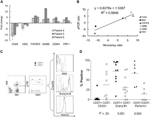
We used real-time PCR to validate the significance of the genes with the greatest differential expression in the microarray results. Three patient samples (patients no. 1-3) with significant TCRVβ expansions that were not used in the microarray analysis were used for this validation. Six genes (GZMH, GZMB, EBI2, CD28, PRF1, and FGFBP2) were selected on the basis of their P value, their significance to the pathways identified by microarray, and their potential biologic functions related to cytotoxic T cells. Real-time quantitative PCR (qPCR) was performed to determine the expression level of each gene normalized to a mean reference value, which was the average of GAPDH and β-ACTIN, and CD57+ relative to CD57− (Figure 3A). For each gene, all 3 replicates had the same direction of expression (CD28 and EBI2 down-regulated, and the other 4 genes up-regulated). Thus the expression levels of these genes by qPCR were highly associated with expected levels as determined by the microarray (Figure 3B; P < .006, R2 = 0.8846), which suggested that microarray-derived differential expression ratio, although compromised by high density, reflected true gene expression changes.
Validation of microarray by real-time PCR. (A) Expression regulation of tested genes across 3 patients (patient nos. 4-6). Relative mRNA levels corresponding to representative genes from CD3+CD4−TCRVβ+CD57+ and CD3+CD4−TCRVβ+CD57− cells were determined by quantitative real-time PCR. The expression level of each gene was normalized to a mean reference value, which was the average of GAPDH and β-ACTIN. Results are presented as mean ± SD of at least triplicates. (B) Dot plot of the expression values in fold change ratio between microarray and qPCR from all validated genes and patient samples. Linear regression demonstrates a significant correlation (R2 = 0.8846; P < .006). (C) Protein expression as demonstrated by flow cytometry assay of CD28, GZMB, and perforin in the CD57+ (clonal) and CD57− (polyclonal) TCRVβ3-restricted CD8 cells in a representative patient. (D) Significant up-regulated perforin and granzyme-B expression and down-regulated CD28 expression in the CD57+ subpopulation was present in all 11 TCRVβ expansions studied but results have been recorded only when more than 50 cells were able to be analyzed by flow cytometry in each subpopulation.
Validation of microarray by real-time PCR. (A) Expression regulation of tested genes across 3 patients (patient nos. 4-6). Relative mRNA levels corresponding to representative genes from CD3+CD4−TCRVβ+CD57+ and CD3+CD4−TCRVβ+CD57− cells were determined by quantitative real-time PCR. The expression level of each gene was normalized to a mean reference value, which was the average of GAPDH and β-ACTIN. Results are presented as mean ± SD of at least triplicates. (B) Dot plot of the expression values in fold change ratio between microarray and qPCR from all validated genes and patient samples. Linear regression demonstrates a significant correlation (R2 = 0.8846; P < .006). (C) Protein expression as demonstrated by flow cytometry assay of CD28, GZMB, and perforin in the CD57+ (clonal) and CD57− (polyclonal) TCRVβ3-restricted CD8 cells in a representative patient. (D) Significant up-regulated perforin and granzyme-B expression and down-regulated CD28 expression in the CD57+ subpopulation was present in all 11 TCRVβ expansions studied but results have been recorded only when more than 50 cells were able to be analyzed by flow cytometry in each subpopulation.
At the level of protein expression, we investigated the expression of CD28, granzyme-B, and perforin using flow cytometry on CD57+ versus CD57− cells in 11 different TCRVβ expansions. Representative flow plots of patient no. 6 who had a TCRVβ3 expansion are shown in Figure 3C. When at least 50 cells were able to be analyzed by flow cytometry for each subpopulation (Figure 3D), the results of all studies confirmed the microarray and qPCR results: that CD28 is down-regulated, and perforin and granzyme-B are up-regulated in CD57+ relative to CD57− T cells in the CD3+CD8+TCRVβ+-restricted population.
Signaling pathways involved in T-cell clone
We have previously shown that multiple myeloma patients with monoclonally expanded T cells have a significantly longer survival.7 However the mechanisms underlying this response remain unclear. In our previous studies, it was demonstrated that clonal T-cell expansions in patients with multiple myeloma can persist for many years.7,14 To investigate whether these findings are recapitulated in WM patients, we performed pathway-based analysis using GSEA.
Pathway-based analyses that detect changes in the expression of genes in entire pathways are far more robust than analyses based upon individual genes. Because some genes have many probe sets, we determined the best probe set for each gene based on the probe set with the largest absolute moderated t test. This reduced the set of all probe sets to 20 552 unique gene symbols. We arranged representative annotated genes into selected functional categories according to gene families derived from the molecular signature database (GSEA). Gene set size filters (minimum = 15, maximum = 500) resulted in filtering out 463 from 1892 gene sets, and the remaining 1429 gene sets were used for further GSEA analysis. In turn, we identified 331 up- and 118 down-regulated gene sets in CD57+ relative to CD57− T cells (FDR < 0.25). If we restricted our attention to the 529 gene sets relating only to pathways, we identified 55 up- and 20 down-regulated pathways.
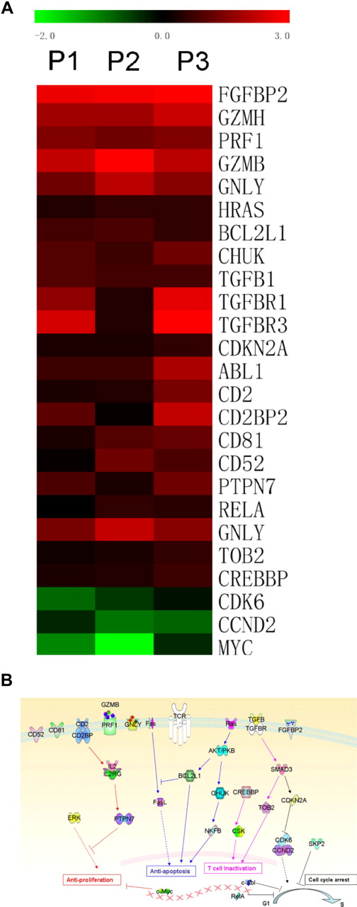
GSEA identified several gene sets and pathways not specifically related to cytotoxic T cells that were up-regulated in the CD3+CD4−TCRVβ+CD57+ samples (Figure 4A and supplemental figures). These included Ras, BCL2L1 (BCL-XL), and CHUK (IKK) in the RAS pathway; CREBBP and CSK in the CSK pathway; TGFB and SMAD in the TAB pathway; TGFBR, CDKN2A (p16), CCND2, and CDK6, which lead to cell cycle arrest; and PTPN7 (HePTP), which can inactivate ERK. We have summarized the contribution of these genes and the possible pathways involved in Figure 4B.
Schematic of signaling pathways and individual differentially expressed genes that contribute to CD3+CD4−TCRVβ+CD57+ T-cell behavior. (A) Heat map of differentially expressed genes shows significant difference in gene expression sets between CD3+CD4−TCRVβ+CD57+ and CD3+CD4−TCRVβ+CD57− samples. P1-3 indicates patient nos. 1 through 3. (B) Signaling pathways contribute to low proliferation index, cytotoxicity, and low rate of turnover of clonal T cells. Patient nos. 1 through 3 were used in this analysis.
Schematic of signaling pathways and individual differentially expressed genes that contribute to CD3+CD4−TCRVβ+CD57+ T-cell behavior. (A) Heat map of differentially expressed genes shows significant difference in gene expression sets between CD3+CD4−TCRVβ+CD57+ and CD3+CD4−TCRVβ+CD57− samples. P1-3 indicates patient nos. 1 through 3. (B) Signaling pathways contribute to low proliferation index, cytotoxicity, and low rate of turnover of clonal T cells. Patient nos. 1 through 3 were used in this analysis.
CD3+CD8+TCRVβ+ clonal T cells have reduced proliferative capacities
To investigate the proliferative capacity of the expanded T-cell clones in WM patients, we examined the proliferation of both CD3+CD8+TCRVβ+ expanded T cells (n = 9) and the remaining CD3+CD8+TCRVβ− cells after CFSE labeling and culture for 4 days stimulated with MACSiBead particles (anti-CD3+ anti-CD2+ anti-CD28). Representative CFSE histograms of CD3+CD8+TCRVβ23+ and TCRVβ23− cells stimulated at a bead to cell ratio of 1:10 are shown in Figure 5A. A significantly higher proportion of CD3+CD8+TCRVβ− cells (mean = 60%) than CD3+CD8+TCRVβ+ cells (mean = 18%) responded to stimulation by progressing through at least 1 cell division (t = 7.73; P < .001; Figure 5B). Although the number of proliferating cells in each of the CD3+CD8+TCRVβ+ populations was reduced, both CD57+ (clonal) and a varying number of CD57− (nonclonal) cells were present in this population. The lack of appropriate reagents prevented a simultaneous analysis of CFSE, TCRVβ+, and CD57 and thus it was not possible to perform CFSE tracking of only the clonal cells.
Proliferation of TCRVβ+ versus TCRVβ− cells. (A) Representative histogram plots of CFSE-labeled cells after 4 days of culture stimulated at a bead to cell ratio of 1:10, demonstrating the low number of TCRVβ+ cells that proliferated. (B) Scatterplot of percentage of cells that proliferated in TCRVβ+ versus TCRVβ− cells for 9 different expansions (t = 7.73; P < .001).
Proliferation of TCRVβ+ versus TCRVβ− cells. (A) Representative histogram plots of CFSE-labeled cells after 4 days of culture stimulated at a bead to cell ratio of 1:10, demonstrating the low number of TCRVβ+ cells that proliferated. (B) Scatterplot of percentage of cells that proliferated in TCRVβ+ versus TCRVβ− cells for 9 different expansions (t = 7.73; P < .001).
Discussion
This study has reported the intriguing observation that monoclonal expansions of cytotoxic T cells are present in 70% of patients with WM, an incidence that is higher than in patients with multiple myeloma (48%).16 As similar clonal expansions in patients with multiple myeloma6-15 and CML3 have prognostic significance, it is likely that these cells have an immunoregulatory function toward the tumor cells, although their specificity has yet to be confirmed. The presence of expanded cytotoxic lymphocytes in CML patients after dasatinib therapy was associated with excellent responses.3 CDR3 length analysis and sequencing has demonstrated in this and other studies that these CD3+CD4−TCRVβ+CD57+ expansions are monoclonal, whereas their CD57− counterparts are polyclonal.10 A significant observation was that expanded T-cell clones were rarely present in patients after nucleoside analog therapy and that the only patient to progress to an aggressive lymphoma was in this cohort. The mechanism of action of nucleoside analogs as cancer drugs is not fully understood, although they are known to have immunosuppressive effects and are not used in patients with multiple myeloma because of a lack of efficacy. Although nucleoside analog therapy is an effective therapy for patients with WM, the resulting lymphopenia may also remove cytotoxic T cells that provide an immunomodulating effect on the tumor, and their removal may be one factor in the increased risk of transformation to a more aggressive lymphoma. The problem of balancing the risk versus benefit of treating WM patients with nucleoside analog therapy has recently received attention after reports that have shown an increased incidence of transformation to non-Hodgkin lymphoma and the development of therapy-related myelodysplasia.17,18
We were particularly interested to assess differences in gene expression between CD57+ and CD57− T cells of the expanded TCRVβ family. To this end, we performed a microarray analysis. As expected, a significant number of genes related to cytotoxic T-cell function occupied the top positions of differentially up-regulated gene list on microarray, namely PRF1, GZMB, FGFBP2, and CD28 negativity. Quantitative PCR and flow cytometry validated the microarray results for mRNA and protein expression of a selection of significant genes. The CD3+CD4−TCRVβ+CD57+ clonal T cells are likely to be tumor-specific cytotoxic lymphocytes with tumor-induced dysfunction.
We also performed GSEA, which is much more robust than individual gene analysis to search for significant differentially expressed genes and related pathways in the clonally expanded CD3+CD4−TCRVβ+CD57+ cells, searching for functional changes that may be related to these cells. Within the pathways identified by GSEA, some are significantly up-regulated in CD3+CD4−TCRVβ+CD57+ samples, which may contribute to important changes in T-cell function (Figure 3A-B). Gene sets other than those involved with cytotoxicity include the RAS pathway, which consists of a variety of genes including Ras, BCL2L1 (BCL-XL), and CHUK (IKK), which could inhibit expanded T cells from apoptosis and allow long-term survival36-38 ; the CSK pathway may recruit CREBBP to directly activate CSK to inhibit T-cell activation;39,40 and the TOB pathway, in which TGFB could activate SMADs that interact with intracellular Tob to maintain unstimulated T cell.41,42 TGFB may also work with TGFBR, CDKN2A (p16) within a G1/S transition pathway to down-regulate expression of CCND2 and CDK6, which leads to cell cycle G1/S transition arrest of T cells.43-46 Up-regulation of PTPN7 (HePTP) would lead to suppression of T-cell proliferation by inactivating ERK.47,48 The up-regulated transforming growth factor-β (TGFβ) receptor in T-cell clones is interesting as a previous study by our group demonstrated that TGFβ produced by myeloma cells causes dendritic cells to function abnormally. TGFβ may be at least partly responsible for the dysfunction of clonal T cells, but the combined evidence from the microarray data suggests that multiple pathways involved with antiapoptosis, antiproliferation, and arrest of the cell cycle are activated in these clonal T cells. In contrast, Janus kinase/signal transducer and activator of transcription was enriched in the CD3+CD4−TCRVβ+CD57− phenotype. Some regulated individual genes may also contribute certain functions. For example, up-regulated SKP2, c-ABL, and RelA contribute to cell cycle arrest.49,50 Down-regulated c-Myc may lead to antiproliferation and cell cycle arrest.41 These signaling pathways all contribute to the clonal T cells developing anergy.
While investigating the array quality control, it became apparent that the microarrays had lower overall fluorescence than a typical array performed on whole-tissue samples. The microarray was designed to interrogate the entire genome, but because our samples were highly purified T cells, the majority of genes represented on the array were not expressed, and were thus dim. The CD57− sample from patient no. 6 had the lowest signal, and consequently had elevated standard error. However, we felt that this was because of the overwhelming contribution of essentially random signals derived from the 82% of probe sets that were not detected on this array. We then moved approximately 60% of the probe sets that were not detected on any array and assessed the differential expression of the remaining genes using strict criteria. However, only 14 genes have a P value less than .001, and supplemental Figure 1D indicates that the number of genes with a P value less than .05 is similar to the proportion expected by chance alone. Indeed, when we corrected for multiple testing, using the positive FDR, we found no genes with FDR less than 0.05. The minimum FDR was 0.747, and we found 5479 genes with FDR between 0.747 and 0.75, indicating that there are 1370 differentially expressed genes in this list, however there is also a high chance that each individual gene is a false positive. The lack of statistical significance in this initial investigation, despite nearly 1000 genes with fold change more than plus or minus 2, is most likely because the data came from 6 arrays that were prepared from 3 patients. The ultimate test for the reliability of a set of microarray results is in the ability to replicate these findings in a series of independent samples, which we were subsequently able to do when we validated the differential expression of a selection of the genes with the smallest P value by real-time PCR and protein assay.
We have thus searched for differentially expressed genes and signaling pathways in expanded T-cell clones to seek evidence of genes that may be involved with mechanisms other than those involved within T-cell cytotoxicity-related pathways. In particular, we have searched for genes that may be involved with mechanisms such as tumor-induced immunosuppression and functional changes in the activation and proliferation of cytotoxic T cells. Blocking the dysregulation of genes suppressed by the tumor cells may provide the basis for a regeneration of cytotoxic T cells and an effective cellular therapeutic. Cytotoxic assays have demonstrated either minimal or no ex vivo activity by the expanded T-cell clones against the autologous malignant plasma cells of patients with myeloma (R.D.B. and D.M.-Y.S., unpublished data, August 27, 2009). Future studies should explore agents that can overcome the mechanisms involved with the suppression of cytotoxicity to the tumor cells, including the possible role of the newer immunomodulatory drugs in restoring the T-cell response. The current studies suggest that many signaling pathways may be involved. Recent studies that demonstrate an enhanced expansion of TCRVβ+-restricted T cells in patients with CML treated with dasatinib suggest that immunoregulatory kinases may at least partially reverse the tumor-induced suppression, but no such kinases are clearly defined in patients with WM.3 The current gene expression studies provide data to suggest that these cells do not proliferate and are inactive. The CFSE studies have shown that they are relatively unresponsive and thus can be considered to be almost anergic. Attempts to induce ex vivo expansion of these cells from patients with multiple myeloma with idiotype-primed dendritic cells, monitored using cytokine release and tetramer (idiotypic peptide) analysis, have not been successful (R.D.B. and D.M.-Y.S., unpublished data, May 24, 2004). This latter observation provides further evidence to confirm the anergic nature of these cells. The challenge now is to discover how to reverse the anergy and tumor-induced suppression of these cells, either ex vivo or in vivo, and then determine whether they can be used as a tumor-directed cellular therapy. A clear identification of the genes and signaling pathways involved has been an important step forward.
The online version of this article contains a data supplement.
The publication costs of this article were defrayed in part by page charge payment. Therefore, and solely to indicate this fact, this article is hereby marked “advertisement” in accordance with 18 USC section 1734.
Acknowledgments
We thank Helen Spiers at the Ramaciotti Center for her help with microarray processing.
This work was supported by grants from Cancer Institute NSW (D.M.-Y.S. and K.K.) and Sydney Foundation for Medical Research (E.A.).
Authorship
Contribution: J.L. designed experiments, performed research, analyzed/interpreted data, made figures, and wrote the paper; D.M-Y.S. designed experiments and wrote the paper; R.D.B. designed the experiments, analyzed/interpreted data, and wrote the paper; M.J.C. performed microarray analysis and wrote the paper; W.K. performed microarray analysis; S.-L.M. contributed the collection of patient samples and performed research; S.Y., E.A., K.K., Y.S.L., T.Y., and Y.C. performed experiments and made figures; P.J.H. and D.E.J. designed experiments and wrote the paper; and all coauthors reviewed and discussed the paper.
Conflict-of-interest disclosure: The authors declare no competing financial interests.
Correspondence: Ross Brown, Institute of Haematology, Royal Prince Alfred Hospital, Missenden Rd, Camperdown, NSW 2050, Australia;e-mail: ross.brown@sswahs.nsw.gov.au.


