In this issue of Blood, Kase and colleagues demonstrate that αB-crystallin controls stress-induced intraocular neovascularization via regulation of VEGF secretion.1
Intraocular neovascularization caused by retinopathy is the primary cause of blindness. Retinopathies, such as retinopathy of prematurity (ROP), diabetic retinopathy (DR), or age-related macular degeneration (AMD), typically occur from an initial stress or injury inducing a hypoxic response with resultant neovascularization.2 The mechanisms controlling neovascularization and stress responses in these retinopathies remain to be elucidated. In this issue of Blood, Kase et al demonstrate a role for αB-crystallin and VEGF in the intraocular neovascularization process.1
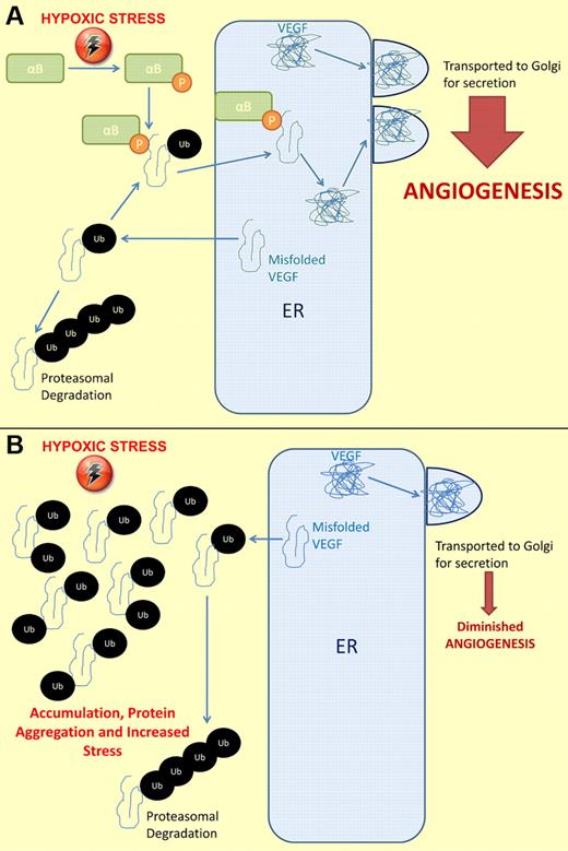
Hypoxic stress results in increased expression of vascular endothelial growth factor (VEGF), a known stimulator of angiogenesis. Increases in expression result in both properly folded and misfolded VEGF proteins. Misfolded VEGF is exported from the endoplasmic reticulum (ER) into the cytoplasm and ubiquitinated. (A) Hypoxic stress also stimulates phosphorylation of αB-crystallin (αB). Phosphorylated αB-crystallin binds misfolded, monoubiquitinated VEGF and returns it to the ER, where it is folded correctly and transported to the Golgi apparatus for secretion. Thus, the secretion of VEGF is up-regulated, angiogenesis occurs, and hypoxic stress may be reduced. (B) Properly folded VEGF is still transported to the Golgi and secreted. In the absence of αB-crystallin, misfolded, monoubiquitinated VEGF accumulates in the cytoplasm. Some of this will become polyubiquitinated and degrade. The rest of the misfolded VEGF can aggregate, leading to increased stress on the cell. Because misfolded VEGF cannot be transported back to the ER to be refolded, decreased secretion of VEGF occurs and hypoxic stress continues.
Hypoxic stress results in increased expression of vascular endothelial growth factor (VEGF), a known stimulator of angiogenesis. Increases in expression result in both properly folded and misfolded VEGF proteins. Misfolded VEGF is exported from the endoplasmic reticulum (ER) into the cytoplasm and ubiquitinated. (A) Hypoxic stress also stimulates phosphorylation of αB-crystallin (αB). Phosphorylated αB-crystallin binds misfolded, monoubiquitinated VEGF and returns it to the ER, where it is folded correctly and transported to the Golgi apparatus for secretion. Thus, the secretion of VEGF is up-regulated, angiogenesis occurs, and hypoxic stress may be reduced. (B) Properly folded VEGF is still transported to the Golgi and secreted. In the absence of αB-crystallin, misfolded, monoubiquitinated VEGF accumulates in the cytoplasm. Some of this will become polyubiquitinated and degrade. The rest of the misfolded VEGF can aggregate, leading to increased stress on the cell. Because misfolded VEGF cannot be transported back to the ER to be refolded, decreased secretion of VEGF occurs and hypoxic stress continues.
The α crystallins represent half of the lens protein content, are expressed in the retinal tissues, and protect cells from thermal and metabolic stress. The α crystallins comprise 2 family members, αA and αB. Both are small heat shock proteins, although only αB-crystallin is stress-inducible. αB-crystallin/HSPB5 functions as a chaperone protein for partially unfolded proteins and regulates cytoskeletal integrity. Under physiologic conditions, αB-crystallin is a cytoplasmic protein, colocalizes with vimentin, and can associate with the Golgi apparatus. During cell stress and pathologic conditions, αB-crystallin protects against apoptosis and prevents protein aggregation. Accordingly, α crystallins are responsible for lens transparency and play an important function preventing protein aggregation that would lead to opacity of the lens.3 Correspondingly, αB-crystallin–deficient mice display severe lens degeneration after chemical hypoxia due to excessive protein aggregation.4 Further, under hypoxia, αB-crystallin is phosphorylated at serine 59, resulting in enhanced chaperone activity.5 In previous studies, the authors and others demonstrated that αB-crystallin protects retinal pigment epithelium (RPE) cells from apoptosis induced by oxidative stress.6,7 Thus, αB-crystallin is important in retinal stress responses and the inhibition of RPE apoptosis. Levels of αB-crystallin increase in the eye during retinal degeneration and cataract formation; however, the chaperone activity of αB-crystallin is reduced with aging. This decrease in function may result in the accumulation of misfolded proteins and, thus, leave the retina unable to cope with various stresses, such as hypoxia, oxidative stress, and injury.
Neovascularization is one response of retinal vessels to hypoxic stress. Several recent studies have demonstrated roles for αB-crystallin in angiogenesis. αB-crystallin displays increased expression and phosphorylation in endothelial cells during tube formation in vitro.8 Tumors undergo neovascularization as they grow and the inner mass becomes hypoxic. Tumor vasculature in αB-crystallin–deficient mice displays high levels of apoptosis and decreased vessel formation.8 Thus, there is a precedence of αB-crystallin stimulating neovascularization in hypoxic tissues. In this issue of Blood, Kase et al study the role of αB-crystallin in retinal neovascularization using oxygen-induced retinopathy (OIR) and laser-induced choroidal neovascularization (CNV).1 In CNV, a laser is used to rupture the Bruch membrane, resulting in hypoxic stress and choroidal neovascularization within 7 to 12 days. In OIR, mice are exposed to a high oxygen (75%) environment for the first 7 to 10 days of life during which the retinal vasculature completes development. Five days after a return to normoxia (21%), neovascularization develops due to changes in oxygen tension, resulting in oxidative stress.9 In agreement with previous studies on αB-crystallin in neovascularization, OIR- and CNV-injured retinas from αB-crystallin–null mice demonstrate increased endothelial apoptosis and diminished intraocular angiogenesis.1 Thus, αB-crystallin supports neovascularization in stressed retinas.
αB-crystallin interacts with several important human growth factors, including vascular endothelial growth factor (VEGF). It is interesting to note that the interaction of αB-crystallin with VEGF occurs in the same region αB-crystallin uses to recognize and bind misfolded proteins.10 This implies that αB-crystallin may chaperone and stabilize misfolded VEGF. Interestingly, VEGF stimulated αB-crystallin phosphorylation and function in microcapillary endothelial cells during angiogenesis.8 Thus, a positive feedback loop may exist between the 2 proteins. In the retina, VEGF is secreted by a majority of the retinal cell types, is important in retinal development, and is expressed at high levels in retinopathies.2 In the current study, Kase et al demonstrate that VEGF protein expression decreases in αB-crystallin–deficient mice compared with wild-type mice after injury. The decreased VEGF protein levels in αB-null mice are caused not by changes in mRNA expression, but by altered protein secretion and ubiquitination (see figure). Interestingly, VEGF is polyubiquitinated equally in control and αB-crystallin knockdown RPE cells. However, mono-tetra-ubiquitinated VEGF increases in αB-crystallin–deficient RPE cells. Additionally, OIR and CNV injury increase the levels of phosphorylated αB-crystallin in retinal tissues,1 which has been associated with increased αB-crystallin chaperone activity.5 αB-crystallin binds and colocalizes with VEGF in the endoplasmic reticulum (ER), which may stimulate VEGF secretion and subsequent angiogenesis. Thus, in the absence of αB-crystallin chaperone function, VEGF accumulates and degrades, resulting in decreased secretion. Inhibition of the 26S proteasome in αB-crystallin–deficient mice partially rescues VEGF secretion resulting in increased retinal angiogenesis. Thus, Kase et al demonstrate the novel finding that phosphorylated αB-crystallin chaperones VEGF protein to the ER in hypoxic cells, resulting in proper VEGF folding and secretion. In the absence of αB-crystallin, misfolded, monoubiquitinated VEGF may be exported to the cytoplasm becoming ubiquitinated and degraded (see figure).1
This novel regulation of VEGF folding and secretion by the chaperone αB-crystallin represents an important control mechanism in the development of retinopathies. Although VEGF is important in physiologic retinal maintenance, it is up-regulated in pathologic retinopathies. In this regard, the stabilization of VEGF by phosphorylated αB-crystallin may contribute to the development of retinopathies. Currently, VEGF antagonists are a main treatment for retinopathies, although VEGF inhibition may lead to atrophy of the retina, particularly in AMD.2 Further, systemic inhibition of VEGF may result in various adverse side effects. Thus, phosphorylated αB-crystallin may represent an alternative therapeutic target for controlling intraocular neovascularization. This new mechanism of VEGF regulation may also function in other cases of hypoxia- or trauma-induced neovascularization, including ischemic pathologies, tumor progression, and wound healing. It remains to be seen whether αB-crystallin is involved in normal homeostasis of endothelium, known to be dependent on VEGF.11 However, because αB-crystallin–null mice display no major problems with vasculature during development or endothelial dysfunction, it appears that αB-crystallin regulates VEGF secretion primarily during postnatal neoangiogenesis. Therefore, manipulation of αB-crystallin phosphorylation and function could be used to inhibit VEGF secretion in pathologic neovascularization or to stimulate VEGF secretion in ischemic tissues. The role of chaperoning of VEGF by αB-crystallin in other tissues and pathologies remains to be revealed, but will lead to exciting advances in the field of angiogenesis.
Conflict-of-interest disclosure: The authors declare no competing financial interests. ■


This feature is available to Subscribers Only
Sign In or Create an Account Close Modal