Abstract
Abstract 2164
Poster Board II-141
Little is known on the functional and genetic integrity of Ph-negative hematopoietic cells (HC) repopulating the bone marrow after successful chronic myeloid leukemia (CML) treatment, although the frequent detection of cytogenetic abnormalities (CA), reminiscent of those seen in myelodysplastic syndromes (MDS), suggests the presence of functional and genetic defects. Telomere attrition represents a useful marker of proliferative and oxidative stress and might provide useful insights to monitor the genetic integrity of the hematopoietic compartment. This approach has been used in combination with a functional study of short and long term progenitors.
We investigated 78 CML patients with persistent (>12 months) complete cytogenetic remission (CCR). Median age was 64 (23-88), M/F ratio was 1.5. Median time from diagnosis and CCR were 64 months (25-915) and 39 months (12-150) respectively. Sokal score was low in 36 patients, intermediate in 28, and high in 14. Six patients were received IFN only, 45 Imatinib only, while 27 were currently on Imatinib, but received previous treatment with INF and/or chemotherapy. Complete and partial molecular responders were 35 and 28 respectively. Fifteen patients had acquired CA (del7: 4 patients, + 8: 5 patients, del5q: 2 patients, del or +Y: 2 patients, and 2 patients had other CA). Short term progenitors (CFU-GM, BFU-E CFU-Mix) and long-term culture-initiating cells (LTC-ICs)(Sutherland HJ et al Blood 1994) have been performed on 30 patients (requiring bone marrow examination for clinical purposes). Telomere length (TRF-L) analysis was performed by Southern Blotting as previously described (Ladetto M et Al, Blood 2004), both on polymorphonucleates (PMN) and on monocyte-depleted PBMC (MD-PBMC) (Rocci et al Exp Hematol 2007) to monitor both the myeloid and lymphoid compartment. Sixty four patients were assessed on repeated samples to monitor the kinetics of telomeric loss (median time 8 months, range 6-20). A control database of 109 healthy subjects has been used for comparison.
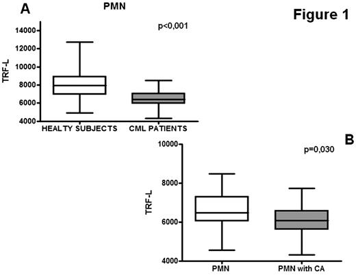
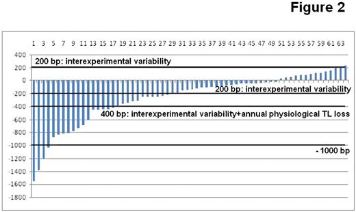
Ph-negative HC of CML patients were functionally impaired compared to controls, with reduced number of CFU-Mix (median 2,62 vs 4, p=0,010), CFU-GM (median 99,5 vs 181, p<0,001) and particularly of LTC-IC (median 88 vs 198, p<0,001). PMN from CML patients showed a major erosion of their telomeric DNA (median telomeric loss 1536 bp p<0.001, figure 1A). This finding was even more striking in patients with acquired CA, who showed a median TRF-L loss of 1900 bp (p<0.001) compared to healthy subjects, and 500 bp compared to other CML patients (p=0.030, figure 1B). Interestingly telomere attrition was less pronounced in the 4 patients with del or +Y and del5q, compared to those with other CA, such as del7 and +8. Telomeric erosion is more severe in younger CML patients, resulting in loss of the association between TRF-L and age, typically seen in healthy subjects. Telomere shortening was observed regardless of the use of TK inhibitors and chemotherapy. We found no correlation between TRF-L and clinical and demographic parameters. When a multivariate analysis on patients and healthy controls was performed, the presence of CML resulted a stronger predictor of telomeric damage compared to age. Analysis of TRF-L kinetics on the whole population over time showed substantial stability or modest physiological shortening in the majority of patients. In none of the patients a relevant recovery of TRF-L over time was noticed. However in 16 (25%) patients a non-physiological telomeric loss was observed (>400bp obtained by considering maximal physiological loss plus technical variability of the assay) (Figure 2). Interestingly the four patients with the most extreme telomeric loss (>1000 bp/year) showed evidence of either CA or impaired hematopoietic fuction by colony assays. Moreover one of these patients progressed to an overt MDS six month after the second determination.
Ph-negative HC repopulating the bone marrow after successful CML treatment: i) have major defects in their functional performances; ii) display severe telomeric loss (roughly comparable to 31 years of physiological aging), which is more pronounced in patients with CA. Moreover the lack of telomeric recovery over time and the presence of a subgroup of patients with ongoing accelerated non-physiological telomeric attrition suggest the need of strict monitoring of the long-term performances and genetic stability of Ph-negative hematopoiesis in CML patients.
No relevant conflicts of interest to declare.
Author notes
Asterisk with author names denotes non-ASH members.


This feature is available to Subscribers Only
Sign In or Create an Account Close Modal