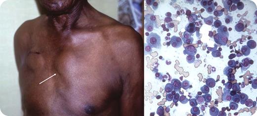
A 67-year-old man had a number of vague cardiac symptoms. When syncope and an arrhythmia occurred, a temporary pacemaker was promptly placed. At the time of the implantation of a permanent pacemaker, physical examination showed a parasternal mass on the left side of the upper sternum (see arrow); the site of the recently implanted pacemaker is medial to the right axilla. He admitted to having lethargy and generalized bony aches, but no fever, sweats, or weight loss. The mass was firm and fixed to the lateral margin of the sternum. He had no adenopathy, hepatosplenomegaly, or prostate nodules. Chest x-ray showed no pulmonary masses but did reveal osteoporosis and suspicious lytic bone lesions. On laboratory evaluation, there was mild anemia and azotemia, a normal prostatic specific antigen, and an elevated total protein.
A needle aspiration, culture, and smear of the parasternal mass were performed. The touch preparation smear (see aspirate photo) showed abundant plasma cells. Cultures were negative. On serum electrophoresis, a monoclonal protein was present; subsequently, it was identified as IgA lambda.
An isolated plasmacytoma presents infrequently. In this patient, the plasmacytoma appeared concurrently with the presence of multiple myeloma. The finding of a cardiac arrhythmia coupled with a lambda chain myeloma abnormality raised the possibility of cardiac amyloidosis. This possibility was assessed by cardiac ultrasound, but it was not present.
Many Blood Work images are provided by the ASH IMAGE BANK, a reference and teaching tool that is continually updated with new atlas images and images of case studies. For more information or to contribute to the Image Bank, visit www.ashimagebank.org.


This feature is available to Subscribers Only
Sign In or Create an Account Close Modal