Abstract
We analyzed 21 children with leukemia receiving haploidentical hematopoietic stem cell transplantation (haplo-HSCT) from killer immunoglobulin (Ig)–like receptors (KIR) ligand-mismatched donors. We showed that, in most transplantation patients, variable proportions of donor-derived alloreactive natural killer (NK) cells displaying anti-leukemia activity were generated and maintained even late after transplantation. This was assessed through analysis of donor KIR genotype, as well as through phenotypic and functional analyses of NK cells, both at the polyclonal and clonal level. Donor-derived KIR2DL1+ NK cells isolated from the recipient displayed the expected capability of selectively killing C1/C1 target cells, including patient leukemia blasts. Differently, KIR2DL2/3+ NK cells displayed poor alloreactivity against leukemia cells carrying human leukocyte antigen (HLA) alleles belonging to C2 group. Unexpectedly, this was due to recognition of C2 by KIR2DL2/3, as revealed by receptor blocking experiments and by binding assays of soluble KIR to HLA-C transfectants. Remarkably, however, C2/C2 leukemia blasts were killed by KIR2DL2/3+ (or by NKG2A+) NK cells that coexpressed KIR2DS1. This could be explained by the ability of KIR2DS1 to directly recognize C2 on leukemia cells. A role of the KIR2DS2 activating receptor in leukemia cell lysis could not be demonstrated. Altogether, these results may have important clinical implications for the selection of optimal donors for haplo-HSCT.
Introduction
T cell–depleted hematopoietic stem cell transplantation (HSCT) from a human leukocyte antigen (HLA)–haploidentical donor (haplo-HSCT) has been reported to benefit from the graft-versus-leukemia effect mediated by natural killer (NK) cells when the donor displays NK alloreactivity versus the recipient.1-3 The molecular basis for NK alloreactivity is represented by NK receptors, namely killer immunoglobulin (Ig)–like receptors (KIRs), which are specific for allotypic determinants that are shared by different HLA-class I alleles (referred to as KIR ligands).4 KIR2DL1 recognizes HLA-C alleles characterized by Lys at position 80 (HLA-CLys80, also named C2 group), KIR2DL2/3 recognizes HLA-C alleles characterized by Asn at position 80 (HLA-CAsn80, C1 group), KIR3DL1 recognizes HLA-B alleles sharing the Bw4 supertypic specificity (HLA-BBw4, Bw4 group), and KIR3DL2 recognizes HLA-A3 and HLA-A11 alleles.5-7 KIR2DL/3DL are inhibitory receptors that, upon engaging the cognate ligand, inhibit lysis.8 Activating KIRs, which are highly homologous in the extracellular domain to their inhibitory counterparts, include KIR2DS1, KIR2DS2, and KIR3DS1, but only KIR2DS1 has been shown to specifically recognize the C2 group of alleles expressed in Epstein-Barr virus–transformed B-lymphocyte (B-EBV) cells.9,10 Additional receptors are the inhibitory CD94/NKG2A and the activating CD94/NKG2C heterodimers, both lectin-like molecules specific for HLA-E, a class Ib molecule that binds leader peptides of most HLA-class I molecules, thus displaying broad reactivity.11 The NK-cell repertoire of each person is primarily determined by KIR genotype, derived from the sum of maternal and paternal haplotypes. The combination of KIR genes define group A haplotype, which has few genes, most of which encoding for inhibitory KIRs, while group B, in addition to inhibitory KIRs, has several genes encoding for activating KIRs.12 Another important feature of the repertoire is the clonal distribution of the HLA-specific receptors.5,13,14 Thus, if a certain KIR gene is expressed, the corresponding KIR protein may be present on the cell surface by variable (but unpredictable) proportions of NK cells. In addition, at the single cell level, this KIR may or may not be coexpressed with other KIRs or NKG2A. Although KIRs and HLA genes segregate independently, the NK cell repertoire is influenced by the HLA genotype. Indeed, to ensure self-tolerance, each individual NK cell expresses at least one inhibitory receptor specific for autologous HLA-class I molecules. In addition, as recently described, rare NK cells lacking inhibitory receptors for self-HLA molecules may be generated but are hyporesponsive.15-17
This kind of receptor repertoire can lead to alloreactivity in haplo-HSCT through the mechanism of “missing self” recognition,18 provided that the donor (1) expresses a KIR-ligand that is missing in the recipient's HLA genotype and (2) expresses the specific KIR, leading to a KIR/KIR-ligand mismatch in graft-versus-host (GVH) direction.3 Thus, to assess the presence of NK alloreactivity in haplo-HSCT, it is necessary to analyze both donor and recipient HLA-class I, as well as donor KIR genotypes. In addition, the phenotypic analysis of KIR and CD94/NKG2A expressed by donor NK cells can further define the size of the alloreactive NK subset.19,20 Finally, functional assays, based on the ability to kill appropriate target cells, provide precise information on the degree of alloreactivity of given NK-cell populations.21
In this study, we analyzed donor-derived alloreactive NK cells in pediatric patients with leukemias given haplo-HSCT. We show that alloreactive NK cells are generated and can persist for years in the recipient. In addition, we provide new information on KIR specificity for HLA-class I molecules and demonstrate that the activating KIR2DS1 is involved in lysis of leukemia cells.
Methods
Donor/recipient pairs in haplo-HSCT
Twenty-one children with hematologic malignancies were included in the study. Details on patient and donor characteristics are reported in Table 1. Informed consent to this study, approved by Fondazione Istituto di Ricovero e Cura a Carattere Scientifico (IRCCS) Policlinico San Matteo (Pavia) review board, was obtained from patients, donors, or parents in accordance with the Declaration of Helsinki. Hematopoietic stem cells were mobilized in the donor through the use of granulocyte colony-stimulating factor (G-CSF) and collected by a cell separator. CD34+ cells were positively selected using the CliniMacs one-step procedure (Miltenyi Biotec, Bergisch Gladbach, Germany). Flow cytometric analysis quantified CD34+ and CD3+ cells before and after selection. The median number of CD34+ cells and of CD3+ cells infused per kilogram of recipient body weight was 23 × 106 (range 10.6-41.2) and 5 × 10³ (range 0.5-35), respectively (see Table 1). All patients were given anti-thymocyte globulin before the allograft to minimize the risk of graft failure. No patient was given posttransplantation pharmacologic immune suppression. Children with evidence of donor engraftment who survived more than 14 days and more than 90 days from transplantation were evaluated for occurrence of acute graft-versus-host disease (GVHD) and chronic GVHD, respectively, according to established criteria.22,23 Donor/recipient human cytomegalovirus serology was determined by enzyme-linked immunosorbent assay, using previously reported methods.24
Patient, donor, and graft characteristics
| Case . | Sex . | Age at Tx . | Diagnosis . | Disease status at Tx . | Cytogenetic/molecular abnormalities . | Donor . | No. of CD34+ infused (×106/kg) . | No. of CD3+ infused (×103/kg) . | CMV serology D/R . | Acute GVHD . | Chronic GVHD . | Relevant infections . | Outcome . |
|---|---|---|---|---|---|---|---|---|---|---|---|---|---|
| 1 | F | 16 y, 3 mo | BCP CD10+ ALL | 3rd CR | t(9;22) | mother | 20 | 12 | +/+ | Grade II | No | No | Alive, CR (+48 mo) |
| 2 | F | 3 y, 8 mo | BCP CD10+ ALL | 2nd CR | / | father | 29.1 | 6.8 | +/+ | No | No | CMV | Relapsed (+7 mo) dead |
| 3 | M | 20 y, 2 mo | AML (M2) | 3rd CR | t(8;21) del.9q | sister | 10.6 | 13 | +/+ | Grade I | No | BK cystitis, EBV, adenovirus, measles, fusarium | Alive, CR (+28 mo) |
| 4 | M | 10 y, 8 mo | AML (M0-M1) | 1st CR | / | mother | 12.2 | 35 | +/+ | No | No | No | Relapsed (+5 mo) dead |
| 5 | M | 4 y, 9 mo | BCP CD10+ ALL + | 2nd CR | / | mother | 21.9 | 17 | +/+ | No | No | No | Alive, CR (+14 mo) |
| 6 | F | 3 y, 9 mo | JMML | Disease present | PTPN11 mutation | mother | 23.7 | 30 | +/+ | Grade II | Limited | No | Alive, CR (+14 mo) |
| 7 | M | 14 y, 1 mo | AML (M0) | 1st CR | FTL3-ITD | sister | 16.1 | 2.9 | +/− | No | No | No | Relapsed (+3 mo) dead |
| 8 | M | 17 y, 2 mo | BCP CD10+ ALL | 2nd CR | / | brother | 23.0 | 4.5 | +/+ | No | No | CMV | Alive, CR (+11 mo) |
| 9 | M | 5 y, 7 mo | BCP CD10+ ALL | 2nd CR | t(9;22) | mother | 15.1 | 16 | +/+ | Grade II | No | No | Alive, CR (+54 mo) |
| 10 | F | 6 y, 4 mo | AML (M1) | Disease present | FTL3-ITD | mother | 31.7 | 14 | +/+ | No | No | No | Relapsed (+5 mo) dead |
| 11 | M | 4 y, 8 mo | T-ALL | 4th CR | / | father | 41.23 | 14 | −/+ | No | No | Candida spp, HHV6, EBV | Alive, CR (+24 mo) |
| 12 | F | 7 y, 1 mo | AML (M2) | 2nd CR | / | mother | 35.0 | 3 | +/+ | No | No | No | Alive, CR (+25 mo) |
| 13 | F | 5 y, 4 mo | BCP CD10+ ALL | 2nd CR | / | mother | 37.0 | 3 | +/+ | Grade II | Limited | No | Alive, CR (+23 mo) |
| 14 | M | 8 y, 5 mo | BCP CD10+ ALL | 2nd CR | / | sister | 11.6 | 19 | +/+ | No | No | No | Relapsed (+12 mo) alive |
| 15 | F | 4 y, 4 mo | BCP CD10+ ALL | 1st CR | / | mother | 19.5 | 1 | +/+ | Grade I | No | CMV, BK cystitis | Alive, CR (+17 mo) |
| 16 | F | 4 y, 1 mo | AML (M7) | 2nd CR | / | father | 22.3 | 2 | +/+ | No | n.e. | No | Relapsed (+3 mo) dead |
| 17 | M | 5 y | T-ALL | 4th CR | / | father | 32,1 | 5 | +/+ | No | No | No | Alive, CR (+24 mo) |
| 18 | M | 8 y, 2 mo | AML (M2) | 2nd CR | t(8;21) | mother | 38.6 | 3 | +/+ | No | No | No | Alive, CR (+22 mo) |
| 19 | M | 16 y, 1 mo | BCP CD10+ ALL | 3rd CR | / | mother | 18.4 | 0.5 | +/− | No | No | No | Relapsed (+4 mo) dead |
| 20 | F | 12 y, 10 mo | BCP CD10+ ALL | 2nd CR | t(9;22) | brother | 20.6 | 4 | +/+ | No | No | VZV | Alive, CR (+16 mo) |
| 21 | M | 11 y,11 mo | Secondary AML | 2nd CR | Complex karyotype | mother | 15.5 | 0.008 | +/+ | No | No | No | Alive, CR (+16 mo) |
F indicates female; M, male; Tx, transplantation; BCP, B-cell precursor; ALL, acute lymphoblastic leukemia; AML, acute myeloid leukemia; JMML, juvenile myelomonocytic leukemia; CR, complete remission; FTL3-ITD, internal tandem duplication; D/R, donor/recipient; GVHD, graft-versus-host disease; n.e., not evaluable; EBV, Epstein-Barr virus; CMV, cytomegalovirus; HHV-6, human herpesvirus 6; BK, papillomavirus type BK; and VZV, varicella-zoster virus.
HLA typing
Donor/recipient alleles of the loci HLA-A, -B, -C, -DRB1, and -DQB1 were identified by polymerase chain reaction single-strand polymorphism (PCR-SSP) and sequence-based typing. Amplification and sequencing of HLA class I and class II genes were performed as previously described.25-27 Alleles were assigned according to DNA sequences.28 Table 2 shows in detail KIR-ligands expressed according to the donor's and patient's HLA-class I genotypes.
KIR-ligand, KIR, NK alloreactive subsets, and lytic capability in the various donor/recipient pairs
| Case . | KIR-ligands (donor)* . | KIR-ligands (patient)† . | Permissive inhibitory KIR (donor)‡ . | Activating KIR (donor)§ . | Alloreactive subset, % (donor)¶ . | Alloreactive subset, % (recipient post-HSCT)¶ . | Lysis vs B-EBV, % (donor)# . | Lysis vs B-EBV, % (recipient post-HSCT)# . |
|---|---|---|---|---|---|---|---|---|
| 1 | Bw4, C1, C2 | Bw4, C2 | 2DL2/3 3DL2 | 2DS1 2DS2 3DS1 | 26 | 54 (7 mo) | 31 (60) | 57 (68) |
| 2 | Bw4, C1, C2 | Bw4, C2 | 2DL2/3 3DL2 | 2DS2 | 6 | 8 (3 mo) | 1 (57) | 1 (71) |
| 3 | Bw4, C1, C2 | Bw4, C2 | 2DL2/3 3DL2 | 2DS2 | 36 | 23 (2 mo) | 57 (83) | 27 (76) |
| 4 | A3, Bw4, C1, C2 | Bw4, C2 | 2DL2/3 3DL2 | 2DS2 3DS1 | 15 | 3 (2 mo) | 2 (36) | Nd |
| 5 | Bw4, C1, C2 | Bw4, C2 | 2DL2/3 3DL2 | 2DS1 2DS2 3DS1 | 36 | 34 (2 mo) | 52 (42) | 45 (55) |
| 6 | Bw4, C1, C2 | A11, Bw4, C2 | 2DL2/3 | 2DS1 2DS2 3DS1 | 9 | 69 (3 mo) | 19 (51) | 7 (40) |
| 7 | Bw4, C1, C2 | Bw4, C2 | 2DL3 3DL2 | NO | 2 | 3 (2 mo) | 6 (53) | 0 (59) |
| 8 | Bw4, C1, C2 | Bw4, C2 | 2DL3 3DL2 | 2DS1 | 5 | 17 (3 mo) | 12 (74) | 39 (83) |
| 9 | Bw4, C1, C2 | Bw4, C1 | 2DL1 3DL2 | 2DS2 | (92) | (2) (3 mo) | 80 (82) | 32 (90) |
| 10 | Bw4, C1, C2 | Bw4, C1 | 2DL1 3DL2 | 2DS2 | (3) | (0.5) (3 mo) | 11 (76) | 27 (80) |
| 11 | C1, C2 | Bw4, C1 | 2DL1 3DL2 | NO | 9 | 10 (5 mo) | 20 (83) | 26 (91) |
| 12 | Bw4, C1, C2 | Bw4, C1 | 2DL1 3DL2 | 2DS1 3DS1 | 11 | 8 (3 mo) | 58 (100) | 42 (97) |
| 13 | Bw4, C1, C2 | Bw4, C1 | 2DL1 3DL2 | 2DS2 | (1) | (0.5) (3 mo) | 25 (96) | 10 (62) |
| 14 | Bw4, C1, C2 | Bw4, C1 | 2DL1 3DL2 | 2DS1 3DS1 | 6 | 3 (7 mo) | 43 (71) | 26 (84) |
| 15 | A11, Bw4, C1, C2 | Bw4, C1 | 2DL1 3DL2 | 2DS1 3DS1 | 13 | 14 (6 mo) | 27 (83) | 14 (88) |
| 16 | Bw4, C1 | C1, C2 | 3DL1 3DL2 | NO | 3 | 3 (2 mo) | 15 (61) | 13 (66) |
| 17 | Bw4, C1, C2 | C1, C2 | 3DL1 3DL2 | NO | 5 | 5 (3 mo) | 26 (83) | 43 (81) |
| 18 | Bw4, C1, C2 | C1, C2 | 3DL1 3DL2 | NO | 8 | 4 (6 mo) | 34 (95) | 23 (66) |
| 19 | Bw4, C1, C2 | A3, C1, C2 | NO | 2DS1 3DS1 | 2 | 2 (3 mo) | n.d. | 10 (56) |
| 20 | Bw4, C1 | C1, C2 | NO 3DL2 | 2DS1 3DS1 | 2 | 21 (6 mo) | 26 (59) | 66 (78) |
| 21 | A3, Bw4, C2 | Bw4, C1, C2 | 3DL2 | NO | 0 | 7 (13 mo) | 14 (43) | 27 (75) |
KIR-ligands expressed in donor HLA-class I genotype; the KIR ligand-mismatch in GVH direction is in bold.
KIR-ligands expressed in patient HLA-class I genotype.
Expressed donor KIR permissive for alloreactivity (which do not recognize patient's HLA-class I alleles); in bold is the KIR specific for KIR ligand-mismatch. Cases no. 19 and 20 were missing KIR3DL1, which is the KIR specific for Bw4-mismatch.
KIR2DS1, 2DS2 and 3DS1 are indicated when expressed in donor KIR genotype. For expression of the other activating KIR (KIR2DS4, 2DS3 and 2DS5) see Table S1.
The percentage was evaluated in polyclonal NK-cell populations by staining with an appropriate combination of anti-KIR and anti-NKG2A mAb followed by appropriate fluorochrome-conjugated second reagents using the methodology described in Figure 2A. Percentages that could not be precisely calculated due to the presence of activating KIR (undistinguishable from the inhibitory form, such as KIR2DS2 and KIR2DL2) are indicated in parentheses. The percentage of alloreactive subset in the recipient (patient after HSCT) was evaluated at the indicated time point (months after HSCT).
Percent-specific lysis, either in the absence or in the presence (in parentheses) of anti-HLA class I mAb, was evaluated in polyclonal NK-cell populations derived from the donor or the recipient. Their cytolytic assays were performed in parallel (ie, at the same time) to phenotypic evaluations. Target cells were represented by appropriate B-EBV cell lines expressing the same KIR-ligands of the patient (E:T of 20:1).
KIR genes profile analysis
Donor DNA was extracted using QIAamp DNA Blood Mini kit (QIAGEN, Hilden, Germany). The KIR genes profiles were performed using Olerup SSP KIR genotyping kit (GenoVision, Saltsjoebaden, Sweden) following the manufacturer's instruction. Table 2 shows the presence of selected KIR genes, while the complete profiles are shown in Table S1 (available on the Blood website; see the Supplemental Materials link at the top of the online article).
Production of KIR soluble receptors
Constructs encoding KIR2DL1*001-Fc, KIR2DL2*001-Fc, KIR2DL3*001-Fc, and KIR2DS1*002-Fc soluble receptors were obtained keeping in frame the cDNA coding for the first 245 amino acids of the indicated KIRs with the cDNA coding for the Fc portion of human IgG1 present in pRB1vector. These KIR-Fc/pRB1 plasmids were transiently transfected into HEK-293T, and cells were cultured using Dulbecco modified Eagle medium (DMEM) supplemented with 10% ultra-low IgG fetal calf serum (FCS; Gibco BRL, Invitrogen, Carlsbad, CA). Soluble chimeric proteins were purified from the supernatants by Protein A-Sepharose affinity column (Amersham Biosciences, Piscataway, NJ), and protein concentrations were determined using a Bradford protein assay (Bio-Rad Laboratories, Hercules, CA). Sodium dodecyl sulfate polyacrylamide gel electrophoresis (SDS-PAGE) and silver staining established the purity of the soluble receptors. Binding of soluble receptors was tested on 721.221 cell line transfected with HLA-Cw6 or -Cw7 (kindly provided by C. Falk, University of Heidelberg, Heidelberg, Germany). Briefly, 105 cells were incubated with 10 μg soluble receptor for 30 minutes at 4°C, washed once, and stained with phycoerythrin (PE)–conjugated goat anti–human second reagent (Southern Biotechnology Associates, Birmingham, AL).
Isolation and culture of NK cells
NK cells from donors and recipients at various time points after HSCT were purified using the RosetteSep method (StemCell Technologies, Vancouver, BC). NK cells were also cultured on irradiated feeder cells in the presence of 2 μg/mL phytohemagglutinin (PHA; Life Technologies, Paisley, United Kingdom) and 100 U/mL recombinant interleukin-2 (rIL-2, proleukin; Chiron, Emeryville, CA) to obtain proliferation of polyclonal or, after limiting dilution, clonal NK cells.21
Monoclonal antibodies and cytofluorimetric analysis
The monoclonal antibodies (mAbs) used in this study reacting with the HLA-specific NK receptors are listed in Table 3. Other mAbs, produced in our laboratory, were c218 (IgG1, anti-CD56), BAB281 (anti-NKp46), Z231 (IgG1, anti-NKp44), A76 (IgG1 anti-NKp30), 6A4 and A6-136 (IgG1 and IgM, respectively, anti-HLA class I).5,29-31 The anti-HLA class I mAb W6/32 was also used. The anti–CD56-PC7 (N901, IgG1) was provided by Beckman-Coulter (Marseille, France); the anti–CD3-APC (UCHT1, IgG1) and anti-CD19 (HD37, IgG1) by DakoCytomation (Glostrup, Denmark); the anti-CD10 (SN5c, IgG1) by Ancell (Bayport, MN); the anti-CD34 (8G12, IgG1) and anti-CD19 (Leu12, IgG1) by Becton Dickinson (Mountain View, CA).
List of mAb against HLA class I–specific NK receptors evaluated in this study
| Clone . | Source* . | Isotype . | Specificity . |
|---|---|---|---|
| 11PB6 | Our laboratory, Miltenyi Biotec | IgG1 | KIR2DL1/S1 |
| EB6B | Our laboratory, Beckman Coulter | IgG1 | KIR2DL1/S1 |
| #143211 | R&D Systems | IgG1 | KIR2DL1 |
| HP-3E4 | M. Lopez-Botet, BD Pharmingen | IgM | KIR2DL1/S1/S4 |
| GL-183 | Our laboratory, Beckman Coulter | IgG1 | KIR2DL2/L3/S2 |
| CH-L | S. Ferrini, BD Pharmingen | IgG2b | KIR2DL2/L3/S2 |
| ECM-41 | Our laboratory | IgM | KIR2DL3 |
| Z27 | Our laboratory, Beckman Coulter | IgG1 | KIR3DL1/S1 |
| DX9 | Miltenyi Biotec | IgG1 | KIR3DL1 |
| AZ158 | S. Parolini | IgG2a | KIR3DL1/L2/S1 |
| Q66 | Our laboratory | IgM | KIR3DL2 |
| Z199 | Our laboratory, Beckman Coulter | IgG2b | NKG2A |
| Z270 | Our laboratory | IgG1 | NKG2A |
| #1381 | R&D Systems | IgG2b | NKG2C |
| Y9 | Our laboratory | IgM | CD94 |
| FES172 | Our laboratory, Beckman Coulter | IgG2a | KIR2DS4 |
Miltenyi Biotec (Bergish Gladbach, Germany); Beckman Coulter (Marseille, France); BD Pharmingen (San Diego, CA.); R&D Systems (Minneapolis, MN); Dr M. Lopez-Botet (IMIM-Hospital del Mar, Barcelona, Spain); Dr S. Ferrini (Istituto Scientifico Tumori, Genova, Italy); Dr S. Parolini (University of Brescia, Brescia, Italy).
To analyze the surface markers of the activated NK cells and leukemia cells, samples were stained with the various mAbs followed by PE-conjugated AffiniPure F(ab′)2 goat anti–mouse IgG (Jackson Immunoresearch Laboratories, West Grove, PA).21 To define the alloreactive NK-cell subset, we performed either 4-color fluorescence analysis of peripheral blood mononuclear cell (PBMC) gating on CD3−CD56+ lymphocytes (using anti–CD3-APC and anti–CD56-PC7 mAb) or double fluorescence analysis of purified activated NK-cell populations. The alloreactive NK-cell subset was evaluated using appropriate mAb combinations. Briefly, cells were incubated with mAbs followed by fluorescein isothiocyanate (FITC)– or PE-conjugated isotype-specific goat anti–mouse second reagents (Southern Biotechnology Associates), or as specified in the figure legends, commercially available fluorochrome-labeled mAbs were used (Table 3). To compare the surface densities, we calculated the ratio between the mean fluorescence intensity (MRFI) of cells stained with the selected mAbs or KIR-Fc and that of cells stained with the second reagent alone.
Samples were analyzed by cytofluorimetric analysis on a FACSCalibur and with the CellQuest program (both from Becton Dickinson).31
Cytolytic assays
NK cells were tested for cytolytic activity in a 4-hour 51Cr-release assay, as previously described.21 Here, we analyzed the susceptibility to lysis of leukemia cells derived from patients pre-HSCT (cryopreserved) and also B-EBV cell lines derived from HLA-typed donors, characterized by the combination of informative KIR-ligands. We also used as target cells 721.221 transfected with HLA-Cw3 or -Cw4 alleles.32 For masking experiments, NK cells were preincubated with mAbs specific to the various NK receptors, while the anti–HLA-class I mAbs were added as the last reagent in the cytolytic assay; mAb concentration was 10 μg/mL. For redirected killing assays, P815 were used as target cells in the presence of mAbs of the IgG isotype at a concentration of 0.5 μg/mL.
Results
Selection of donor/recipient pairs in haplo-HSCT
Among the 21 pediatric patients analyzed, 12 had acute lymphoblastic leukemia (ALL; 10 with a B-cell precursor phenotype and 2 with T-ALL), 8 had acute myeloid leukemia (AML; 7 of de novo origin and 1 secondary to previous cytotoxic/radiant treatment), and 1 had juvenile myelomonocytic leukemia (JMML), shown in Table 1). All these cases were selected on the basis of the expression in the donor of a given KIR ligand (HLA allele) that was missing in the patient. Table 2 shows the KIR ligand-mismatch present in the donor/recipient pairs including C1, C2, Bw4, and HLA-A3/-A11. When the donor expresses the relevant KIR gene specific for the KIR ligand-mismatch, NK-cell alloreactivity in GvH direction may be expected. Such prediction is based on the analysis of donor KIR genotype (Tables 2, S1). Moreover, in 2 cases (no. 19 and no. 20), donors were found to lack the relevant KIR (KIR3DL1) specific for the KIR ligand-mismatch (Bw4). Accordingly, these donors could not be considered as “classical” alloreactive donors. On the other hand, as shown below, in such donors, the presence of activating KIR may considerably impact on NK alloreactivity.
Identification of alloreactive NK subsets in HSC donors and their persistence in recipients after transplantation
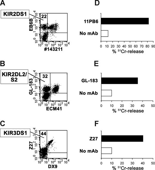
As previously reported, the alloreactive NK subset can be defined by the presence of receptors that are permissive for target cell lysis and by the absence of receptors that would block lysis.21 Accordingly, alloreactive subsets will include NK cells expressing inhibitory KIR(s) specific for KIR ligand(s) absent in the recipient. In contrast, NK cells expressing NKG2A or inhibitory KIRs specific for the KIR-ligand(s) expressed by the recipient are nonalloreactive. However, since in most instances the available anti-KIR mAbs do not distinguish between inhibitory and activating forms, a limitation of phenotypic analyses may be the underestimation of the alloreactive subsets. A more precise quantification of alloreactive versus nonalloreactive subsets is now allowed by the combined use of novel mAbs (listed in Table 3) that can distinguish inhibitory from activating forms of certain KIR. This is true for KIR2DL1 versus S1 (C.C., E.V., manuscript in preparation; see also Figure S1) and for KIR3DL1 versus S1,33-35 but not for KIR2DL2 versus S2.36 As shown in Figure 1, double fluorescence analysis using appropriate mAb combinations allows the identification of the NK cell subset expressing exclusively a given activating KIR. In addition, the use of a redirected killing assay against P815 target cells and of appropriate mAbs recognizing both forms of KIR, makes it possible to obtain a functional demonstration of the presence of activating KIRs within the NK cell pool.
Identification of NK subpopulations expressing the activating form of KIRs. Double fluorescence (A-C) and reverse-antibody dependent cellular cytotoxicity (R-ADCC) analyses (D-F) were performed using NK cell populations derived from the following informative donors: no. 20 (A,D) and no. 4 (B,E and C,F). Dot plots identify the subset expressing the activating KIR in the upper left quadrant with the percentage of cells included. For staining, the following mAb were used in combination: #143211-FITC (anti-KIR2DL1) and EB6B-PE (anti-KIR2DL1/S1) (A); ECM41 (anti-KIR2DL3) and GL-183 (anti-KIR2DL2/L3/S2) followed by isotype-specific PE- and FITC-conjugated, respectively, second reagents (B); DX9-FITC (anti-KIR3DL1) and Z27-PE (anti-KIR3DL1/S1) (C). For R-ADCC, 51Cr-labeled P815 were incubated with polyclonal NK cell populations in the absence (□) or presence (■) of the indicated mAb. Effector to target (E:T) ratio of 4:1 was used. The results are representative of 3 independent experiments; the SD of the mean of the triplicates was less than 5%.
Identification of NK subpopulations expressing the activating form of KIRs. Double fluorescence (A-C) and reverse-antibody dependent cellular cytotoxicity (R-ADCC) analyses (D-F) were performed using NK cell populations derived from the following informative donors: no. 20 (A,D) and no. 4 (B,E and C,F). Dot plots identify the subset expressing the activating KIR in the upper left quadrant with the percentage of cells included. For staining, the following mAb were used in combination: #143211-FITC (anti-KIR2DL1) and EB6B-PE (anti-KIR2DL1/S1) (A); ECM41 (anti-KIR2DL3) and GL-183 (anti-KIR2DL2/L3/S2) followed by isotype-specific PE- and FITC-conjugated, respectively, second reagents (B); DX9-FITC (anti-KIR3DL1) and Z27-PE (anti-KIR3DL1/S1) (C). For R-ADCC, 51Cr-labeled P815 were incubated with polyclonal NK cell populations in the absence (□) or presence (■) of the indicated mAb. Effector to target (E:T) ratio of 4:1 was used. The results are representative of 3 independent experiments; the SD of the mean of the triplicates was less than 5%.
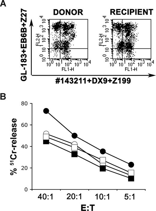
In Figure 2A, case no. 5 properly illustrates the phenotypic approach used to define the alloreactive subset. The patient's HLA-I genotype contained KIR ligands Bw4 and C2, while the donor's HLA-I genotype contained Bw4 and C1/C2 (Table 2). Thus, in this case, C1 represented the KIR ligand-mismatch in GVH direction, and KIR2DL2/3 represented the permissive inhibitory KIR. The donor's KIR genotype revealed the presence of all the activating forms of the KIRs evaluated in this study as well as their inhibitory counterparts: KIR2DL1+/S1+, KIR2DL2+/L3+/S2+, 3DL1+/S1+, and KIR3DL2+ (Tables 2, S1). A suitable way to detect the alloreactive subset was to stain cells using a mixture of GL-183 (anti-KIR2DL2/L3/S2), EB6B (anti-KIR2DL1/S1), and Z27 (anti-KIR3DL1/S1) mAbs conjugated with the fluorochrome PE and a mixture of #143211 (anti-KIR2DL1), DX9 (anti-KIR3DL1), and Z199 (anti-NKG2A) mAbs conjugated with FITC. By this approach, the alloreactive subset (confined in the upper left quadrant of Figure 2A) included cells expressing KIR2DL2/L3 (permissive inhibitory KIR), KIR2DS1, KIR2DS2, and KIR3DS1 (activating KIR), but not KIR2DL1, KIR3DL1, and NKG2A (other inhibitory receptors). This subset corresponded to 36% in the donor and 34% in the patient after transplantation (subsequently referred to as the recipient). Using this technique, we were able to determine the size of the alloreactive NK subset present in all donor/recipient pairs included in this study (Table 2). In case no. 5, in agreement with the high percentage of alloreactive NK cells, both donor- and recipient-derived polyclonal populations efficiently killed the patient's leukemia blasts, as well as B-EBV cell lines (eg, BOB), expressing Bw4 and C2 (ie, the same KIR ligands expressed by the patient; Figure 2B).
Identification of the alloreactive subset at the population level. Case no. 5 was characterized by C1 KIR-ligand mismatch, because the patient expressed HLA-A*2402,*2601, -B*3502,*5301 (Bw6/Bw4), -C*0401,*0602 (C2/C2) while the donor expressed HLA-A*24,*26, -B*18,*53 (Bw6/Bw4), -C*1203,*0602 (C1/C2). (A) The alloreactive subset was evaluated in the donor and in the recipient (2 months after transplantation). NK cells were incubated with the mixture of GL-183-PE (anti-KIR2DL2/L3/S2), EB6B-PE (anti-KIR2DL1/S1), Z27-PE (anti-KIR3DL1/S1), DX9-FITC (anti-KIR3DL1), no.143211-FITC (anti-KIR2DL1) and Z199 (anti-NKG2A) followed by anti–IgG2b-FITC. The alloreactive subset resides in the top left quadrant: 36% in the donor and 34% in the recipient. (B) Donor (filled symbols)– and recipient (open symbols)–derived polyclonal NK-cell populations were tested in cytolytic assays against the patient's leukemia blasts (CD10+ ALL, squares) and the B-EBV BOB (circles) expressing the homozygous haplotype HLA-A*2402, -B*5101 (Bw4), -C*1502 (C2). Both effector cells efficiently lysed these target cells at different E:T ratios as shown. The results are representative of 2 independent experiments; the standard deviation of the mean of the triplicates was less than 5%.
Identification of the alloreactive subset at the population level. Case no. 5 was characterized by C1 KIR-ligand mismatch, because the patient expressed HLA-A*2402,*2601, -B*3502,*5301 (Bw6/Bw4), -C*0401,*0602 (C2/C2) while the donor expressed HLA-A*24,*26, -B*18,*53 (Bw6/Bw4), -C*1203,*0602 (C1/C2). (A) The alloreactive subset was evaluated in the donor and in the recipient (2 months after transplantation). NK cells were incubated with the mixture of GL-183-PE (anti-KIR2DL2/L3/S2), EB6B-PE (anti-KIR2DL1/S1), Z27-PE (anti-KIR3DL1/S1), DX9-FITC (anti-KIR3DL1), no.143211-FITC (anti-KIR2DL1) and Z199 (anti-NKG2A) followed by anti–IgG2b-FITC. The alloreactive subset resides in the top left quadrant: 36% in the donor and 34% in the recipient. (B) Donor (filled symbols)– and recipient (open symbols)–derived polyclonal NK-cell populations were tested in cytolytic assays against the patient's leukemia blasts (CD10+ ALL, squares) and the B-EBV BOB (circles) expressing the homozygous haplotype HLA-A*2402, -B*5101 (Bw4), -C*1502 (C2). Both effector cells efficiently lysed these target cells at different E:T ratios as shown. The results are representative of 2 independent experiments; the standard deviation of the mean of the triplicates was less than 5%.
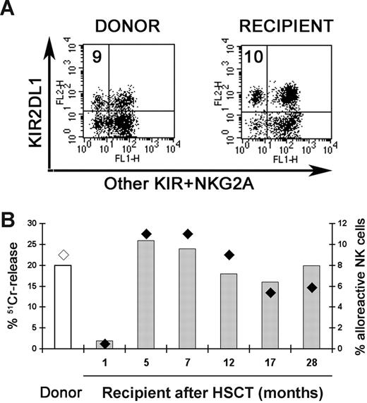
Remarkably, the presence of alloreactive NK cells could be demonstrated in the majority of patients even a long time after transplantation. A representative case (no. 11) is shown in Figure 3; the size of the alloreactive subset and its cytolytic activity was evaluated at various time points for up to 28 months.
Alloreactive NK cells at various time points after transplantation. In case no. 11, characterized by C2 KIR-ligand mismatch and no activating KIR, the alloreactive NK cell subset was represented by cells expressing only KIR2DL1. (A) Staining with a mixture of EB6B-PE, DX9-FITC, CH-L-FITC, and Z199 followed by anti-IgG2b-FITC identified alloreactive NK cells, derived from the donor and the recipient (5 months after HSCT), in the top left quadrant (percent is indicated). (B) The specific lysis of BM15 B-EBV cell line expressing the homozygous haplotype HLA-A*0101, -B*4901 (Bw4), -C*0701 (C1) (columns) and the size of alloreactive NK subset (diamonds) were evaluated analyzing the NK cells derived from the donor (◇) and the recipient (♦) at different time points after transplantation. E:T ratio of 20:1 is shown.
Alloreactive NK cells at various time points after transplantation. In case no. 11, characterized by C2 KIR-ligand mismatch and no activating KIR, the alloreactive NK cell subset was represented by cells expressing only KIR2DL1. (A) Staining with a mixture of EB6B-PE, DX9-FITC, CH-L-FITC, and Z199 followed by anti-IgG2b-FITC identified alloreactive NK cells, derived from the donor and the recipient (5 months after HSCT), in the top left quadrant (percent is indicated). (B) The specific lysis of BM15 B-EBV cell line expressing the homozygous haplotype HLA-A*0101, -B*4901 (Bw4), -C*0701 (C1) (columns) and the size of alloreactive NK subset (diamonds) were evaluated analyzing the NK cells derived from the donor (◇) and the recipient (♦) at different time points after transplantation. E:T ratio of 20:1 is shown.
Role of KIR2DS1 in the induction of alloreactivity against C2+ leukemia cells
Previous data demonstrated that KIR2DS1 selectively recognizes C2 alleles.9,10 The use of KIR2DS1-Fc soluble receptor confirmed this specific binding (Figure 4A).10 In the representative case no. 5 described above (Figure 2), double staining, using mAb combinations as in Figure 1A, revealed that polyclonal NK populations derived from the donor or from the recipient expressed 27% and 26% of KIR2DS1+/L1− cells, respectively. We investigated whether killing of the patient's leukemia blasts or Bw4+C2+ B-EBV cell lines involve, at least in part, KIR2DS1 (as assessed by mAb-mediated blocking experiments). NK cell clones, characterized by the KIR2DS1+/L2+ or KIR2DS1+/NKG2A+ phenotype, were derived from both donor and recipient. Clones were highly cytolytic against the above target cells, and lysis was strongly inhibited by blocking KIR2DS1. Two representative clones derived from the patient after transplantation; 5R-13 clone (KIR2DS1+/L2+) and 5R-4 clone (KIR2DS1+/NKG2A+) are shown in Figure 4B and C and in Figure 4D and E, respectively. These data provide the first experimental evidence that KIR2DS1 is directly involved in the process of recognition and killing of leukemia blasts. Interestingly, in the same experiments, mAb-mediated masking of the inhibitory receptor NKG2A had only a marginal effect, while blocking of KIR2DL2 highly incremented lysis of both leukemia blasts and B-EBV cell line. This effect was unexpected, since target cells were lacking the C1 KIR ligand. Although not shown, the same clones did not kill PHA-blasts nor a B-EBV cell line derived from donor no. 5 (C1/C2), thus demonstrating that they were not autoreactive.
Positive role of KIR2DS1 in allorecognition. (A) 721.221 cells transfected with HLA-Cw6 (C2) or -Cw7 (C1) were stained with either W6/32 anti-HLA class I mAb followed by PE-conjugated isotype-specific goat anti–mouse mAb or with KIR2DL1-Fc or KIR2DS1-Fc soluble receptors followed by PE-conjugated goat anti–human second reagents. Empty profiles indicate staining with second reagent only. Numbers within histograms indicate MRFI. The representative 5R-13 (KIR2DS1+/L2+; B,C) and 5R-4 (KIR2DS1+/NKG2A+; D,E) clones, derived from recipient no. 5, 6 months after transplantation were tested against patient leukemia blasts (B,D), BOB (C; see Figure 2 legend for HLA-I genotypes) and LM expressing HLA-A*3,*29, -B*44,*35 (Bw4/Bw6), -Cw*4,*5 (C2/C2) (E) B-EBV cell lines. The cytolytic activity was evaluated in the absence (□) and in the presence (■) of mAb to the indicated molecules, such as 11PB6 (KIR2DS1), GL-183 (KIR2DL2), Y9 (CD94/NKG2A), and A6-136 (HLA-class I). Similar data were obtained also with HP-3E4 (KIR2DS1) and CH-L (KIR2DL2) mAb. The E:T ratio used was 10:1. The results are representative of 2 independent experiments; the standard deviation of the mean of the triplicates was less than 5%.
Positive role of KIR2DS1 in allorecognition. (A) 721.221 cells transfected with HLA-Cw6 (C2) or -Cw7 (C1) were stained with either W6/32 anti-HLA class I mAb followed by PE-conjugated isotype-specific goat anti–mouse mAb or with KIR2DL1-Fc or KIR2DS1-Fc soluble receptors followed by PE-conjugated goat anti–human second reagents. Empty profiles indicate staining with second reagent only. Numbers within histograms indicate MRFI. The representative 5R-13 (KIR2DS1+/L2+; B,C) and 5R-4 (KIR2DS1+/NKG2A+; D,E) clones, derived from recipient no. 5, 6 months after transplantation were tested against patient leukemia blasts (B,D), BOB (C; see Figure 2 legend for HLA-I genotypes) and LM expressing HLA-A*3,*29, -B*44,*35 (Bw4/Bw6), -Cw*4,*5 (C2/C2) (E) B-EBV cell lines. The cytolytic activity was evaluated in the absence (□) and in the presence (■) of mAb to the indicated molecules, such as 11PB6 (KIR2DS1), GL-183 (KIR2DL2), Y9 (CD94/NKG2A), and A6-136 (HLA-class I). Similar data were obtained also with HP-3E4 (KIR2DS1) and CH-L (KIR2DL2) mAb. The E:T ratio used was 10:1. The results are representative of 2 independent experiments; the standard deviation of the mean of the triplicates was less than 5%.
KIR2DL2/3+ NK clones display poor alloreactivity against C2/C2 leukemia cells
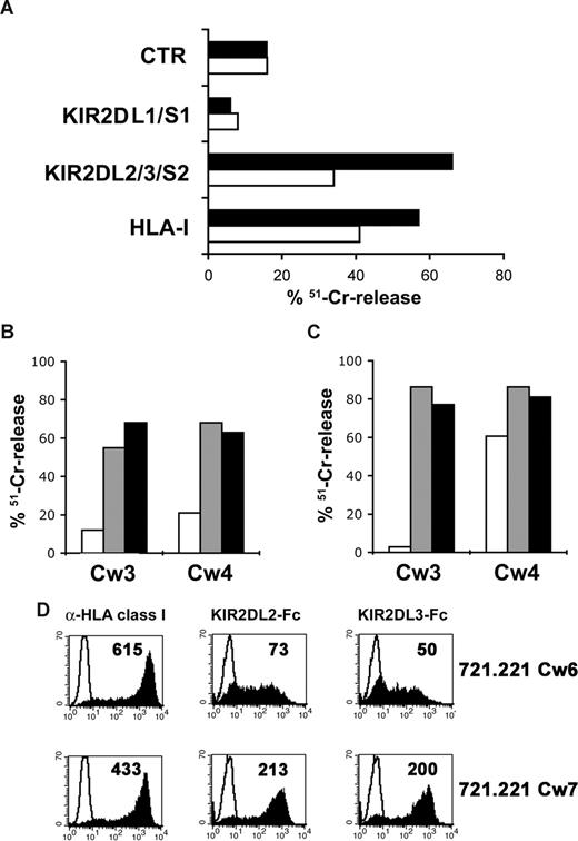
The results reported above suggested that KIR2DL2/3 could recognize not only the expected group of HLA-C alleles (ie, C1), but also alleles belonging to C2 group. This possibility was analyzed both at population and clonal levels, using NK cells derived from different donors and recipients after transplantation. Leukemia blasts, B-EBV cell lines, and cell transfectants were used as target cells. At 3 months after HSCT, the representative case no. 6 displayed a high percentage of KIR2DL2/L3/S2 (82%) and a low percentage of KIR2DS1 (7%). Remarkably, although the alloreactive subset (phenotypically defined in Figure 2A) was highly represented in the polyclonal NK-cell population (ie, 69%), the cytolytic activity against the patient's leukemia blasts or BOB B-EBV cell line was low (Figure 5A). The cytolytic activity against both target cells could be restored by mAb-mediated masking of KIR2DL2/L3/S2 or HLA class I, which suggested that KIR2DL2/L3 in these cells, inhibited lysis through recognition of C2. Analysis of NK-cell clones revealed that only 2 of 16 clones, expressing KIR2DL2/L3 as the only inhibitory receptor, displayed alloreactivity. In this case, however, KIR2DS1 was not expressed in the analyzed panel of NK cell clones. It is worth noting that nonalloreactive NK clones (see the representative 6R-30 clone in Figure 5B) displayed low cytotoxicity not only against Cw3-transfected target cells (C1 specificity) but also against Cw4+ transfectants (C2 specificity). On the other hand, alloreactive NK clones (see the representative 6R-14 clone in Figure 5C) in line with the expected specificity of KIR2DL2/3+ NK cells, discriminated between the 2 C groups. These data may imply that in haplo-HSCT pairs in which C1 represents the KIR-ligand mismatch, phenotypically defined alloreactive NK cells (based on the expression of KIR2DL2/3 alone) may actually display a limited capability of killing C2+ leukemia blasts. This is likely due to the inability of a fraction of KIR2DL2/L3+ NK cells to kill not only C1+ but also C2+ target cells. These results have been further substantiated by the observation that KIR2DL2-Fc and KIR2DL3-Fc soluble receptors could bind not only to 721.221/Cw7 (ie, C1+), but also to 721.221/Cw6 (ie, C2+) cell transfectants (Figure 5D). In all instances, binding to the C2+ transfectant was lower as compared with the C1+ transfectant.
KIR2DL2/3 recognize both C1 and C2 groups of alleles. Case no. 6 was characterized by C1 KIR-ligand mismatch, because the patient expressed HLA-A*1101,*3202, -B*3508,*1517 (Bw6/Bw4), -C*1504,*1602 (C2/C2), while the donor expressed HLA-A*33,*32, -B*14,*15 (Bw6/Bw4), -C*0802,*1602 (C1/C2). (A) Polyclonal activated NK cells derived at the third month after transplantation were tested for cytolytic activity against patient's leukemia blasts (JMML; ■) and the B-EBV BOB (□) in the absence and presence of mAb to the indicated molecules, such as 11PB6 (KIR2DL1/S1), GL-183 (KIR2DL2/L3/S2), and A6-136 (HLA-class I). The E:T ratio used was 10:1. The representative KIR2DL1/S1− KIR2DL2/3+ 6R-30 (B) and 6R-14 (C) clones were tested against 221/Cw3 and 221/Cw4 cell transfectants in the absence (□) and in the presence of the anti-KIR2DL2/3 ( ) CH-L mAb, or the anti–HLA-class I (■) 6A4 (in case of Cw4 transfectant) or A6-136 mAb (in case of Cw3 transfectant). The E:T ratio used was 5:1. (D) 721.221 cells transfected with HLA-Cw6 or -Cw7 were stained with either W6/32 anti–HLA-class I mAb or with KIR2DL2-Fc, KIR2DL3-Fc soluble receptors. Empty profiles indicate staining with second reagent alone. Numbers within histograms indicate MRFI.
KIR2DL2/3 recognize both C1 and C2 groups of alleles. Case no. 6 was characterized by C1 KIR-ligand mismatch, because the patient expressed HLA-A*1101,*3202, -B*3508,*1517 (Bw6/Bw4), -C*1504,*1602 (C2/C2), while the donor expressed HLA-A*33,*32, -B*14,*15 (Bw6/Bw4), -C*0802,*1602 (C1/C2). (A) Polyclonal activated NK cells derived at the third month after transplantation were tested for cytolytic activity against patient's leukemia blasts (JMML; ■) and the B-EBV BOB (□) in the absence and presence of mAb to the indicated molecules, such as 11PB6 (KIR2DL1/S1), GL-183 (KIR2DL2/L3/S2), and A6-136 (HLA-class I). The E:T ratio used was 10:1. The representative KIR2DL1/S1− KIR2DL2/3+ 6R-30 (B) and 6R-14 (C) clones were tested against 221/Cw3 and 221/Cw4 cell transfectants in the absence (□) and in the presence of the anti-KIR2DL2/3 ( ) CH-L mAb, or the anti–HLA-class I (■) 6A4 (in case of Cw4 transfectant) or A6-136 mAb (in case of Cw3 transfectant). The E:T ratio used was 5:1. (D) 721.221 cells transfected with HLA-Cw6 or -Cw7 were stained with either W6/32 anti–HLA-class I mAb or with KIR2DL2-Fc, KIR2DL3-Fc soluble receptors. Empty profiles indicate staining with second reagent alone. Numbers within histograms indicate MRFI.
Killing of C1/C1 leukemia blasts by KIR2DL1+ NK cells is not influenced by the coexpression of KIR2DS2
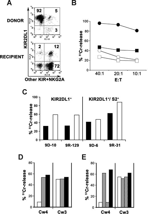
As stated above (Figure 1B), at present, no mAbs are available that distinguish NK cells expressing KIR2DL2 from those expressing KIR2DS2. This may impair the phenotypic assessment of the alloreactive subset, when this is coexpressing KIR2DS2. Indeed, KIR2DS2 was expressed in 3 of 7 cases in which C2 was the KIR ligand missing in the recipient (Table 2). In these 3 cases, the size of the alloreactive subset, defined phenotypically by the presence of KIR2DL1 and the absence of other inhibitory KIRs, may be underestimated due to the exclusion of cells coexpressing KIR2DS2 and KIR2DL1, as these cells would be stained by the antibody that recognizes KIR2DL2/L3/S2. Because KIR2DL1+/2DS2+ cells should be included in the alloreactive population, we performed a clonal analysis to define their actual frequency. While in 2 cases (no. 10 and no. 13) no KIR2DL1+/2DS2+ alloreactive NK clones could be isolated, a substantial number of such clones could be obtained in a third case (no. 9 in Figure 6). In this instance, freshly derived donor NK cells exhibited a high percentage of KIR2DL1 single positive cells (52%). After culture in the presence of IL-2, these cells represented the majority of NK cells (92%). Notably, the combined expression of KIR2DL1 with other receptors was confined to a minor subset (5%; Figure 6A top panel). The pattern of receptor expression changed dramatically in recipient NK cells; indeed, single KIR2DL1+ cells represented only 2%, whereas 9% of NK cells coexpressed KIR2DL1 and other receptors. This pattern of receptor expression remained substantially unchanged (2% and 12%, respectively) after culture in the presence of IL-2 (Figure 6A bottom panel). Importantly, recipient NK cells were characterized by this phenotype both at early (2 and 3 months) and at late time points (1 and 3 years) after HSCT. Notably, data were similar in peripheral blood and in bone marrow-derived NK cells (at the third month). These discrepancies between donor and recipient mainly reflected a decrease in KIR2DL1+ cell subset (14%) and an increment of KIR2DL2/L3/S2+ cells (74%) in the recipient. Consistent with their phenotype, donor NK cells displayed a higher alloreactivity as compared with NK cells obtained from the recipient (Figure 6B). Nevertheless, the cytolytic activity of recipient NK cells could still be demonstrated against autologous leukemia blasts, as well as against the KIR ligand-related BM15 B-EBV cell line (Figure 6B). Analysis at the clonal level revealed that alloreactivity was detectable in all clones expressing KIR2DL1 alone or in combination with KIR2DS2. The cytolytic activity of 4 representative NK clones, derived from the donor (no. 9D) and from the recipient (no. 9R), belonging to these phenotypes are shown in Figure 6C. Importantly, all KIR2DL1+ clones displayed comparable cytolytic activity against leukemia blasts irrespective of the presence or absence of KIR2DS2. In addition, all these clones killed the BM15 B-EBV cell line. In an attempt to better evaluate the possible contribution of KIR2DS2 to the cytolytic activity of KIR2DL1+/2DS2+ clones, we further analyzed cell transfectants expressing HLA-Cw3 or -Cw4. As shown in Figure 6, both KIR2DL1+/2DS2− (Figure 6D) and KIR2DL1+/2DS2+ (Figure 6E) clones displayed a clear-cut ability to discriminate between the 2 types of transfectants. Indeed, they killed Cw3+ (C1) but not Cw4+ (C2) transfectants. Blocking of KIR2DL1 by specific mAbs efficiently restored lysis of Cw4+ transfectants, while it had no effect on killing of Cw3+ transfectants. In addition, blocking of KIR2DS2 did not modify killing of Cw3+ transfectants by KIR2DL1+/2DS2+ NK clones. Taken together, these data show that both groups of KIR2DL1+ NK clones are alloreactive and that KIR2DS2 does not contribute to lysis.
Alloreactivity versus C2 missing target in case of expression of KIR2DS2. Case no. 9 was characterized by C2 KIR-ligand mismatch, because the patient expressed HLA-A*0201,*2402, -B*4001,*5101 (Bw6/Bw4), -C*0102,*0304 (C1), while the donor expressed HLA-A*02,*04, -B*40,*44 (Bw6/Bw4), -C*03,*02 (C1/C2). Thus KIR2DL1 represents the relevant inhibitory KIR. KIR2DS2 was found to be present by KIRtyping analysis, but no mAb can discriminate it from its inhibitory counterparts, KIR2DL2/3, which were also present. Dot plots (A) using 11PB6 (recognizing here KIR2DL1 only) followed by anti–IgG1-PE in combination with CH-L (anti-KIR2DL2/L3/S2), AZ158 (anti-KIR3DL1/2), and Z199 (anti-NKG2A), followed by anti-IgG2b/2a-FITC, are not completely informative to assess the size of the alloreactive subset. Activated NK-cells derived from the donor and the recipient (3 months after HSCT) are shown. (B) Cytolytic activity of activated NK cell populations derived from the donor (filled symbols) and from the recipient at the third month after HSCT (open symbols) was tested against patient leukemia blasts (CD10+ ALL, squares) and BM15 B-EBV cell line (Bw4+ and C1+) expressing the homozygous haplotype HLA-A*0101, -B*4901 (Bw4), -C*0701 (C1) (circles) at different E:T ratios as indicated. (C) representative NK cell clones, derived from the donor (no. 9D) and the recipient (no. 9R), characterized by KIR expression of either KIR2DL1 alone (9D-10 and 9R-129 clones) or KIR2DL1 in combination with KIR2DS2 (9D-6 and 9R-31 clones), were tested against patient leukemia blasts (■) or BM15 (□) at E:T ratio of 10:1. 9R-129 (D) and 9R-31 (E) were tested against 221-Cw4 and 221-Cw3 cell transfectants in the absence (□) or in the presence of anti-KIR2DL1 (HP-3E4,  ), anti-KIR2DS2 (GL-183, ▫), or anti-HLA-I (a mixture of A6-136 and 6A4, ■) mAb, at E:T ratio of 5:1. Representative experiments are shown; the SD of the mean of the triplicates was less than 5%.
Alloreactivity versus C2 missing target in case of expression of KIR2DS2. Case no. 9 was characterized by C2 KIR-ligand mismatch, because the patient expressed HLA-A*0201,*2402, -B*4001,*5101 (Bw6/Bw4), -C*0102,*0304 (C1), while the donor expressed HLA-A*02,*04, -B*40,*44 (Bw6/Bw4), -C*03,*02 (C1/C2). Thus KIR2DL1 represents the relevant inhibitory KIR. KIR2DS2 was found to be present by KIRtyping analysis, but no mAb can discriminate it from its inhibitory counterparts, KIR2DL2/3, which were also present. Dot plots (A) using 11PB6 (recognizing here KIR2DL1 only) followed by anti–IgG1-PE in combination with CH-L (anti-KIR2DL2/L3/S2), AZ158 (anti-KIR3DL1/2), and Z199 (anti-NKG2A), followed by anti-IgG2b/2a-FITC, are not completely informative to assess the size of the alloreactive subset. Activated NK-cells derived from the donor and the recipient (3 months after HSCT) are shown. (B) Cytolytic activity of activated NK cell populations derived from the donor (filled symbols) and from the recipient at the third month after HSCT (open symbols) was tested against patient leukemia blasts (CD10+ ALL, squares) and BM15 B-EBV cell line (Bw4+ and C1+) expressing the homozygous haplotype HLA-A*0101, -B*4901 (Bw4), -C*0701 (C1) (circles) at different E:T ratios as indicated. (C) representative NK cell clones, derived from the donor (no. 9D) and the recipient (no. 9R), characterized by KIR expression of either KIR2DL1 alone (9D-10 and 9R-129 clones) or KIR2DL1 in combination with KIR2DS2 (9D-6 and 9R-31 clones), were tested against patient leukemia blasts (■) or BM15 (□) at E:T ratio of 10:1. 9R-129 (D) and 9R-31 (E) were tested against 221-Cw4 and 221-Cw3 cell transfectants in the absence (□) or in the presence of anti-KIR2DL1 (HP-3E4,  ), anti-KIR2DS2 (GL-183, ▫), or anti-HLA-I (a mixture of A6-136 and 6A4, ■) mAb, at E:T ratio of 5:1. Representative experiments are shown; the SD of the mean of the triplicates was less than 5%.
Discussion
The present study provides new relevant data on haplo-HSCT, a procedure in which donor NK alloreactivity is crucial for the successful eradication of high-risk leukemias. In particular, we show that: (1) donor-derived alloreactive NK cells are generated and persist for many years in haplo-HSCT recipients; (2) KIR2DL2/3+ NK cells can barely distinguish between C1+ or C2+ target cells, and their alloreactivity is in most cases dependent on the coexpression of KIR2DS1, which can directly recognize C2 alleles on leukemia cells; and (3) new mAbs make it possible to distinguish between certain activating and inhibitory KIR, thus allowing a better phenotypic identification of alloreactive NK subsets.
Recent experimental evidences support the notion that, during maturation, NK cells require recognition of major histocompatibility complex (MHC) class I molecules to acquire full effector function, a phenomenon defined as “licensing” or “education.”15-17 Lack of proper signaling may lead to hyporesponsiveness of NK cells lacking MHC-specific inhibitory receptors. Thus, a question arises: why donor NK cells undergoing maturation in the mismatched recipient can develop into alloreactive NK cells capable of killing leukemia cells, since they express KIR that are not engaged by HLA-class I alleles of the patient? A possible explanation is that in haplo-HSCT, due to infusion of particularly high numbers of cells, the bone marrow (BM) microenvironment in which NK cells undergo maturation may be predominantly of donor type. This would be sufficient to support a process of NK education, which includes alloreactive NK cells.
NK alloreactivity can be predicted to occur when analysis of HLA-class I genotypes reveals that a KIR ligand is expressed in the donor but is missing in the recipient (KIR ligand-mismatch). This evaluation must be complemented by KIR genotypic analysis of the donor to assess the presence of the relevant KIR, displaying specificity for the KIR-ligand mismatch. However, the genetic evidence of a KIR/KIR-ligand mismatch is not sufficient to predict the size of the alloreactive NK-cell subset. Indeed, in a given donor, the proportion of NK cells expressing a certain KIR, as well as the possible coexpression of other KIRs or NKG2A, is unpredictable. The definition of the size of the alloreactive NK cell subset is important, because it has been shown to directly correlate with the magnitude of lysis of KIR-mismatched target cells.21,37 In this context, in a previous study, we showed that it is possible to determine the percentage of alloreactive NK cells using appropriate mAb combinations.21 Alloreactive NK cells are characterized by the expression of KIRs specific for the KIR-ligand mismatch and by the lack of other inhibitory receptors specific for the HLA-class I alleles of the patient. In the past, a major limitation was that the available anti-KIR mAbs could not distinguish between inhibitory and activating KIRs. This could result in an underestimation of the actual size of the alloreactive NK-cell subset. In the present study, we took advantage of new anti-KIR mAbs that distinguish inhibitory versus activating KIRs when used in appropriate combinations. Thanks to this approach, in most instances, it is now possible to precisely identify NK subpopulations that uniquely express activating KIRs. A limitation is still represented by the lack of mAbs able to distinguish KIR2DS2+ from KIR2DL2+ cells when both these forms are expressed in the KIR genotype. Phenotypic analysis using appropriate anti-KIR mAbs can even be performed directly in PBMCs, thus allowing a rapid screening to identify the most appropriate donor for HSCT. Through this improved approach, in most instances, it is now possible to precisely identify the alloreactive NK subset, since this will include not only cells expressing the relevant inhibitory KIR, but also those coexpressing activating KIRs. Notably, activating KIRs should be considered as “permissive KIRs,” irrespective of their ability to participate or not in the recognition of leukemia cells. In this context, the specificity of activating KIRs for HLA ligands remained at least in part poorly defined. A previous study reported that soluble KIR2DS1 tetramers specifically bind to peptide-HLA-C2 complexes in B-EBV cells.10 In agreement with this finding, here we show that KIR2DS1-Fc binds to B-EBV cell lines or cell transfectants (Figure 4A), thus providing evidence that KIR2DS1, similarly to KIR2DL1, can discriminate between C1 and C2 groups. Binding of KIR2DS1, however, was weaker than that of KIR2DL1. In addition, an alloreactive NK-cell clone has been described to coexpress KIR2DS1 and NKG2A, and KIR2DS1-mediated recognition of C2 was able to override NKG2A-mediated inhibition in cytotoxicity experiments against C2+ EBV targets.38 In our present study, we provide the first evidence that KIR2DS1 plays a crucial role in the process of inducing lysis of C2/C2 leukemia blasts and not only of EBV-transformed cell lines (Figure 4). Indeed, a series of NK-cell clones, coexpressing KIR2DS1 together with either NKG2A or KIR2DL2/3, were derived from both donor and recipient after HSCT. These clones efficiently killed the patient leukemia blasts, and their cytolytic activity was inhibited by mAb-mediated blocking of KIR2DS1. On the other hand, the B-EBV cell line derived from the donor (C1/C2) was resistant to lysis by the same clones (either autologous or derived from the patient after HSCT). In this case, lysis could be restored only upon mAb-mediated blocking of HLA-class I molecules. The lack of autoreactivity of KIR2DS1+ NK clones in a person carrying the ligand for this receptor is likely related to the fact that the donor was heterozygous for C2, while the patient was homozygous. This suggests that, in these clones, a balance between triggering via KIR2DS1 and inhibition via HLA-specific inhibitory receptors can control alloreactivity versus self-tolerance at the single cell level. Chewing et al described that the “missing self” alloresponse to C2 could rarely be observed in NK cells from donors lacking KIR2DS1.39 We have now clarified the molecular basis of this phenomenon by demonstrating that KIR2DL2/3 can recognize not only C1, but also C2. Although the strength of the interaction between KIR2DL2/3 and C2 is weaker as compared with that between KIR2DL2/3 and C1 (as documented by KIR-Fc binding experiments), it is sufficient to inhibit the NK-cell cytotoxicity at the polyclonal and clonal levels (Figure 5). These data are in line with a recent paper by Moesta et al providing evidence that KIR2DL2/L3 (KIR2DL2 with higher efficiency) could interact not only with C1, but also with C2.40 This issue was addressed both by lysis inhibition experiments using NKL cell line transduced with either KIR2DL2*001 or KIR2DL3*001 as effector cells and by KIR-Fc binding to beads coated with different HLA alleles.40 Our data indicate that, in case of C1-mismatched HSCT, most but not all KIR2DL2/3+ NK cells displayed weak alloreactivity against C2/C2 target cells. This variability in alloreactivity is likely to reflect not only the presence of KIR2DS1, but also the relative density of other activating NK receptors such as natural cytotoxicity receptors (NCRs).31,41 Other factors that may influence the alloreactivity of these clones may be represented by the surface density of KIR2DL2/3 inhibitory receptors, as well as their allelic variability. In addition, the level of inhibition may be influenced by allelic differences within the C2 subgroup as suggested by Moesta et al.40 The reason why the low alloreactivity of KIR2DL2/3+ clones was not documented in previous studies may be because no extensive analysis of these clones was performed on C2/C2 targets. Notably, analysis was focused on clones selected on the basis of alloreactivity.3-5,32,42-44 Remarkably, among C1-mismatched pairs, all patients given haplo-HSCT from donors expressing KIR2DS1 (ie, 4 of 8) are alive and in complete remission, while fatal relapses occurred in 3 of 4 recipients transplanted from donors lacking this activating KIR, thus suggesting a clinical relevance of KIR2DS1-mediated recognition of leukemia. We also addressed the question of whether KIR2DS2 could directly recognize and induce killing of leukemia blasts. This was not documented, at least in the cases analyzed so far. Analysis of NK cell clones allowed us to confirm that KIR2DL1 recognizes exclusively C2 group of alleles and that alloreactivity against C1/C1 target cells is mediated by NK cells expressing KIR2DL1 as the only inhibitory receptor. These cells may or may not coexpress activating KIRs (eg, KIR2DS2 in Figure 6) that do not influence their alloreactivity.
In conclusion, our data demonstrate that NK alloreactivity against leukemia blasts is regulated by integrated signals delivered by both inhibitory and activating KIRs. The importance of a precise phenotypic analysis allowing the definition of the size of the alloreactive subset is documented by the demonstration that not only inhibitory, but also certain activating KIRs (eg, KIR2DS1) play a crucial role in the process of leukemia blast recognition and killing. On the other hand, the expected alloreactivity against C2/C2 leukemia blasts of KIR2DL2/3+ NK cells is often impaired, because this receptor recognizes not only C1 but also C2. Taken together, these findings provide new information that may greatly improve the understanding of the mechanisms at the basis of successful haplo-HSCT in patients with high-risk leukemias.
The online version of this article contains a data supplement.
The publication costs of this article were defrayed in part by page charge payment. Therefore, and solely to indicate this fact, this article is hereby marked “advertisement” in accordance with 18 USC section 1734.
Acknowledgments
This work has been supported by grants awarded by Fondazione Berlucchi; Associazione Italiana per la Ricerca sul Cancro (AIRC); Istituto Superiore di Sanità (ISS); Ministero dell'Istruzione, dell'Università e della Ricerca (MIUR-PRIN 2005, project 2005063024_004, MIUR-PRIN 2006 project 2006061378_003, FIRB-MIUR project-RBNE017B4); European Union FP6, LSHB-CT-2004-503319-Allostem (the European Commission is not liable for any use that may be made of the information contained); Fondazione Compagnia di San Paolo; Ministero della Salute, Ricerca Finalizzata 2006 Programma Integrato Oncologia.
Authorship
Contribution: D.P. designed and performed research, interpreted data, and wrote the paper; S. Marcenaro and M.F. performed research and analyzed data; S. Martini performed research and collected data; M.E.B. took care of patients and provided selected samples; D.M. provided samples of leukemia blasts; E.R. performed research; C.C. contributed vital new analytical tools; M.M. performed HLA typing; R.M. designed research; M.C.M. supervised research; E.V. contributed vital new analytical tools and discussed data; L.M. designed research and revised the paper; F.L. directs the transplantation program and was responsible for patient care; and A.M. designed research, interpreted data, and wrote the paper.
Conflict-of-interest disclosure: A.M. and E.V. are founders and shareholders of Innate-Pharma (Marseille, France). The remaining authors declare no competing financial interests.
Correspondence: Daniela Pende, Istituto Nazionale per la Ricerca sul Cancro, L go R Benzi 10, 16132 Genova, Italy; e-mail: daniela.pende@istge.it.


This feature is available to Subscribers Only
Sign In or Create an Account Close Modal