Abstract
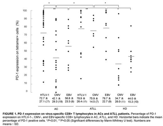
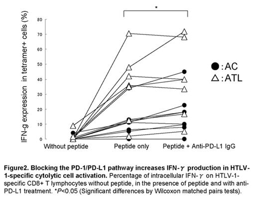
Adult T-cell leukemia/lymphoma (ATLL) develops after infection with human T-cell leukemia virus-1 (HTLV-1) with a long latent period. The negative regulatory programmed death-1/programmed death-ligand 1 (PD-1/PD-L1) pathway has been implicated in the induction of CTL exhaustion during chronic viral infection and tumor escape from host immunity. We have previously reported decreased frequency and function of HTLV-1 Tax-specific CD8+ T-cell in ATLL patients due to insufficient cytolytic effector molecule. To determine whether the PD-1/PD-L1 pathway is involved in the immunosuppressive state in HTLV-1-infected individuals and immune evasion in ATLL patients, we examined PD-1/PD-L1 expression in asymptomatic HTLV-1 carriers (ACs) and ATLL patients. PD-1 expression was significantly up-regulated on lymphocytes in ACs and ATLL patients compared to non-HTLV-1-infected individuals. The rates of PD-1 expression on HTLV-1-specific CTL in ACs and ATLL patients were similarly high (p = 0.32) although PD-1 expression was significantly up-regulated on total lymphocytes, cytomegalovirus- and EBV-specific CTL in ATLL patients compared to ACs and non-HTLV-1-infected individuals (Figure1, p < 0.01). This may be one reason why HTLV-1-infected cells are not eradicated even in the presence of anti-HTLV-1 antigen-specific CTL in the carrier state. Primary ATLL cells in 15.0% of ATLL patients and an HTLV-1-infected cell line expressed PD-L1, while few lymphocytes from ACs were positive for this ligand. Anti-PD-L1 antagonistic antibody restored HTLV-1-specific CTL functions (IFN-γ, TNF-α, CD107a) ex vivo (Figure2). These observations suggest that the immunoregulatory PD-1/PD-L1 pathway is operative during persistent viral infection, and the pathway may induce the exhausted phenotype of CTL in HTLV-1 infected individuals, which may lead to immunodeficiency and immune evasion in ACs and ATLL patients. Furthermore, reactivation of exhausted T cells by blocking the PD-1/PD-L1 may be an effective immunological strategy for the clearance of persistent HTLV-1 infection and the treatment of ATLL.
PD-1 expression on virus-specific CD8+ T lymphocytes in ACs and ATLL patients. Percentage of PD-1 expression on HTLV-1-, CMV-, and EBV-specific CD8+ lymphocytes in AC, ATLL, and HD. Horizontal bars indicate the mean percentage of PD-1 positive cells. *P < 0.01; **P < 0.05 (Significant differences by Mann-Whitney U test). Numbers are means ± SD.
PD-1 expression on virus-specific CD8+ T lymphocytes in ACs and ATLL patients. Percentage of PD-1 expression on HTLV-1-, CMV-, and EBV-specific CD8+ lymphocytes in AC, ATLL, and HD. Horizontal bars indicate the mean percentage of PD-1 positive cells. *P < 0.01; **P < 0.05 (Significant differences by Mann-Whitney U test). Numbers are means ± SD.
Blocking the PD-1/PD-L1 pathway increases INF-γ production in HTLV-1-specific cytolytic cell activation. Percentage of intracellular IFN-γ on HTLV-1-specific CD8+ T lymphocytes without peptide, in the presence of peptide and with anti-PD-L1 treatment. *P < 0.05 (Significant differences by Wilcoxon matched pairs tests).
Blocking the PD-1/PD-L1 pathway increases INF-γ production in HTLV-1-specific cytolytic cell activation. Percentage of intracellular IFN-γ on HTLV-1-specific CD8+ T lymphocytes without peptide, in the presence of peptide and with anti-PD-L1 treatment. *P < 0.05 (Significant differences by Wilcoxon matched pairs tests).
Author notes
Disclosure: No relevant conflicts of interest to declare.


This feature is available to Subscribers Only
Sign In or Create an Account Close Modal