Abstract
The class IA subgroup of phosphoinositide 3-kinase (PI3K) is activated downstream of antigen receptors, costimulatory molecules, and cytokine receptors on lymphocytes. Targeted deletion of individual genes for class IA regulatory subunits severely impairs the development and function of B cells but not T cells. Here we analyze conditional mutant mice in which thymocytes and T cells lack the major class IA regulatory subunits p85α, p55α, p50α, and p85β. These cells exhibit nearly complete loss of PI3K signaling downstream of the T-cell receptor (TCR) and CD28. Nevertheless, T-cell development is largely unperturbed, and peripheral T cells show only partial impairments in proliferation and cytokine production in vitro. Both genetic and pharmacologic experiments suggest that class IA PI3K signaling plays a limited role in T-cell proliferation driven by TCR/CD28 clustering. In vivo, class IA–deficient T cells provide reduced help to B cells but show normal ability to mediate antiviral immunity. Together these findings provide definitive evidence that class IA PI3K regulatory subunits are essential for a subset of T-cell functions while challenging the notion that this signaling mechanism is a critical mediator of costimulatory signals downstream of CD28.
Introduction
Assembly of membrane signaling complexes in lymphocytes is directed in part by the phospholipid products of phosphoinositide 3-kinase (PI3K) enzymes that are activated following receptor engagement.1 In T cells, antigen recognition is followed by rapid and sustained accumulation of the PI3K product phosphatidylinositol-3,4,5-trisphosphate (PIP3) at the plasma membrane, with particular concentration at the immunologic synapse.2-5 The class IA enzymes are thought to be the main subgroup that produces PIP3 and mediates signals downstream of antigen receptors and costimulatory receptors.1 Genetic manipulations that enhance PI3K pathway activity cause lymphoproliferation in mice.6-9 Conversely, pharmacologic inhibitors of PI3K, such as wortmannin and LY294002, potently block T- and B-cell proliferation.10-13 These observations have supported an essential role for PI3K signaling in lymphocyte activation.1 The clearest link between T lymphocyte signaling and PI3K activation thus far has been through the costimulatory molecule CD28. Phosphorylation of its YXXM motif is thought to be a key means to recruit PI3K enzymes to the cell membrane, and the function of primary T cells is impaired by mutation of this motif.14-16
PI3K enzymes constitute a multigene family, and most members of this family are ubiquitously expressed and comparably sensitive to inhibition by wortmannin and LY294002.17,18 In addition, wortmannin and LY294002 inhibit other cellular enzymes, including the kinase mTOR that is essential for T-cell proliferation.18-20 Therefore, a precise understanding of PI3K signaling in T cells requires examination of the roles of individual isoforms and subgroups. The 3 class IA catalytic isoforms (p110α, p110β, p110δ) exist as heterodimers with 1 of 5 regulatory subunits (p85α, p55α, p50α, p85β, or p55γ), each possessing conserved Src homology-2 (SH2) domains and other modular domains thought to mediate association with signaling complexes. Class IA regulatory isoforms are essential for stability and localization of the catalytic subunits but possess additional adapter functions independent of their role in regulating class IA PI3K catalytic subunits.21 Mouse gene-targeting experiments have identified essential functions for p85α in B cells and mast cells.11,22-24 However, T-cell development and function are unimpaired in mice lacking either p85α, p85α/p55α/p50α, or p85β.11,24,25 Mice lacking p85α have impaired T-helper differentiation, but this appears to be due to T-cell–extrinsic defects.22,26 p85β-deficient T cells show no differences in PI3K signaling responses but have enhanced survival following suboptimal stimulation, suggesting a possible adapter function for p85β in a T-cell survival pathway.25
T cells express all 3 class IA PI3K isoforms (p110α, p110β, and p110δ). T cells lacking p110α or p110β have not been studied, owing to early embryonic lethality in the gene-targeted mice.27,28 Mice with a knock-in point mutation in p110δ that abolishes kinase activity (denoted p110δKI herein) exhibit selective impairments in T-cell signaling, including reduced T-cell receptor (TCR)–mediated Ca2+ mobilization as well as reduced proliferation in vitro.29 p110δKI and p110δ-null (p110δKO) mice exhibit impaired T-dependent antibody responses29-31 ; however, this could be the result of B-cell–intrinsic defects. Other T-cell–mediated responses have not been tested in p110δKI or p110δKO mice. Further, residual T-cell function in mice lacking p110δ activity could be mediated by signaling through p110α and p110β. Because of these factors, a more complete deletion of class IA PI3K is needed to determine the role of this subclass in T cells.
In this study we assessed the general function of class IA in T cells by deletion of genes that encode all 4 class IA regulatory isoforms normally expressed in T cells (p85α, p55α, p50α, p85β). Using conditional gene targeting, we ensured that class IA PI3K signaling would be abrogated specifically in T cells. The results establish that class IA PI3K regulatory subunits are essential for PI3K signaling output and do contribute to T-cell proliferation and function under certain conditions. However, even in the absence of detectable Akt phosphorylation these cells are able to proliferate under costimulatory conditions and mount effective antiviral responses in vivo. These results indicate that in comparison with B cells, T cells are less dependent on PI3K signaling for proliferation and immune function.
Materials and methods
Mice
The generation of Pik3r1f mice is described elsewhere.32 These mice were bred with Pik3r2n (p85β-null) mice33 and to Lck-Cre transgenic mice34 or CD4-Cre transgenic mice.35 Mice were maintained in a mixed background (C57BL/6 × 129SvEv) for the experiments shown here and were studied at 2 to 4 months of age. Animals were housed in autoclaved microisolator cages with sterilized food and water. All procedures were approved by the institutional animal care and use committee of the University of California-Irvine.
Fluorescence-activated cell sorter analysis
Single-cell suspensions of red blood cell (RBC)–depleted spleens, lymph nodes, thymi, or purified T cells were stained with different combinations of the following reagents: anti-CD8 PerCP (BD Biosciences, San Diego, CA); anti-Thy1.2 PE, anti-CD62L PE, anti-CD25 PE, anti-CD44 FITC, anti-CD69 FITC, and anti-CD4 APC (eBioscience, San Diego, CA); and annexin V PE (Caltag, Burlingame, CA). Cell divisions were tracked following staining of cells with carboxyfluorescein diacetate succinimidyl ester (CFSE; Molecular Probes, Eugene, OR). Samples were acquired on a FACScalibur (BD Biosciences) and data analyzed by CellQuest (BD Biosciences) and FlowJo (Tree Star, San Carlos, CA) software.
Immunoblotting
Purified T cells (1 × 10⁶) were lysed in a detergent buffer (1% Triton X-100, 50 mM Tris [pH 7.4], 10% glycerol, 150 mM NaCl) containing a protease inhibitor cocktail (Sigma, St Louis, MO) and proteins resolved by 7.5% or 10% sodium dodecyl sulfate–polyacrylamide gel electrophoresis (SDS-PAGE). Proteins were transferred to nitrocellulose membranes, which were then blocked in 5% milk in Tris-buffered saline (TBS) for 1 hour. To analyze PI3K isoform expression, primary antibodies used were purified rabbit antisera specific for p110β (H-198, Santa Cruz Biotechnology, Santa Cruz, CA) and p110δ (kindly provided by Bart Vanhaesebroeck), a mixture of hybridoma supernatants specific for p110α (clones U3A and I1A, kind gift from Anke Klippel), an mAb specific for an N-terminal epitope in p85α (clone AB6, Upstate, Charlottesville, VA), and a rabbit antiserum that recognizes all class IA PI3K regulatory isoforms (anti-pan-p85; no. 06-195, Upstate). Rabbit polyclonal or monoclonal antibodies specific for Bcl-XL, phosphorylated S6 (phospho-S6) (Ser235/236), phospho-IκBα (Ser32), and phospho-Akt (Ser473) (nos. 2762, 2211, 9241 and clone 193H12, Cell Signaling Technology, Beverly, MA) were also detected to measure T-cell activation by immunoblot. As loading controls, mAbs specific for β-actin (clone AC15, Sigma, St Louis, MO) were used.
Purification and culture of T cells
T cells were purified via negative selection on magnet-assisted cell separation (MACS) columns (Miltenyi Biotec, Auburn, CA) as described,11 using the Pan T Cell Isolation Kit or the CD8 T cell Isolation Kit. Purity was verified to be comparable between wild-type (WT) and knockout cells and above 95% by fluorescence-activated cell sorter (FACS) analysis. For all assays, cells were cultured in RPMI 1640 supplemented with 10% heat-inactivated FCS, 5 mM HEPES, 2 mM l-glutamine, penicillin (100 U/mL), streptomycin (100 μg/mL), and 50 μM β-mercaptoethanol. T cells were stimulated with platebound anti-CD3 (clone 2C11, Southern Biotechnology, Birmingham, AL), anti-CD28 (clone 37.51, BD Biosciences), and IL-2 (R&D Systems, Minneapolis, MN). For earlier time points, T cells or lymphocytes were stimulated by coating with anti-CD3 (1 μg/mL) and then crosslinking with goat antihamster (Vector Laboratories, Burlingame, CA) at 20 μg/mL for 2 or 5 minutes. To inhibit PI3K pharmacologically, wortmannin or LY294002 (Calbiochem, San Diego, CA) was added at final concentrations of 50 nm and 10 μM, respectively. To activate or inhibit the Erk pathway, cells were treated with PMA at 20 ng/mL or U0126 at 10 μM (Promega, Madison, WI). For thymidine incorporation assays, 5 × 10⁴ T cells were stimulated in triplicate wells of 96-well plates (Falcon, Bedford, MA) with various amounts of the listed stimuli, and they were allowed to proliferate for 48 hours. Then, 1 μCi (0.037 MBq) 3H-thymidine was then added, and 16 hours later the cells were harvested onto filter mats with a Tomtec (Hamden, CT) harvester. Filters were counted with a BetaPlate system (Wallac, Turku, Finland). Cells were labeled with CFSE, as previously described.25
Superantigen proliferation assays
T cells were purified as described in “Purification and culture of T cells” and mixed in round-bottom 96-well plates (Falcon) with various doses of staphylococcal enterotoxin B (SEB; Sigma) and splenocytes that were treated with 50 μM mitomycin C for 30 minutes prior to mixing. This was performed using a 1:1 T-cell–splenocyte ratio, with 1 × 10⁵ T cells per well. To measure proliferation, cells were pulsed with 3H-thymidine, as described in “Purification and culture of T cells.”
Immunizations, antibody ELISAs, and germinal center analysis
Mice were injected intraperitoneally with 100 μL nitrophenol (NP)–Ficoll or NP-OVA (Biosearch Technologies, Novato, CA), which was precipitated using Imject Alum (Pierce, Rockford, IL). Serum was obtained from vena cava bleeds 8 days after injection. To measure levels of serum IgM, IgG1, IgG2a, or IgG3, enzyme-linked immunosorbent assay (ELISA) was conducted on duplicate samples of sera using platebound NP-BSA (Biosearch Technologies) as a capture reagent. Antibodies specific for NP were detected with anti–mouse IgM, IgG1, IgG2a, or IgG3 using horseradish peroxidase (HRP)–conjugated antibodies (Zymed, South San Francisco, CA). Absorbance was read at 405 nm, and dilutions that yielded signals within a linear range were used for data analysis. Germinal center formation was assessed in spleens from the mice used for antibody ELISAs by staining frozen sections with antibodies to B220 and peanut agglutinin (PNA) and imaging using immunofluorescence microscopy, as previously described.29
Intracellular detection of phosphorylated proteins and p85α
For early time points, purified T cells were stimulated with platebound anti-CD3, anti-CD3 and IL-2, or with anti-CD3 and anti-CD28 as described above. To detect phospho-S6 or p85α, cells were fixed and permeabilized using the Cytofix/Cytoperm kit from BD Biosciences. A rabbit polyclonal antibody specific to Ser235/236 phospho-S6 (no. 2211, Cell Signaling Technology) was then added, followed by an FITC-conjugated goat anti–rabbit IgG antibody (F0382, Sigma). After washing, the cells were analyzed via FACS. For single-cell analysis of p85α, mAb specific to p85α (clone AB6, Upstate Biotechnology) was used in conjunction with goat anti–mouse IgG-FITC (F8624, Sigma). To detect phosphorylated ZAP-70 (Tyr493), p44/42 MAPK/Erk (Thr202/Tyr204), or Akt (Ser473), cells were fixed and permeabilized using paraformaldehyde and methanol and stained with rabbit mAbs per the manufacturer's suggested protocol (clones 136F12, 197G2, and 193H12; Cell Signaling Technology). As secondary antibodies, these reagents were followed with goat anti–rabbit IgG antibodies that were coupled to Alexa-488 or Alexa-647 (nos. 11070 or 21246, respectively; Molecular Probes).
Calcium flux assay
Lymphocytes were loaded with the calcium indicator dye Fluo-3 and stimulated as in previous reports.29 Briefly, after acquiring a baseline level of Fluo-3 for 1 minute, T cells were then stimulated with anti-CD3 plus goat antihamster, and Fluo-3 intensity was measured using flow cytometry for 7 minutes. At the end of the stimulation, cells were treated with ionomycin at 1 μM (Calbiochem).
Cytokine ELISAs
Supernatants from T cells stimulated with platebound anti-CD3, anti-CD3 and IL-2, or with anti-CD3 and anti-CD28 were taken 48 hours after stimulation, and the levels of IL-2 and IFN-γ were measured using the eBioscience Ready-Set-Go kits (nos. 88-7024 and 88-7314, respectively).
Mouse hepatitis virus infections
Mice were injected intraperitoneally with 2 × 10⁵ plaque-forming units (PFU) of mouse hepatitis virus (MHV) (strain DM) suspended in 500 μL Hanks balanced salt solution (HBSS) and killed 5, 7, and 12 days after infection. Splenocytes were isolated and stained for flow cytometric analysis with the following reagents: anti-CD8 PerCP (BD Biosciences), anti-CD4 APC (eBioscience), anti-IFN-γ PE (BD Biosciences), and PE-conjugated Db/S510-518 major histocompatibility complex (MHC) class I tetramer, which is used for the identification of CD8 T cells specific for viral spike protein antigen.36 Cytotoxic T-lymphocyte (CTL) assays were conducted using target cells loaded with relevant (S510-518) or irrelevant (OVA) peptides, as previously described.37 Ex vivo IFN-γ production was measured using intracellular cytokine staining (ICS) after stimulation with immunodominant peptides, as previously described.37 For intracranial infections, mice were infected with 1000 PFU MHV (strain 275). Brain and liver tissue were isolated at 5 and 12 days after infection and subjected to a titer assay for replicative virus, as previously described.37
Statistical analysis
For all assays, a 2-tailed paired Student t test was conducted to determine statistical significance.
Results
Reduced class IA PI3K catalytic subunit expression in T cells lacking regulatory subunits
To generate mice with combined deletion of regulatory isoforms in T cells, we bred Pik3r1f mice bearing floxed alleles of Pik3r1 (p85α, p55α, p50α)32 with Pik3r2n (p85β-null) mice and Lck-Cre transgenic mice. In this strain the Cre transgene is expressed starting in the double-negative (DN) thymocyte stage. We use the nomenclature “r1ΔT/r2n” and “r1f/r2n” to refer to Cre+ and Cre− mice, respectively. Previously we determined that T cells from Pik3r2n (p85β-null) mice develop normally and have no detectable defects in PI3K signaling or compensatory changes in PI3K isoform expression, so the r1f/r2n (Cre−) littermates serve as controls.25 No defects have been observed in other leukocyte cell types in Pik3r2n mice.25,38
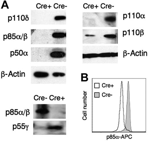
Purified T cells from r1ΔT/r2n mice contained undetectable levels of p85α, p55α, p50α, and p85β (Figure 1A). Intracellular staining using an mAb specific for p85α revealed uniform decreases in r1ΔT/r2n T cells (Figure 1B). This indicated that the Cre/lox system generates efficient deletion of p85α and its alternative transcripts in T cells in these mice. Intracellular stains of thymocytes revealed that this deletion was complete by the double-positive (DP) stage of T-cell development (data not shown). Expression of class IA catalytic subunits was either undetectable (p110α and p110δ) or greatly reduced (p110β) in r1ΔT/r2n T cells (Figure 1A). p110α monomers are unstable,39 providing a likely explanation for the disappearance of this isoform and other catalytic isoforms in double-knockout T cells. In the absence of Pik3r1 and Pik3r2 gene products, the remaining regulatory isoform p55γ encoded by Pik3r3 did show compensatory up-regulation (Figure 1A), and this isoform might mediate the residual catalytic isoform expression.
Complete deletion of major class IA PI3K regulatory subunits causes greatly reduced expression of catalytic isoforms. (A) Immunoblot analysis of catalytic and regulatory isoform expression in purified T cells from r1f/r2n (Cre−) mice or r1ΔT/r2n (Cre+) mice. p55γ could only be detected in the Cre+ lysates using high-sensitivity enhanced chemiluminescence (ECL) reagents (Pierce SuperSignal Femto ECL kit). Data are representative of 4 sets of littermates. (B) Intracellular detection of p85α in T cells of Cre− or Cre+ mice. Fluorescent staining was also observed in T cells from p85α-null (Pik3r1tm1) mice24 and is likely due to cross-reacting proteins, because this antibody detects one other strong band on immunoblots (data not shown). Data are representative of 4 sets of littermates.
Complete deletion of major class IA PI3K regulatory subunits causes greatly reduced expression of catalytic isoforms. (A) Immunoblot analysis of catalytic and regulatory isoform expression in purified T cells from r1f/r2n (Cre−) mice or r1ΔT/r2n (Cre+) mice. p55γ could only be detected in the Cre+ lysates using high-sensitivity enhanced chemiluminescence (ECL) reagents (Pierce SuperSignal Femto ECL kit). Data are representative of 4 sets of littermates. (B) Intracellular detection of p85α in T cells of Cre− or Cre+ mice. Fluorescent staining was also observed in T cells from p85α-null (Pik3r1tm1) mice24 and is likely due to cross-reacting proteins, because this antibody detects one other strong band on immunoblots (data not shown). Data are representative of 4 sets of littermates.
Loss of class IA PI3K does not impair T-cell development
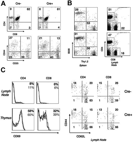
Staining with antibodies to CD4, CD8, CD25, and CD44 revealed no reproducible differences in thymocyte development between r1ΔT/r2n, wild-type (WT), and r1f/r2n mice (Figure 2A). The numbers of DP, CD4 single-positive (CD4SP), and CD8 single-positive (CD8SP) cells were as follows (mean ± SEM, n = 5 mice; no significant differences of P < .05): 186×106 ± 25×106 versus 130×106 ± 13×106 (DP), 18×106 ± 3×10⁶ versus 22×106 ± 3×10⁶ (CD4SP), 9×10⁶ ± 2×10⁶ versus 14×106 ± 5×10⁶ (CD8SP). Peripheral lymphoid organs of young mice showed normal ratios of T cells and B cells as well as CD4/CD8 ratios (Figure 2B). The numbers of CD4 cells (38×106 ± 6×10⁶ versus 32×106 ± 6×10⁶) and CD8 cells (21×106 ± 5×10⁶ versus 15×106 ± 4×10⁶) in the spleen were not significantly different. Expression of activation and memory markers was also unaltered on double-knockout T cells (Figure 2C). Postponing deletion to the DP thymocyte stage using a CD4-Cre strain yielded comparable results (data not shown).
Normal development of lymphocytes in r1ΔT/r2n mice. (A) Flow cytometry of thymi stained with antibodies to CD4 and CD8. CD4−/CD8− (DN) gated thymocytes were also stained with CD44 and CD25 (lower panel). (B) B220 and Thy1.2 expression on splenocytes and CD4/CD8 staining on lymph node cells. Top panels, Cre−; bottom panels, Cre+. (C) Histograms of CD69 expression in CD4+ and CD8+ single-positive thymocytes and lymph node cells as well as expression of memory markers CD44 and CD62L in CD4− or CD8− gated lymph node cells. Numbers represent percentages of cells in the quadrants or regions indicated. Data are representative of 3 sets of littermates.
Normal development of lymphocytes in r1ΔT/r2n mice. (A) Flow cytometry of thymi stained with antibodies to CD4 and CD8. CD4−/CD8− (DN) gated thymocytes were also stained with CD44 and CD25 (lower panel). (B) B220 and Thy1.2 expression on splenocytes and CD4/CD8 staining on lymph node cells. Top panels, Cre−; bottom panels, Cre+. (C) Histograms of CD69 expression in CD4+ and CD8+ single-positive thymocytes and lymph node cells as well as expression of memory markers CD44 and CD62L in CD4− or CD8− gated lymph node cells. Numbers represent percentages of cells in the quadrants or regions indicated. Data are representative of 3 sets of littermates.
Loss of class IA PI3K causes selective impairments in T-cell signaling
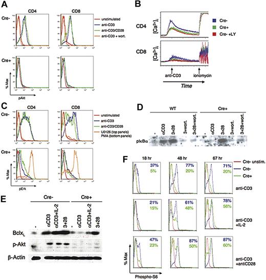
To determine the biochemical effects of class IA PI3K depletion on signal transduction, we measured changes in intracellular Ca2+, protein phosphorylation, and gene transcription following TCR engagement in the presence or absence of CD28 signaling. Where possible, flow cytometric assays were used to obtain single-cell signaling data rather than population measurements. Figure 3A,E shows that Akt phosphorylation was essentially undetectable at both early (5-minute) and late (18-hour) time points in CD4+ or CD8+ T cells lacking Pik3r1 and Pik3r2 gene products. Surface expression of TCR/CD3 was normal, and Zap-70 phosphorylation was not impaired, suggesting that the earliest elements of TCR signaling were intact in double-knockout T cells (data not shown). However, loss of PI3K activation was associated with blunted Ca2+ mobilization and markedly reduced phosphorylation of Erk and IκB following TCR crosslinking (Figure 3B-D). The defects in TCR-mediated Ca2+ flux and IκB phosphorylation were similar to those seen in control T cells treated with PI3K inhibitors. Together with the complete loss of Akt phosphorylation, a standard readout of PI3K signaling output, these data indicate that in regulatory subunit–deficient T cells, TCR-mediated PI3K activation is abolished. Thus, residual class IA proteins and class IB PI3K do not signal efficiently under these conditions.
Biochemical defects in r1ΔT/r2n T cells. (A-D) Activation of biochemical pathways at early time points downstream of the TCR, IL-2R, and CD28 in r1ΔT/r2n (Cre+) mice compared with either r1f/r2n (Cre−) or WT mice. Lymph node cells were stimulated for 5 minutes, and phosphorylation of Akt (A), Erk (C), and IκB (D) was measured. (B) Kinetics of calcium mobilization was assessed using cells loaded with Fluo-3. Where indicated, CD4 and CD8 subsets are distinguished by flow cytometry. The general PI3K inhibitors wortmannin (wort) or LY294002 (LY) were used where indicated. In panel C, the MEK inhibitor U0126 was used as a specificity control in Cre− cells, and the phorbol ester PMA was used as a positive control to show that the Erk pathway can be activated in Cre+ cells. (E-F) Activation of biochemical pathways at later time points downstream of the TCR, IL-2R, and CD28. (E) Immunoblot analysis of Bcl-XL expression and Akt phosphorylation at 18 hours after stimulation of purified T cells (CD4 and CD8). Data are representative of at least 3 experiments, except in the case of 18-hour phospho-Akt analysis (n = 2). (F) Overlays of intracellular phosphorylation of S6 at various time points are shown. Numbers indicate the percentage of cells positive for phospho-S6.
Biochemical defects in r1ΔT/r2n T cells. (A-D) Activation of biochemical pathways at early time points downstream of the TCR, IL-2R, and CD28 in r1ΔT/r2n (Cre+) mice compared with either r1f/r2n (Cre−) or WT mice. Lymph node cells were stimulated for 5 minutes, and phosphorylation of Akt (A), Erk (C), and IκB (D) was measured. (B) Kinetics of calcium mobilization was assessed using cells loaded with Fluo-3. Where indicated, CD4 and CD8 subsets are distinguished by flow cytometry. The general PI3K inhibitors wortmannin (wort) or LY294002 (LY) were used where indicated. In panel C, the MEK inhibitor U0126 was used as a specificity control in Cre− cells, and the phorbol ester PMA was used as a positive control to show that the Erk pathway can be activated in Cre+ cells. (E-F) Activation of biochemical pathways at later time points downstream of the TCR, IL-2R, and CD28. (E) Immunoblot analysis of Bcl-XL expression and Akt phosphorylation at 18 hours after stimulation of purified T cells (CD4 and CD8). Data are representative of at least 3 experiments, except in the case of 18-hour phospho-Akt analysis (n = 2). (F) Overlays of intracellular phosphorylation of S6 at various time points are shown. Numbers indicate the percentage of cells positive for phospho-S6.
Although the PI3K/Akt pathway is thought to contribute to CD28-mediated NFκB pathway activation in T cells, CD28 engagement augmented TCR-mediated IκB phosphorylation in both Cre− and Cre+ T cells (Figure 3D). Surprisingly, wortmannin pretreatment blocked IκB phosphorylation under all conditions in both genotypes.
Analyzing later time points in T-cell activation, we found no detectable Akt phosphorylation, impaired Bcl-XL up-regulation, and reduced S6 phosphorylation in r1ΔT/r2n T cells in response to all stimuli after 18 hours (Figure 2E-F). However, in contrast to the Akt phosphorylation and in agreement with the IκB phosphorylation, the other defects were partially corrected when exogenous IL-2 or anti-CD28 antibodies were included.
Loss of class IA PI3K causes selective impairments in T-cell proliferation
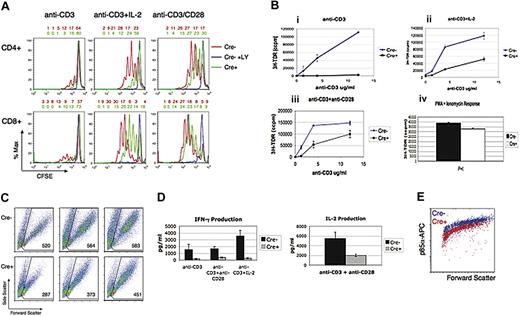
We measured proliferation using thymidine incorporation or cell-division–tracking (CFSE dilution) assays. Both methods showed that the mitogenic effect of TCR crosslinking (anti-CD3 alone) was greatly reduced in r1ΔT/r2n T cells compared with r1f/r2n (Figure 4A-B). Consistent with the biochemical analyses, addition of IL-2 or anti-CD28 partially corrected this defect, though responses were never as robust as those seen in r1f/r2n (Figure 4) or wild-type cells (data not shown). These defects were found in both CD4 and CD8 T cells (Figure 3A) and were also observed in r1ΔT/r2n T cells from CD4-Cre mice (data not shown). We did not observe any differences in apoptosis when comparing r1ΔT/r2n and r1f/r2n T cells cultured in media alone, anti-CD3, or anti-CD3 plus anti-CD28 (data not shown).
Defective TCR-dependent proliferation and cytokine production in r1ΔT/r2n T cells. (A) Shown 60 hours after stimulation, CFSE-labeled purified T cells were gated on CD4 or CD8, and the histograms were overlaid between genotypes. Above each FACS plot are numbers showing the percentage of cells in each division, with the rightmost number indicating the nondivided population. (B) Thymidine incorporation assays of stimulated r1f/r2n (Cre−) or r1ΔT/r2n (Cre+) purified T cells. Stimuli used were anti-CD3 alone (i), anti-CD3 plus IL-2 (ii), anti-CD3 plus anti-CD28 (iii), and PMA plus onomycin (iv). Data are shown as the mean ± SEM of triplicate wells from a representative experiment. (C) Cell-size profile of 60-hour–stimulated Cre− or Cre+ cells. The mean forward scatter of live cells is included on each dot plot. (D) ELISA quantification of IL-2 and IFN-γ in supernatants of r1f/r2n (Cre−) or r1ΔT/r2n (Cre+) CD4+ T cells stimulated for 48 hours. Data are shown as the mean ± SEM of 3 independent experiments comparing Cre− and Cre+ T cells. (E) Intracellular stain for p85α in anti-CD3/anti-CD28–stimulated Cre− and Cre+ purified T cells. Dot plot of forward scatter versus p85α is shown. Data are representative of at least 3 independent experiments.
Defective TCR-dependent proliferation and cytokine production in r1ΔT/r2n T cells. (A) Shown 60 hours after stimulation, CFSE-labeled purified T cells were gated on CD4 or CD8, and the histograms were overlaid between genotypes. Above each FACS plot are numbers showing the percentage of cells in each division, with the rightmost number indicating the nondivided population. (B) Thymidine incorporation assays of stimulated r1f/r2n (Cre−) or r1ΔT/r2n (Cre+) purified T cells. Stimuli used were anti-CD3 alone (i), anti-CD3 plus IL-2 (ii), anti-CD3 plus anti-CD28 (iii), and PMA plus onomycin (iv). Data are shown as the mean ± SEM of triplicate wells from a representative experiment. (C) Cell-size profile of 60-hour–stimulated Cre− or Cre+ cells. The mean forward scatter of live cells is included on each dot plot. (D) ELISA quantification of IL-2 and IFN-γ in supernatants of r1f/r2n (Cre−) or r1ΔT/r2n (Cre+) CD4+ T cells stimulated for 48 hours. Data are shown as the mean ± SEM of 3 independent experiments comparing Cre− and Cre+ T cells. (E) Intracellular stain for p85α in anti-CD3/anti-CD28–stimulated Cre− and Cre+ purified T cells. Dot plot of forward scatter versus p85α is shown. Data are representative of at least 3 independent experiments.
Using forward scatter as a measure of cell size at the time of CFSE analysis, we observed defective growth in r1ΔT/r2n T cells (Figure 4C). Similar to the pattern of proliferative defects, the most severe growth defects were apparent in response to anti-CD3, with lesser impairments when IL-2 or anti-CD28 was included. Production of IL-2 and IFN-γ was greatly diminished in r1ΔT/r2n T cells under various stimulation conditions (Figure 4D). Further, modulation of surface levels of CD25, CD44, CD69, and CD62L was defective in these T cells (data not shown).
Because our model employed a conditional gene-targeting strategy, it was important to determine whether any responding T cells represented rare clones that escaped deletion of Pik3r1. To this end, we used intracellular p85α staining on stimulated r1f/r2n and r1ΔT/r2n T cells. Figure 4E demonstrates that while background staining increased in proportion to changes in forward scatter, stimulated Cre+ cells did not express p85α as compared with Cre− mice. Thus, the response to anti-CD3 plus anti-CD28 was not due to expansion of any subset of T cells that had failed to excise the floxed allele.
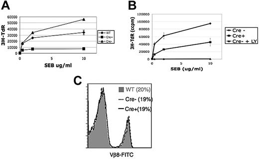
To study a more physiological, antigen-presenting cell–dependent system of T-cell proliferation, we mixed T cells with mitomycin C–treated autologous splenocytes and stimulated them using the SEB. Similar to our observations using crosslinking antibodies to CD3 and CD28, we found that whereas LY294002 treatment completely abolished SEB responses, r1ΔT/r2n T cells were markedly impaired but not totally unresponsive (Figure 5A-B). The percentage of Vβ8+ responder T cells before stimulation was unaltered in the r1ΔT/r2n mice (Figure 5C).
Partially defective superantigen-mediated proliferation in r1ΔT/r2n T cells. (A-B) Thymidine incorporation assay based on titrations of superantigen (SEB) on WT T cells, Cre− T cells (± LY), and Cre+ T cells mixed with autologous splenocytes are shown. (C) Histogram of Vβ8 TCR staining in WT (gray shading), Cre− (dotted), or Cre+ (solid black) T cells. Data are representative of 3 experiments.
Partially defective superantigen-mediated proliferation in r1ΔT/r2n T cells. (A-B) Thymidine incorporation assay based on titrations of superantigen (SEB) on WT T cells, Cre− T cells (± LY), and Cre+ T cells mixed with autologous splenocytes are shown. (C) Histogram of Vβ8 TCR staining in WT (gray shading), Cre− (dotted), or Cre+ (solid black) T cells. Data are representative of 3 experiments.
Loss of class IA PI3K impairs T-cell help to B cells but not CD8 function or viral immunity
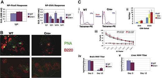
To investigate the in vivo consequences of T cell-specific class IA PI3K ablation, we first assessed T-cell help for B-cell antibody responses to hapten NP. In r1ΔT/r2n mice, both T-independent and T-dependent IgM production were equivalent to control mice, whereas titers of NP-specific IgG1, IgG2a, and IgG3 were reduced but not absent (Figure 6A). Consistent with the reduced amount of class-switched antibody, there was a significant reduction in germinal center number and size (Figure 6B). These findings indicate that T cells lacking class IA PI3K signaling have reduced helper function for B cells.
Defective T-cell help to B cells and germinal center formation following immunization, but normal antiviral responses in r1ΔT/r2n mice. (A) Serum antibody levels in mice injected with NP-Ficoll (left panel) or NP-OVA (right panel) are shown from WT versus r1ΔT/r2n (Cre+) mice. Data are shown as means ± SEM of 4 or 5 mice per genotype using serum dilutions in the linear range of detection. (B) Representative spleen sections of NP-OVA–immunized WT or Cre+ mice stained with B220-PE to identify B cells and PNA-FITC to identify germinal centers. Quantification of 4 WT and 7 Cre+ sections revealed a significant reduction in germinal center number (mean ± SEM of WT versus Cre+, 6.5 ± 0.6 versus 4.4 ± 0.6; P < .05) despite equivalent numbers of follicles (8.5 ± 1 versus 11.3 ± 1.2; P > .1). Germinal center size, measured by counting total green pixels and dividing by germinal center number, was also significantly reduced (2897 ± 513 versus 1463 ± 271; P < .01). Images were visualized using a Nikon Eclipse TE1000-S microscope equipped with a Nikon 4×/0.1 numerical aperture Plane objective (Nikon, Melville, NY). Images were captured using a Phonometrics CoolSnap ES camera (Roper Scientific, Ottobrunn, Germany) and Media Cybernetics ImagePro Express software version 5.1.1.14 (Media Cybernetics, Silver Spring, MD). Images were overlaid in Adobe Photoshop CS2 (Adobe, San Jose, CA); measurements were made using ImageJ software version 1.37 (National Institutes of Health, Bethesda, MD). (C) Flow cytometric tetramer staining of splenic CD8+ T cells (i), intracellular measurement of IFN-γ production (ii), and CTL assay (iii) conducted 7 days after intraperitoneal infection of MHV in WT or r1ΔT/r2n (Cre+) mice. Tetramer stains are of sham and infected animals of each genotype and are representative of 5 mice per genotype. The percent of tetramer-positive cells is shown in blue. CTL and ICS data are shown as the mean ± SEM of 3 to 5 mice per genotype (iv, v). Viral titer in WT and Cre+ mice after intracranial infection. Brain (iv) and liver (v) titers were measured at days 5 and 12 after infection. Data are shown as the mean ± SEM of 3 to 5 mice. Similar responses were observed in r1f/r2n mice.
Defective T-cell help to B cells and germinal center formation following immunization, but normal antiviral responses in r1ΔT/r2n mice. (A) Serum antibody levels in mice injected with NP-Ficoll (left panel) or NP-OVA (right panel) are shown from WT versus r1ΔT/r2n (Cre+) mice. Data are shown as means ± SEM of 4 or 5 mice per genotype using serum dilutions in the linear range of detection. (B) Representative spleen sections of NP-OVA–immunized WT or Cre+ mice stained with B220-PE to identify B cells and PNA-FITC to identify germinal centers. Quantification of 4 WT and 7 Cre+ sections revealed a significant reduction in germinal center number (mean ± SEM of WT versus Cre+, 6.5 ± 0.6 versus 4.4 ± 0.6; P < .05) despite equivalent numbers of follicles (8.5 ± 1 versus 11.3 ± 1.2; P > .1). Germinal center size, measured by counting total green pixels and dividing by germinal center number, was also significantly reduced (2897 ± 513 versus 1463 ± 271; P < .01). Images were visualized using a Nikon Eclipse TE1000-S microscope equipped with a Nikon 4×/0.1 numerical aperture Plane objective (Nikon, Melville, NY). Images were captured using a Phonometrics CoolSnap ES camera (Roper Scientific, Ottobrunn, Germany) and Media Cybernetics ImagePro Express software version 5.1.1.14 (Media Cybernetics, Silver Spring, MD). Images were overlaid in Adobe Photoshop CS2 (Adobe, San Jose, CA); measurements were made using ImageJ software version 1.37 (National Institutes of Health, Bethesda, MD). (C) Flow cytometric tetramer staining of splenic CD8+ T cells (i), intracellular measurement of IFN-γ production (ii), and CTL assay (iii) conducted 7 days after intraperitoneal infection of MHV in WT or r1ΔT/r2n (Cre+) mice. Tetramer stains are of sham and infected animals of each genotype and are representative of 5 mice per genotype. The percent of tetramer-positive cells is shown in blue. CTL and ICS data are shown as the mean ± SEM of 3 to 5 mice per genotype (iv, v). Viral titer in WT and Cre+ mice after intracranial infection. Brain (iv) and liver (v) titers were measured at days 5 and 12 after infection. Data are shown as the mean ± SEM of 3 to 5 mice. Similar responses were observed in r1f/r2n mice.
To assess CD8+ T-cell function in vivo, we employed a model in which mice are infected intracranially or intraperitoneally with a replicating virus, MHV.40,41 In r1ΔT/r2n mice infected with MHV, the generation and differentiation of virus-specific T cells was unimpaired, viral clearance was comparable to control mice, and ex vivo stimulation with immunodominant viral peptides showed normal CTL activity and IFN-γ production (Figure 6C). Thus, class IA PI3K function is not required for CD8+ differentiation and effector function in this model and also suggests a normal CD4+ T-cell contribution to these responses.
Effects of pharmacologic inhibitors of PI3K and mTOR
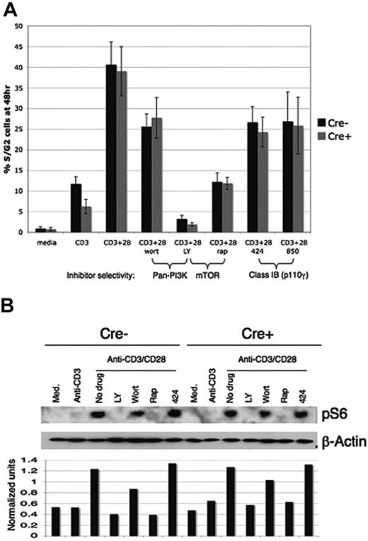
Our finding of robust T-cell function in PI3K-deficient T cells seemed paradoxic considering that proliferation under all conditions was abolished by 10 μM LY294002 (Figure 4A-B). However, at this concentration LY inhibits not only class IA PI3K but other PI3K classes as well as the mammalian target of rapamycin (mTOR).18-20,42 The specific mTOR inhibitor rapamycin also blocked proliferation (Figure 7A). When 50 nM wortmannin was used, a concentration that inhibits PI3K catalytic function without potently affecting mTOR,19,20 there were modest reductions in proliferation of both r1ΔT/r2n and r1f/r2n T cells (Figure 7A). Two specific inhibitors of class IB PI3K, AS604850 and AS252424,43,44 showed a similar ability to reduce TCR/CD28-mediated proliferation of T cells of both genotype when included at 10 μM (Figure 7A). These findings indicate that residual proliferation in class IA–deficient T cells is partially dependent on class IB PI3K and strongly dependent on mTOR. Consistent with PI3K-independent mTOR signaling, T cells stimulated for 18 hours with anti-CD3 plus anti-CD28 exhibited robust phosphorylation of the S6 protein that was more strongly inhibited by rapamycin and LY294002 than by wortmannin or a class IB inhibitor (Figure 7B).
Proliferation and mTOR pathway activation are partially resistant to PI3K inhibition in r1ΔT/r2n T cells. (A) Purified T cells were treated for 15 minutes with the indicated inhibitors and then stimulated for 48 hours with platebound anti-CD3 alone or anti-CD3 plus CD28. Proliferation was measured by cell-cycle analysis, with the percentage of cells in S and G2 phases combined in the analysis. The mean ± SD of 4 experiments is shown. Inhibitor concentrations were 50 nM wortmannin; 10 μM LY294002, 424 (AS252424), and 850 (AS604850) (GE Healthcare Life Sciences, Piscataway, NJ); and 10 ng/mL rapamycin. (B) Immunoblot analysis for phosphorylation of S6 in purified T cells after 18 hours with indicated stimulation conditions. “Normalized units” refers to the relative band intensities of phospho-S6 compared with β-actin, calculated using ImageQuant. To compensate for its short half-life, wortmannin was added every 6 to 12 hours in both the proliferation and phospho-S6 experiments.
Proliferation and mTOR pathway activation are partially resistant to PI3K inhibition in r1ΔT/r2n T cells. (A) Purified T cells were treated for 15 minutes with the indicated inhibitors and then stimulated for 48 hours with platebound anti-CD3 alone or anti-CD3 plus CD28. Proliferation was measured by cell-cycle analysis, with the percentage of cells in S and G2 phases combined in the analysis. The mean ± SD of 4 experiments is shown. Inhibitor concentrations were 50 nM wortmannin; 10 μM LY294002, 424 (AS252424), and 850 (AS604850) (GE Healthcare Life Sciences, Piscataway, NJ); and 10 ng/mL rapamycin. (B) Immunoblot analysis for phosphorylation of S6 in purified T cells after 18 hours with indicated stimulation conditions. “Normalized units” refers to the relative band intensities of phospho-S6 compared with β-actin, calculated using ImageQuant. To compensate for its short half-life, wortmannin was added every 6 to 12 hours in both the proliferation and phospho-S6 experiments.
Discussion
Class IA PI3K heterodimers are encoded by multiple genes for catalytic and regulatory subunits. The combined function of class IA PI3K in T cells has been difficult to define. General PI3K inhibitors are not selective for class IA enzymes18 and also show differing effects on calcium flux, cytokine production, and proliferation depending on the type of T-cell and stimulation conditions.1 Mice lacking p85α, p85α/p55α/p50α, or p85β show normal or enhanced T-cell proliferation.1,11,24,25 Mice lacking p110δ function (p110δKI) do exhibit impaired T-cell signaling and antigen-driven proliferation29 ; however, some T-cell function remains that might be mediated by other catalytic subunits. Furthermore, the T-cell–intrinsic functions of p110δ in vivo have not been reported (see “Note added in proof”). The findings of the present study are based on a novel mouse model in which T cells exhibit the most thorough deletion of class IA PI3K activity reported thus far. Although we disrupted genes encoding the regulatory subunits, at the protein level we also observed dramatic loss of catalytic isoform expression. This was associated with nearly complete ablation of TCR-initiated PI3K activation, as judged by single-cell and population measurements of Akt phosphorylation and by reductions in Ca2+ flux and Erk phosphorylation that were similar to control cells treated with wortmannin. Blockade of PI3K signaling in regulatory subunit–deficient T cells appears more complete than what was observed in a study of p110δKI T cells, in which TCR-mediated Erk phosphorylation was partially preserved and proliferative responses to anti-CD3 plus anti-CD28 were unaffected.29
Several reports have identified possible scaffolding functions of class IA regulatory subunits as well as GTPase-activating protein (GAP) activity toward small GTPases of the Rac and Rab families.21,45-47 Although we did not examine these interactions in this system, the data indicate that loss of putative adapter functions of p85α, p55α, p50α, and p85β does not severely impair T-cell development or function. However, we cannot exclude the possibility that some of the greater defects in these cells compared with p110δKI T cells are related to adapter functions of the regulatory subunits.
A previous study demonstrated that a constitutively active class IA PI3K transgene can promote positive selection of thymocytes.48 We did not observe dramatic differences in T-cell development, implying that whereas increased class IA PI3K signaling can promote differentiation, this pathway is not essential for positive selection. Crossing of the mutant strain onto a TCR transgenic background is necessary to confirm these conclusions. It is possible that residual p85α protein in DN cells is sufficient to mediate signals for the TCRβ selection checkpoint. In addition, the class IB PI3K isoform p110γ has been implicated in T-cell development and might be able to compensate for loss of class IA PI3K in thymocytes in vivo.48-50
Analysis of r1ΔT/r2n mice showed that some T-cell responses are absolutely dependent on class IA PI3K signaling, whereas other responses are partially dependent or independent of this pathway. Our data agree with analyses of p110δKI T cells in showing that TCR-triggered increases in intracellular Ca2+ are attenuated in cells with reduced class IA PI3K activity. Our results also confirm and extend the conclusion of the p110δKI study in demonstrating an unexpectedly limited role for class IA PI3Ks and Akt in CD28-mediated responses. This costimulatory molecule has long been known to recruit PI3K via a classic pYXXM motif, and it is generally assumed that PI3K binding and activation are integral to CD28 signaling function. Given the fact that Itk is dispensable for CD28 function51 and activated Akt can replace some CD28 functions,52 a favored model is that Akt is an important PI3K effector downstream of CD28.53 Our data show that CD28 ligation augments T-cell growth and proliferation without appreciable activation of PI3K and Akt. Moreover, CD28 signaling restores some biochemical events in double-knockout T cells that are considered to be downstream of PI3K and Akt, such as phosphorylation of IκB and induction of Bcl-XL.14,15,53 It is possible that other CD28-linked signaling proteins such as Grb2 could initiate these downstream responses. However, these responses are wortmannin sensitive in the r1ΔT/r2n T cells. These results imply that CD28 or the combination of TCR/CD3 and CD28 can signal via other wortmannin-sensitive enzymes without appreciable Akt activation. Alternatively, residual low levels of PI3K signaling might be sufficient to promote NFκB activation via PKCθ, which has been implicated downstream of CD28 via class IA PI3K.16 It is also worth investigating whether class II and III PI3Ks, which are generally wortmannin sensitive but do not generate PIP3, have important roles in T-cell activation. Indeed, class III PI3K was shown recently to regulate mTOR.54,55 It is also possible that class IB-derived PIP3 contributes to some functional responses, as suggested by the partial inhibition of T-cell proliferation by class IB inhibitors. A recent report showed that the formation of the immune synapse recruits G protein–coupled receptors (GPCRs) and enhances their signaling, which may in turn activate the class IB PI3Ks in T cells.56 However, our data suggest that class IB signaling in activated T cells is not sufficient to drive appreciable Akt phosphorylation and is not required for mTOR activation.
Treatment of human T cells with PI3K inhibitors diminishes their ability to help B cells secrete immunoglobulin in vitro.57 Although T-dependent antibody responses are impaired in p110δ-deficient mice,29-31 these defects might not be T-cell intrinsic. Using selective gene targeting in T cells, we have shown that class IA PI3K signaling in T cells contributes to their helper function in B-cell antibody responses to haptenated protein antigens in vivo. The apparent decrease in severity of these defects in our system relative to p110δ-deficient mice might be explained by the fact that the p110δ mutation is present in all cells and decreases the function of B cells and possibly antigen-presenting cells such as dendritic cells.
When we assessed the response to a replicating viral pathogen (MHV), we found that r1ΔT/r2n mice are able to clear virus normally. Antigen-specific IFN-γ–secreting CD8+ T cells are generated efficiently in these mice and are able to lyse target cells with similar potency as control T cells. Ultimately, further experiments using different doses of virus, other viral models, as well as assessment of CD8 memory cell development and function are necessary to determine the full extent of the importance of class IA PI3K in the T-cell response to viral pathogens. In addition, it will be important to determine the nature and source of the stimuli that allow for CD8 effector function to develop in these mice that exhibit impaired T-cell cytokine production in vitro.
Several novel compounds targeting one or more class IA catalytic isoforms have been described and are under evaluation as possible therapies for various disease states, including allergy, leukemia, and inflammation.44,58-60 One concern with such an approach would be possible suppression of adaptive immunity. Our findings support many studies of B lymphocytes in showing an important role for class IA PI3K in humoral immunity. However, our data are surprising in that they imply that T-cell–mediated immunity to viruses can occur in the absence of class IA PI3K function. Thus, broad inhibition of class IA PI3K might not strongly impair cellular immunity. On the other hand, autoimmune syndromes develop in older r1ΔT/r2n mice61 and in p110δKI mice,29 indicating that reduction of class IA PI3K function can impair self-tolerance. Further studies are required to determine whether r1ΔT/r2n mice have alterations in central tolerance, peripheral tolerance, and/or T helper differentiation that might explain autoimmune susceptibility.
Authorship
Contribution: J.A.D. performed and designed research and wrote the paper; M.G.K. and J.S.O. performed and designed research; L.N.S. and T.I.M. performed research; J.L., H.J., C.R., and L.C.C. contributed vital reagents; and T.E.L. and D.A.F. designed research and wrote the paper.
Conflict-of-interest disclosure: The authors declare no competing financial interests.
Correspondence: David A. Fruman, University of California, Irvine, Department of Molecular Biology and Biochemistry, 3242 McGaugh Hall, Irvine, CA 92697-3900; e-mail: dfruman@uci.edu.
The publication costs of this article were defrayed in part by page charge payment. Therefore, and solely to indicate this fact, this article is hereby marked “advertisement” in accordance with 18 USC section 1734.
Acknowledgments
This work was supported by National Institutes of Health grants AI50831 (D.A.F.), NS018146 (T.E.L.), PO1-CA089021 (L.C.C.), T32 AI060573 (J.S.O.), and T32 CA9054 (M.G.K.) and a Howard Hughes Medical Institute Predoctoral Fellowship (J.L.).
We thank Ankit Nayyar and Sookhee Chun for technical assistance, Chris Schaumburg for help with MHV infections, and Craig Walsh and Aimee Edinger and their laboratories for helpful discussions.

This feature is available to Subscribers Only
Sign In or Create an Account Close Modal