Abstract
Using recombinant human glycoprotein VI (GPVI), we evaluated the effect of N-linked glycosylation at the consensus site Asparagine92-Glycine-Serine94 (N92GS94) on binding of this platelet-specific receptor to its ligands, human type I collagen, collagen-related peptide (CRP), and the snake venom C-type lectin convulxin (CVX). In COS-7 cells transiently transfected with GPVI, deglycosylation with peptide-N-glycosidase F (PNGase F; specific for complex N-linked glycans) or tunicamycin decreases the molecular weight of GPVI and reduces transfected COS-7 cell binding to both CRP and CVX. In stably transfected Dami cells, the substitutions N92A or S94A, but not L95H, resulted in a 30% to 40% decrease in adhesion to CVX, but a 90% or greater decrease in adhesion to CRP and a 65% to 70% decrease in adhesion to type I collagen. Treatment with PNGase F, but not Endoglycosidase H (Endo H) (specific for high-mannose N-linked glycans), produced an equivalent decrease in molecular weight. Neither N92A nor S94A affected the expression of GPVI, based on the direct binding of murine anti–human GPVI monoclonal antibody 204-11 to transfected Dami cells. These findings indicate that N-linked glycosylation at N92 in human GPVI is not required for surface expression, but contributes to maximal adhesion to type I collagen, CRP and, to a lesser extent, CVX.
Introduction
Glycoprotein VI (GPVI) plays a key role in platelet adhesion to collagens and subsequent platelet activation,1,2 is uniquely expressed by cells of the megakaryocytic lineage, and is a member of the immunoglobulin gene superfamily, closely related to Fc receptor gamma chain (FcRγ) and natural killer receptors.3,4 While GPVI binds specifically to polymerized collagens, it also has a high affinity for convulxin (CVX), a C-type lectin from the venom of the tropical rattlesnake, Crotalus durissus terrificus,5 and the synthetic, collagen-related peptide (CRP) (Gly-Pro-Hyp)10.6,7
Mouse and human GPVI cDNA exhibit a 67.3% similarity in nucleotide sequence.4 One N-linked glycosylation site has been predicted in human GPVI and is located within extracellular domain D1 at residue N92. Mouse GPVI has a comparable consensus site at N93 plus an additional N-linked glycosylation site at N244 within D2.4
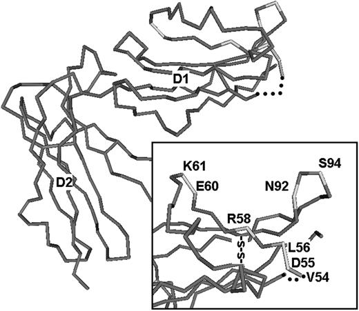
Immunoglobulin (Ig)–like receptor 1 (Lir-1) is another member of the Ig gene superfamily that is composed of 2 Ig-like domains and has 44% sequence identity to GPVI. Figure 1 is an alpha-carbon trace of Lir-1 based on the crystal structure (Protein Database [PDB] catalog no. 1G0X)8 and depicted as a continuous tube. In the upper lefthand corner, the entire Lir-1 molecule is shown, with the extracellular domains D1 and D2 designated. In the enlarged image in the inset at the bottom right, selected residues within 2 predominant hydrophilic loops of D1 are depicted, and the GPVI residues that are positional homologs are indicated.
Certain of these residues are featured in the elegant model of the GPVI binding site for CVX proposed by Batuwangala et al.9 In that model, 2 charged sites on GPVI that span D1 and D2 were proposed to interact with 2 complementary charged sites on CVX. One of the major sites (designated no. 1 in that study) would span the sequence V54 to K61 in D1, and that sequence is a positional homolog to the Lir-1 hydrophilic loop depicted in this figure. The key GPVI residues implicit in this model are D55, R58, E60, and K61.
Additional binding site residues were identified by Lecut et al,10 who mapped the epitope on GPVI that is recognized by murine monoclonal antibody 9O12. Using phage display, they observed that V54 and L56 contribute to this epitope. Since 9O12 also inhibits the binding of collagen, CRP, and CVX to GPVI, while other inhibitory antibodies, such as 3F8 or 9E18, had a differential effect on each ligand, they concluded that the CVX and CRP binding sites are distinct but overlapping. V54 and L56 happen to be located in the putative CVX binding loop proposed by Batuwangala et al.9
The last 2 residues highlighted in Figure 1 (N92 and N94) would be situated on an adjacent hydrophilic loop. These contribute to a consensus N-linked glycosylation site Asparagine92-Glycine-Serine94 (N92GS94) and may influence ligand recognition. In this study, we analyzed the extent of N-linked glycosylation at this site and the effect of deglycosylation on the binding of GPVI to type I collagen, CRP, and CVX.
Materials and methods
Cells and reagents
COS-7 and Dami cells were purchased from the American Type Culture Collection (ATCC, Rockville, MD). CVX was purified from snake venom as previously described.11 Synthetic peptides, including CRP (Gly-Cys-Pro-[Gly-Pro-Hyp]10-Gly-Cys-Pro-Gly), GPA (Gly-Cys-Pro-[Gly-Pro-Ala]10-Gly-Cys-Pro-Gly), and GPP (Gly-Cys-Pro-[Gly-Pro-Pro]10-Gly-Cys-Pro-Gly), were generously provided by Dr Michael Barnes (Cambridge University, United Kingdom).6 Endoglycosidase H (Endo H) and peptide-N-glycosidase F (PNGase F) were purchased from New England Biolabs (Beverly, MA). Rabbit polyclonal anti–human FcRγ was purchased from Upstate Biotechnology (Lake Placid, NY). Acid-insoluble (Horm) bovine tendon collagen (type I) was obtained from Nycomed (Munich, Germany). Murine monoclonal antibody 6F1 (anti–integrin α2β1) was a gift from Dr Barry Coller (The Rockefeller University, New York, NY).
Homology model of GPVI. In the main portion of the figure, an α carbon (Cα) trace of the Lir-1 molecule, a member of the Ig gene superfamily and a close homolog of GPVI, is depicted as a tubular representation. This representation is derived from the crystal structure of Lir-1 (PDB 1G0X) and is rendered using the program Cn3D (version 4.1) produced by the National Center for Biotechnology Information (Bethesda, MD) and distributed freely to the public. In the main figure, the extracellular domains D1 and D2 are labeled. In the enlargement in the bottom right corner (inset), selected amino residues of GPVI that are positional homologs to those in Lir-1 are labeled. These residues are all situated in 2 adjacent hydrophilic loops in the ligand binding region of D1. The first hydrophilic loop contains residues thought to participate in ligand binding: V54, D55, L56, R58, E60, and K61. An adjacent hydrophilic loop contains, at its apex, the consensus N-linked glycosylation site represented by residues N92 and S94.
Homology model of GPVI. In the main portion of the figure, an α carbon (Cα) trace of the Lir-1 molecule, a member of the Ig gene superfamily and a close homolog of GPVI, is depicted as a tubular representation. This representation is derived from the crystal structure of Lir-1 (PDB 1G0X) and is rendered using the program Cn3D (version 4.1) produced by the National Center for Biotechnology Information (Bethesda, MD) and distributed freely to the public. In the main figure, the extracellular domains D1 and D2 are labeled. In the enlargement in the bottom right corner (inset), selected amino residues of GPVI that are positional homologs to those in Lir-1 are labeled. These residues are all situated in 2 adjacent hydrophilic loops in the ligand binding region of D1. The first hydrophilic loop contains residues thought to participate in ligand binding: V54, D55, L56, R58, E60, and K61. An adjacent hydrophilic loop contains, at its apex, the consensus N-linked glycosylation site represented by residues N92 and S94.
Amplification of GPVI cDNA
GPVI cDNA was amplified from platelet RNA by reverse transcriptase–polymerase chain reaction (RT-PCR) using the Superscript one step RT-PCR kit (Gibco BRL, Gaithersburg, MD) with primers GP6F (5′-CTC AGG ACA GGG CTG AGG AA -3′, nucleotide [nt] 4-23) and GP6R (5′-CCA TGA TCC CTC CCT TGG AT -3′, nt 1089-1070). The amplified cDNA was subcloned into pGEM-T (Promega, Madison, WI) and sequenced. One clone with the correct sequence (based on GenBank accession no. NM_016363) was then subcloned into the mammalian expression vector protamine complementary DNA3) pcDNA3 (Invitrogen, Carlsbad, CA).
Synthesis of human FcRγ chain cDNA
FcRγ chain cDNA was amplified from leukocyte RNA by RT-PCR using the forward primer 5′-GCCGATCTCCAAGCCAG-3′ and the reverse primer 5′-TGAGGGCTGGAAGAACCAGA-3′, based on the published sequence.12 The amplified cDNA was subcloned in pGEM-T Easy, sequenced, and subcloned into pcDNA3.
Generation of N92A, S94A, and L95H by site-directed mutagenesis
Site-directed mutagenesis was carried out using the Altered Sites II in vitro Mutagenesis System (Promega). Briefly, human GPVI cDNA was subcloned into the vector pAlter-1 (Promega) in a 3′ to 5′ orientation, and designated pAlter-hGP6. Mutagenic oligonucleotides in reverse-complimentary sequence were synthesized as follows: (1) N92A: 5′-GAGGCTTCCGGCCTGGTAGGAG-3′; (2) S94A: 5′-GGACCAGAGGGCTCCGTTCTGG-3′; and (3) L95H: 5′-CAGGGACCAGTGGCTTCCGTT-3′.
Transient expression in COS-7 cells
COS-7 cells were first transfected with FcRγ cDNA in pcDNA3, and stable COS-7 cell lines expressing FcRγ were selected by Western blot of cell lysate proteins using rabbit anti-FcRγ antibody. A stable FcRγ-expressing clone was then transfected with wild-type or mutant GPVI cDNA in pcDNA3. Transfected cells were grown for 48 hours in the DNA-containing media and then subjected to adhesion and ligand-blotting assays. To normalize for transfection efficiency, luciferase activity was measured using the Luciferase Assay System (Promega).
Production of stable Dami transfectants
Wild-type GPVI, N92A, S94A, and L95H, subcloned in pcDNA3, were separately transfected into Dami cells using the Effectene transfection system (Qiagen, Valencia, CA). Selection was carried out for 2 weeks in Iscove modified Dulbecco medium (IMDM) supplemented with 0.5 mg/mL geneticin (GibcoBRL). The surviving cells were cloned to generate established stable cell lines that would be screened for adhesion to convulxin or CRP using a colorimetric assay.15 The binding of biotin-conjugated convulxin to proteins in sodium dodecyl sulfate (SDS) lysates of Dami or COS-7 (ligand blot) was performed as described.11
Coprecipitation of GPVI with rabbit polyclonal anti-FcRγ
A stable Dami line 1G4 expressing wild-type human GPVI was solubilized in 1% (wt/vol) digitonin at a cell density of 107/mL. Soluble protein was incubated with rabbit anti-FcRγ IgG for 2 hours at ambient temperature, and protein G–Sepharose (Pierce Chemical, Rockford, IL) was then added. After a 1-hour incubation, the beads were washed 4 times in Tris (tris(hydroxymethyl)aminomethane)–buffered saline (TBS) containing 0.1% Tween 20 (Promega), resuspended in NuPAGE loading buffer (Invitrogen) and incubated at 70°C for 10 minutes. The proteins thus eluted were separated by NuPAGE using a 4% to 12% gradient slab gel in MOPS (3-N-morpholino)-propanesulfonic acid)–SDS buffer. Proteins separated in the polyacrylamide gel were transferred electrophoretically to a polyvinylidene difluoride (PVDF) membrane, and the membrane was then probed with either rabbit anti-FcRγ IgG or biotin-CVX. Bound probes were visualized by enhanced chemiluminescence (ECL; Amersham) using horseradish peroxidase (HRP)–conjugated secondary reagents.
Deglycosylation with tunicamycin
Stable Dami cell transfectants or transient COS-7 cell transfectants expressing wild-type GPVI were grown in appropriate media containing 0.5 μg/mL tunicamycin (Sigma, St Louis, MO) or the vehicle dimethyl sulfoxide (DMSO; 0.1% vol/vol), as a control, for 48 hours. Tunicamycin inhibits the transfer of dolichol pyrophosphate precursor to asparagine (Asn) in the consensus sequence for N-linked glycosylation (Asn-X-serine/threonine).
Deglycosylation of GPVI with N-linked glycosidases
Wild-type GPVI expressed by the stable Dami line 1G4 was treated with PNGase F or Endo Hf.16 1G4 cells (107) were washed once in phosphate-buffered saline (PBS) and resuspended in 1 mL of TBS containing 2 mM EDTA (ethylenediaminetetraacetic acid). Triton X-100 was added to a final concentration of 1% (vol/vol), the mixture was gently agitated for 5 minutes at ambient temperature, and the insoluble material was pelleted by centrifugation. The supernatant or TX-100 soluble protein (180 μL) was mixed with denaturing buffer (20 μL) (supplied by the manufacturer, New England Biolabs). For treatment with Endo Hf, 20 μL of G5 buffer (New England Biolabs) and 5 μL of Endo Hf were then added, and the mixture was incubated at 37°C for 18 hours. For treatment with PNGase F, 20 μLof G7 buffer (New England Biolabs), 20 μL of 10% Nonidet P-40 (NP40; vol/vol), and 5 μL of PNGase F were added, and the mixture was incubated at 37°C for 18 hours. Following digestion, the soluble proteins were prepared for 4% to 12% NuPAGE and Western blot analysis as described for the coprecipitation of GPVI. Separated proteins were probed with monoclonal anti–human GPVI antibody LJ6.5.
Cell adhesion to CRP or CVX
Cell adhesion was quantitated by measuring the alkaline phosphatase activity of adherent cells using para-nitrophenylphosphate (PNPP), as described by Bellavite et al.15 Wells of a 96-well microtiter tray were coated with either 5 μg/mL CVX in PBS, pH 7.4, or 10 μg/mL CRP in 10 mM acetic acid at 4°C. The wells were blocked with 2% bovine serum albumin (BSA; Sigma) in TBS at ambient temperature for 60 minutes, then rinsed twice in TBS. COS-7 cell transfectants were detached from culture flasks using 1 mM EDTA in PBS then washed once in TBS and resuspended at 106 cells/mL in TBS containing 0.5% (wt/vol) BSA (TBS-BSA). Dami cell transfectants were spun down and washed once in TBS then resuspended in TBS-BSA at 106 cells/mL. One hundred microliters of either cell suspension was added to each precoated well in triplicate and incubated at ambient temperature for 30 minutes. The tray was gently washed in saline then substrate solution (0.1 M citrate buffer, pH 5.4, containing PNPP and 0.1% Triton X-100; 150 μL/well) was added, and the plates were gently and continuously agitated on a horizontal shaker for 60 minutes at ambient temperature. Color was developed by adding 2N NaOH (100 μL/well), and the absorbance at 405 nm of each well was recorded using an enzyme immunoassay (EIA) reader.
Cell adhesion to type I collagen
Transfected Dami cell adhesion to type I collagen under static conditions was performed as described by Lecut et al17 and Lagrue-Lak-Hal et al.18 Briefly, type I collagen (20 μg/mL) was immobilized in wells of Immulon II microtiter plates (Dynatech Laboratories, Chantilly, VA) by incubation for 1 hour at ambient temperature. The wells were blocked with BSA and washed as described in “Cell adhesion to CRP or CVX.” A 100-μL suspension of Dami cell transfectants (106 cells/mL) was added to each precoated well in triplicate and incubated at ambient temperature for 45 minutes with gentle agitation. Nonadherent cells were removed by gentle aspiration and rinsing with saline, then adherent cells were quantitated based on alkaline phosphatase activity, as described in “Cell adhesion to CRP or CVX.” Nonspecific adhesion was measured using wells coated only with BSA.
Measurement of surface expression of GPVI
Surface expression of GPVI was measured by flow cytometry using mouse anti–human GPVI monoclonal antibody 204-11.19 After the cells were washed in PBS, as described in “Cell adhesion to CRP to CVX”, 1 × 10⁶ cells were resuspended in 19 μL Tyrode buffer (12 mM NaHCO3, 2 mM MgCl2,137.5 mM NaCl, 2.6 mM KCl, pH 7.4) containing 1% (wt/vol) BSA, to which was added 1 μL 204-11, previously diluted to 10 μg/mL in the same buffer. After a 30-minute incubation, 400 μL PBS was added, and the amount of bound antibody was measured within 30 minutes using a FACSCalibur flow cytometer (Becton Dickinson, San Jose, CA). Data were analyzed with BD CellQuestPro software (Becton Dickinson), and the amount of bound antibody was reported as the geometric mean fluorescence intensity (GMFI).
Results
Transient expression of wild-type and N-deglycosylated human GPVI in COS-7 cells
COS-7 cells stably expressing human FcRγ chain were transiently transfected with wild-type GPVI or the mutants N92A or S94A. Transient expression of these constructs in complex with FcRγ permitted an initial assessment of the effect of these amino acid substitutions as well as enzymatic deglycosylation on the electrophoretic properties, expression, and function of GPVI. We must emphasize that the purpose of these experiments in transiently transfected COS-7 cells was to obtain initial qualitative comparisons of the GPVI mutants. These initial studies were then followed by quantitative comparisons between mutants expressed in Dami cells, as described in the next section.
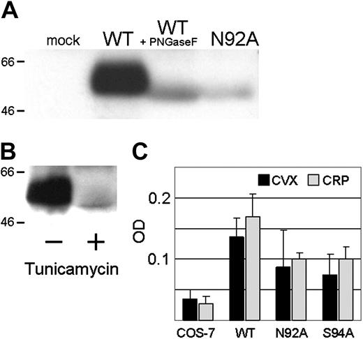
The ligand blot assay (Figure 2A) using biotin-CVX permitted a simultaneous analysis of the electrophoretic properties of each construct and its ability to bind to CVX. In SDS lysates from transfected COS-7 cells, the apparent molecular weight (MWApp) of wild-type GPVI is roughly 59.5 kDa. Both GPVI N92A and S94A exhibit an increased electrophoretic mobility resulting in an MWApp of 52.6 kDa. The same increase in electrophoretic mobility is obtained following enzymatic deglycosylation of wild-type GPVI with PNGase F (Figure 2A) but not Endo H (not shown), suggesting that this increase results from the disruption of N-linked glycosylation at the consensus site N92-G-S94. GPVI was not detectable in lysates of mock-transfected COS-7 cells by this ligand blot assay, as is evident in Figure 2A.
The relative affinity of SDS-denatured N-deglycosylated mutants and wild-type GPVI for CVX was also evident in this ligand blot assay. Based on optical scanning and then normalization for transfection efficiency, the binding of N92A was decreased to about 14%, while the binding of S94A was deceased to about 6%.
Effect of tunicamycin on GPVI
COS-7 cells expressing wild-type GPVI were cultured for 48 hours in the presence of 0.5 μg/mL tunicamycin or an equal volume of the carrier DMSO, as a negative control. As shown in a ligand blot (Figure 2B), tunicamycin treatment resulted in an increase in electrophoretic mobility equivalent to that of the mutant N92A. Moreover, the binding of biotin-CVX was significantly attenuated, as evident in the marked decrease in the intensity of the band.
Transient expression of GPVI by transfected COS-7 cells. (A) The ligand blot assay was used to assess the binding of biotin-CVX to proteins in lysates of (from left to right): mock-transfected cells; cells transfected with wild-type (WT) GPVI; cells transfected with WT GPVI and then digested with the endoglycosidase PNGase F; and cells transfected with the substitution mutant N92A. The electrophoretic mobility of 2 representative marker proteins is indicated to the left of the gel. (B) The ligand blot assay used to assess the binding of biotin-CVX to proteins in lysates of (left) cells transfected with wild-type GPVI (–) and (right) cells transfected with wild-type GPVI then treated for 48 hours with tunicamycin (+). (C) Adhesion of COS-7 transfectants to CVX (▪) or CRP (□). The optical density (OD) is indicated on the x-axis. The identity of the transfectants is indicated on the ordinate and includes (from left to right): mock-transfected cells (COS-7); cells transfected with wild-type GPVI (WT); and cells transfected with N92A or S94A. Each bar represents the mean of 3 experiments. Error bars represent 1 SD.
Transient expression of GPVI by transfected COS-7 cells. (A) The ligand blot assay was used to assess the binding of biotin-CVX to proteins in lysates of (from left to right): mock-transfected cells; cells transfected with wild-type (WT) GPVI; cells transfected with WT GPVI and then digested with the endoglycosidase PNGase F; and cells transfected with the substitution mutant N92A. The electrophoretic mobility of 2 representative marker proteins is indicated to the left of the gel. (B) The ligand blot assay used to assess the binding of biotin-CVX to proteins in lysates of (left) cells transfected with wild-type GPVI (–) and (right) cells transfected with wild-type GPVI then treated for 48 hours with tunicamycin (+). (C) Adhesion of COS-7 transfectants to CVX (▪) or CRP (□). The optical density (OD) is indicated on the x-axis. The identity of the transfectants is indicated on the ordinate and includes (from left to right): mock-transfected cells (COS-7); cells transfected with wild-type GPVI (WT); and cells transfected with N92A or S94A. Each bar represents the mean of 3 experiments. Error bars represent 1 SD.
Adhesion of COS-7 transfectants expressing mutant GPVI
COS-7 cells expressing FcRγ and wild-type GPVI adhered well to either CVX or CRP (Figure 2C). The mean adhesion of transfectants expressing either N92A or S94A was decreased to about 60% that of wild-type GPVI (Figure 2C). However, the level of expression in COS-7 was not particularly strong, and the variability of this transient expression system precluded a more quantitative evaluation of the impact of these mutations on adhesion. Consequently, we opted to establish stably transfected Dami cell lines with a more consistent and quantifiable level of GPVI expression in order to compare more precisely the function of wild-type GPVI versus that of each mutant.
Characterization of stable Dami transfectants
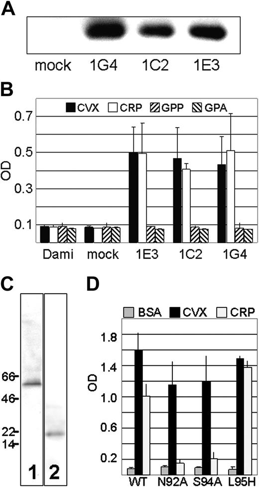
Dami cells express endogenous FcRγ.3 Nevertheless, we first tested the necessity to cotransfect additional exogenous recombinant FcRγ in order to obtain efficient surface expression of GPVI. Of several stably transfected Dami lines that were developed, 3 were evaluated: 1G4 was transfected with wild-type GPVI alone; 1C2 and 1E3 were each cotransfected with wild-type GPVI plus FcRγ. Using the ligand blot assay (Figure 3A), prominent protein bands with the MWApp expected of wild-type GPVI (roughly 59.5 kDa) were detected in SDS lysates of 1G4, 1C2, and 1E3, whereas no band was observed in mock-transfected cells. The intensity of the 1G4 band was actually somewhat greater than that of the other samples, but it is clear that cotransfection of FcRγ was not required for efficient synthesis of GPVI. A comparison of surface expression and adhesive properties of the transfected cell lines (Figure 3B) confirmed the lack of requirement for exogenous FcRγ. Moreover, the specificity of the transfected GPVI for CRP is enforced by the complete absence of adhesion to the control peptides GPP and GPA (Figure 3B).
To confirm that 1G4 expresses GPVI that is associated with endogenous Dami FcRγ, we showed that transfected GPVI was coprecipitated from 1G4 lysates by rabbit polyclonal antibody specific for human FcRγ (Figure 3C). GPVI was not coprecipitated by the same antibody from lysates of mock-transfected cells (not shown).
Comparison of GPVI mutants
Dami cell lines stably transfected with wild-type GPVI and mutants N92A, S94A, or L95H were then generated using the same protocol in the absence of exogenous FcRγ. For comparative functional assays, representative clones that exhibited comparable surface expression were selected, as determined by the binding in flow cytometry of murine monoclonal anti–human GPVI antibody 204-11 (Table 1).
Surface GPVI by flow cytometry
. | . | GMF1, mean ± SD . | . | |
|---|---|---|---|---|
| Cloned Dami cell line . | N . | mAb 204-11 . | Control IgG . | |
| Mock transfected | 2 | 12 ± 8 | 14 ± 5 | |
| GPVI (wild type) | 3 | 84 ± 19 | 13 ± 7 | |
| N92A | 4 | 109 ± 31 | 11 ± 9 | |
| S94A | 3 | 88 ± 23 | 9 ± 7 | |
. | . | GMF1, mean ± SD . | . | |
|---|---|---|---|---|
| Cloned Dami cell line . | N . | mAb 204-11 . | Control IgG . | |
| Mock transfected | 2 | 12 ± 8 | 14 ± 5 | |
| GPVI (wild type) | 3 | 84 ± 19 | 13 ± 7 | |
| N92A | 4 | 109 ± 31 | 11 ± 9 | |
| S94A | 3 | 88 ± 23 | 9 ± 7 | |
The level of total GPVI in each clone was also measured by Western blot using our murine monoclonal anti–human GPVI antibody LJ6.5. The level of GPVI in lysates of cells producing each of these mutants was indistinguishable from that found in lysates of cells producing wild-type GPVI (not shown). Thus, quantitative expression of GPVI does not appear to be affected by any of these 3 substitutions.
A comparison of the adhesion of these cell lines with CVX or CRP (Figure 3D) confirmed that (1) for L95H, adhesion to CVX is unaffected and adhesion to CRP is actually increased by 40%; and (2) for N92A and S94A, adhesion to CVX is decreased to roughly 75%, while adhesion to CRP is decreased to roughly 20%. Thus, these 2 substitutions disrupt the ability of GPVI to bind to CRP and, to a lesser extent, CVX. The fact that the binding of antibody 204-11 to N92A and S94A is equivalent to that of wild-type GPVI (Table 1), even though each exhibits reduced ability to adhere to CVX or CRP (Figure 3), is not an inconsistency. Moroi et al19 reported that 204-11 binds to a region “near,” but not necessarily at, the collagen binding site of GPVI. Our results indicate that neither N92A nor S94A disrupts the epitope(s) recognized by 204-11.
Stable expression of GPVI by Dami cell lines. (A) The requirement for exogenous FcRγ was tested by comparing the expression of transfected wild-type GPVI in cell lines that were not cotransfected with FcRγ (1G4) or cotransfected with FcRγ (1C2 and 1E3). The binding of biotin-CVX to GPVI in lysates from each cell line indicate that 1G4 expresses as much or more total GPVI as do 1C2 or 1E3. (B) Adhesion of Dami cell lines to CVX (▪), CRP (□), control peptide GPP (▧) or control peptide GPA (▨). Optical density (OD) is indicated on the abscissa. The identity of each cell line is indicated on the ordinate. Values represent mean ± SD for 3 experiments. (C) Immunoprecipitation. Rabbit polyclonal anti–human FcRγ was used to precipitate endogenous FcRγ from 1G4. The precipitated proteins were isolated, solubilized, and analyzed by Western blot using either biotin-CVX (lane 1) or the same polyclonal anti–human FcR antibody (lane 2) to visualize the proteins. The electrophoretic mobilities of selected molecular weight marker proteins are indicated to the left of the gel. (D) Adhesion of Dami lines transfected with wild-type GPVI (WT) or GPVI substitution mutants N92A, S94A, and L95H (ordinate). Adhesion to plates coated with BSA (▦), CVX (▪), or CRP (□) was measured. Optical density (OD) is indicated on the abscissa. Error bars represent 1 SD.
Stable expression of GPVI by Dami cell lines. (A) The requirement for exogenous FcRγ was tested by comparing the expression of transfected wild-type GPVI in cell lines that were not cotransfected with FcRγ (1G4) or cotransfected with FcRγ (1C2 and 1E3). The binding of biotin-CVX to GPVI in lysates from each cell line indicate that 1G4 expresses as much or more total GPVI as do 1C2 or 1E3. (B) Adhesion of Dami cell lines to CVX (▪), CRP (□), control peptide GPP (▧) or control peptide GPA (▨). Optical density (OD) is indicated on the abscissa. The identity of each cell line is indicated on the ordinate. Values represent mean ± SD for 3 experiments. (C) Immunoprecipitation. Rabbit polyclonal anti–human FcRγ was used to precipitate endogenous FcRγ from 1G4. The precipitated proteins were isolated, solubilized, and analyzed by Western blot using either biotin-CVX (lane 1) or the same polyclonal anti–human FcR antibody (lane 2) to visualize the proteins. The electrophoretic mobilities of selected molecular weight marker proteins are indicated to the left of the gel. (D) Adhesion of Dami lines transfected with wild-type GPVI (WT) or GPVI substitution mutants N92A, S94A, and L95H (ordinate). Adhesion to plates coated with BSA (▦), CVX (▪), or CRP (□) was measured. Optical density (OD) is indicated on the abscissa. Error bars represent 1 SD.
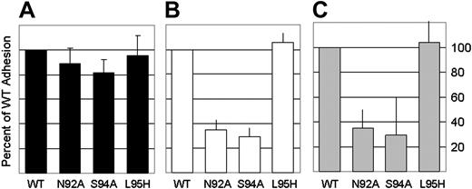
Adhesion to Horm type I collagen was more complex, because Dami cells also express another collagen receptor, the integrin α2β1. However, as reported by Lecut et al17 and Lagrue-Lak-Hal et al,18 conditions can be established to distinguish the relative contributions of GPVI and α2β1 to adhesion under static conditions, since the latter requires divalent cations and is specifically inhibited by the monoclonal antibody 6F1. The results are depicted in Figure 4. In the presence of divalent cations (2 mM CaCl2 and 1 mM MgCl2; Figure 4A), the adhesion of Dami transfected with wild-type GPVI or L95H were equivalent, while modest decreases were seen with adhesion of Dami transfected with N92A or S94A (average decreases equal to 11% and 18%, respectively). In the presence of divalent cations and the monoclonal antibody 6F1 (Figure 4B), the contribution of the integrin α2β1 is inhibited, and the residual adhesion reflects the contribution of GPVI. Again, L95H had no effect, but adhesion mediated by N92A and S94A were decreased, on average, by 65% and 70%, respectively. The results obtained in the presence of 2 mM EDTA (Figure 4C) were comparable with those seen in the presence of 6F1. Average inhibition by N92A and S94A were 65% and 70%, respectively. Adhesion of L95H in the presence of EDTA was again comparable to that of wild-type GPVI. These results confirm that these substitutions in the sequence of GPVI affect GPVI-dependent adhesion to type I collagen without an effect on the concomitant adhesion mediated by integrin α2β1. Moreover, despite the overexpression of GPVI in these Dami cell transfectants, endogenous α2β1 still contributes substantially to total adhesion to type 1 collagen in this static system.
Endoglycosidase treatment of wild-type GPVI
In human GPVI, the tripeptide sequence N92GS94 is a consensus motif for N-glycosylation. Consequently, we next addressed whether the effect of N92A or S94A on adhesion results from the disruption of glycosylation.
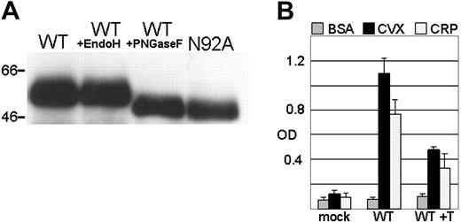
The electrophoretic mobility of wild-type GPVI is increased when isolated from Dami cells and treated with PNGase F (which hydrolyzes nearly all N-glycan chains from glycopeptides) but not Endo H (which cleaves only high mannose–type N-glycans) (Figure 5A). The increase in mobility resulting from PNGase F treatment is equivalent to that observed for transfected Dami N92A (Figure 5A), and identical results were obtained with Dami S94A (not shown). Thus, the increase in electrophoretic mobility of N92A and S94A is consistent with loss of complex N-glycans at this consensus site.
Adhesion of Dami cell transfectants to type I (Horm) fibrillar collagen. Dami cells transfected with WT GPVI, N92A, S94A, or L95H were incubated in wells of microtiter plates coated with type 1 (Horm fibillar collagen). Adherent cells were quantitated as described in “Materials and methods.” The results represent the average of 3 independent experiments. Following the precedent set in previous reports where GPVI-mediated adhesion to collagen was measured,17,18 the adhesion of Dami cells transfected with WT GPVI was set as maximal in each experiment, and the results for each mutant GPVI transfectant were then expressed as percent of maximal WT adhesion. The average percent of WT adhesion (± 1 SD) for all 3 experiments is represented in panels A through C. (A) Adhesion in the presence of 2 mM CaCl2 plus 1 mM MgCl2. (B) Same as panel A, except Dami transfectants were incubated with 6F1 monoclonal antibody (20 μg/mL) for 60 minutes at ambient temperature prior to the onset of the adhesion assay. (C) Adhesion in the presence of 2 mM EDTA. Error bars represent 1 SD.
Adhesion of Dami cell transfectants to type I (Horm) fibrillar collagen. Dami cells transfected with WT GPVI, N92A, S94A, or L95H were incubated in wells of microtiter plates coated with type 1 (Horm fibillar collagen). Adherent cells were quantitated as described in “Materials and methods.” The results represent the average of 3 independent experiments. Following the precedent set in previous reports where GPVI-mediated adhesion to collagen was measured,17,18 the adhesion of Dami cells transfected with WT GPVI was set as maximal in each experiment, and the results for each mutant GPVI transfectant were then expressed as percent of maximal WT adhesion. The average percent of WT adhesion (± 1 SD) for all 3 experiments is represented in panels A through C. (A) Adhesion in the presence of 2 mM CaCl2 plus 1 mM MgCl2. (B) Same as panel A, except Dami transfectants were incubated with 6F1 monoclonal antibody (20 μg/mL) for 60 minutes at ambient temperature prior to the onset of the adhesion assay. (C) Adhesion in the presence of 2 mM EDTA. Error bars represent 1 SD.
Treatment of wild-type GPVI with endoglycosidases or tunicamycin. (A) A shift in the electrophoretic mobility of wild-type GPVI was observed following treatment with endoglycosidases. MAb LJ6.5 was used in a Western blot to visualize GPVI in lysates of (from left to right): cells transfected with wild-type (WT) GPVI; cells transfected with WT GPVI and then digested with the endoglycosidase Endo H, which cleaves high mannose type N-glycans; cells transfected with WT GPVI and then digested with the endoglycosidase PNGaseF, which cleaves complex type N-glycans; and cells transfected with the substitution mutant N92A. The electrophoretic mobility of 2 representative marker proteins is indicated to the left of the gel. (B) The influence of tunicamycin treatment on Dami cell adhesion. The adhesion of Dami lines transfected with vector alone (mock) or WT GPVI was compared to that of same Dami lines transfected with WT GPVI but first exposed to tunicamycin for 48 hours. Adhesion to plates coated with BSA (▦), CVX (▪) or CRP (□) was measured. Optical density (OD) is indicated on the abscissa. Error bars represent 1 SD.
Treatment of wild-type GPVI with endoglycosidases or tunicamycin. (A) A shift in the electrophoretic mobility of wild-type GPVI was observed following treatment with endoglycosidases. MAb LJ6.5 was used in a Western blot to visualize GPVI in lysates of (from left to right): cells transfected with wild-type (WT) GPVI; cells transfected with WT GPVI and then digested with the endoglycosidase Endo H, which cleaves high mannose type N-glycans; cells transfected with WT GPVI and then digested with the endoglycosidase PNGaseF, which cleaves complex type N-glycans; and cells transfected with the substitution mutant N92A. The electrophoretic mobility of 2 representative marker proteins is indicated to the left of the gel. (B) The influence of tunicamycin treatment on Dami cell adhesion. The adhesion of Dami lines transfected with vector alone (mock) or WT GPVI was compared to that of same Dami lines transfected with WT GPVI but first exposed to tunicamycin for 48 hours. Adhesion to plates coated with BSA (▦), CVX (▪) or CRP (□) was measured. Optical density (OD) is indicated on the abscissa. Error bars represent 1 SD.
Tunicamycin treatment of stable Dami transfectants
Tunicamycin treatment of Dami transfected with wild-type GPVI for 48 hours resulted in a 60% decrease in binding to CVX or CRP (Figure 5B). GPVI isolated from these treated cells showed decreased binding to CVX in a ligand blot assay and an increase in electrophoretic mobility characteristic of the N-deglycosylated protein (not shown).
Discussion
Our findings indicate that the consensus site N92GS94 of human GPVI is N-glycosylated and that the presence of complex oligosaccharides at this position is important for maximal binding of GPVI to both CVX and CRP. We also observed that N-glycosylation at this site is important for GPVI-mediated adhesion to type I collagen under conditions where the contribution of other receptors, such as α2β1, can be reduced or eliminated. Although the presence of this consensus site had been acknowledged even in the earliest reports of the GPVI sequence, our study is the first documentation of such N-glycosylation.
There are a number of explanations for the influence of N-glycosylation on the adhesive properties of GPVI. One possibility is that the complex oligosaccharide chain itself makes direct contact with collagens, CRP, or CVX. We have no evidence for or against this hypothesis. A second possibility is that the presence of the N-oligosaccharide chain maintains the binding site of GPVI in an optimal orientation. Based on our own cumulative results, one observation that is not completely consistent with this explanation is the fact that soluble, recombinant human GPVI, produced by Drosophila S2 cells, does not show an inordinate loss of affinity for CVX or CRP, even though the nature of N-glycosylation in insect cells is different and considered to be typically high-mannose type. However, one could argue that the presence of N-glycosylation per se, regardless of the precise nature of the oligosaccharide structure, is sufficient to confer optimal activity to GPVI.
It has been proposed that the binding sites on GPVI for CVX and CRP are not identical. Using HEL cells expressing recombinant human GPVI, Lecut et al10 reported that labeled CVX binds to 65 000 sites with a dissociation constant (Kd) of 0.35 nM and that cell adhesion to collagen (Horm collagen; equine tendon collagen type I) was inhibited by CVX with a 50% inhibitory concentration (IC50) of 0.08 nM. On the other hand, CRP had a modest effect on cell adhesion to collagen, which was inhibited by a maximum of only 40% at CRP concentrations of 2.5 μg/mL or higher. Adhesion of the same cells to solid-phase CVX was efficiently inhibited by collagen (IC50, 10 μg/mL), but an inhibition of only 20% was attained with CRP at concentrations up to 50 μg/mL. Finally, adhesion to CRP was inhibited fully by either collagen (IC50, 2.5 μg/mL) or CVX (IC50, 0.06 nM). These results were interpreted to mean that CVX and CRP bind to distinct but overlapping sites on GPVI, but could also be explained by differences in the relative affinity of each ligand for GPVI.
At the same time, the hypothesis of 2 distinct and overlapping sites received support from the comparative inhibition of 3 monoclonal anti-GPVI antibodies, 9E18, 3F8, and 9O12, which recognize distinct epitopes and do not cross-compete.10 9E18 inhibits binding of human recombinant GPVI to collagen by 45%, and binding to CRP by 85%, but has no effect on binding to CVX. 3F8 inhibits binding to CVX by 55% and to CRP by 50% but has little effect on binding to collagen (10% inhibition). 9O12 inhibits completely the binding of GPVI to CRP, inhibits binding to CVX by up to 80% and binding to collagen by up to 85%.10,17 A synergistic inhibition was observed by the combination of 9O12 and 3F8.
Our findings that deglycosylation of GPVI has a more profound effect on binding to CRP or type I collagen than to CVX give further support to the hypothesis that there are the 2 distinct but overlapping binding sites. We propose that the loss of an N-linked glycan at the N92 is sufficient to alter the conformation of the GPVI molecule. It remains to be determined whether this occurs during intracellular posttranslational processing and/or at the level of the surface-expressed glycoprotein. It also remains to be determined whether the conformational change is localized to the hydrophilic loops within the ligand-binding region of the molecule or is more globalized. Whatever the alteration, it is apparently not sufficient to inhibit processing and surface expression of GPVI. Similar findings have been made with certain other N-glycosylated receptors, such as the human granulocyte-macrophage colony-stimulating factor (GM-CSF) receptor α-subunit20 and the purinogenic receptor P2Y12.21
The influence of N-glycosylation on GPVI function potentially represents another basis for heterogeneity of this receptor and may contribute to the range of expression and/or activity seen between individuals, as reported by our group11 and others.22,23 Moreover, abnormalities in N-glycosylation of GPVI could contribute to acquired defects in GPVI-mediated platelet reactivity to collagen, such as recently reported in 2 patients with malignant hemopathies.24
Prepublished online as Blood First Edition Paper, July 12, 2005; DOI 10.1182/blood-2005-04-1454.
Supported by grant R01 HL46979, awarded by the National Heart, Lung, and Blood Institute (NHLBI) to T.J.K.
The publication costs of this article were defrayed in part by page charge payment. Therefore, and solely to indicate this fact, this article is hereby marked “advertisement” in accordance with 18 U.S.C. section 1734.
The authors thank Dr Barry Coller (Rockefeller University, New York, NY) for his gift of monoclonal antibody 6F1 and the late Dr Michael Barnes (Cambridge University, United Kingdom) for his generous contribution of CRP and the control peptides GPP and GPA.
This is manuscript number 17252-MEM from The Scripps Research Institute.

This feature is available to Subscribers Only
Sign In or Create an Account Close Modal