It has recently been reported that in a xenotransplantation model of human cells injected into nonobese diabetic-severe combined immunodeficient mice, direct intratibial injection of stem cells improves engraftment.1,2 However, the complexities of using this approach clinically are substantial. We sought to determine whether in a syngeneic mouse model the delivery of donor primitive hematopoietic cells via the arterial supply of the host's femur, in which vascular barriers are preserved, could achieve improvement in localization and engraftment efficiency.
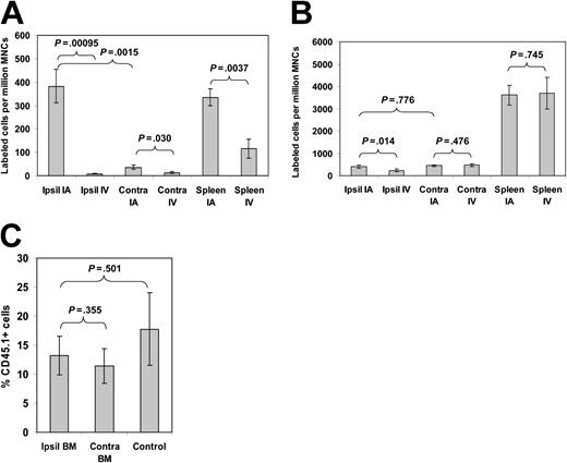
Whole bone marrow mononuclear cells (BM MNCs) or lineage-depleted (lin-) BM cells were isolated from a donor mouse and labeled with either a green fluorescent label (carboxyfluorescein diacetate succinimidyl ester) or a red fluorescent label (seminaphthorhodafluor). Simultaneously, an equal number of labeled cells (5 × 10⁵ lin- BM cells) were injected in the femoral artery through an intra-arterial catheter, and the differentially labeled cells were injected through a peripheral vein, the tail vein. This number of cells was chosen to allow for accurate determination of the number of homed cells without saturation of the assay. One hour following injection, the number of cells homing to the femoral BM “downstream” of the femoral artery injection (ipsilateral BM), the BM in the opposite femur (contralateral BM), and spleen were analyzed. Intra-arterial delivery of BM cells resulted in a 10-fold excess of cells in the ipsilateral BM compared with the contralateral BM (P = .0015) and a 47-fold excess compared with intravenously injected cells (P = .000 95; Figure 1A). These results demonstrate that local arterial injection markedly enhanced the delivery of primitive cells to the local hematopoietic site. Increased localization of cells to the BM has also been noted following isolated limb perfusion of BM MNCs.3 However, in these studies the injection technique severely inhibited the cells' ability to freely circulate to other hematopoietic sites. In our study, free circulation of the cells was observed. Therefore, the difference between the number of “homed” cells in ipsilateral versus contralateral BM suggested that a substantial first-pass effect influenced the homing process.
Homing and engraftment of bone marrow cells following arterial or venous injection. Equal numbers of differentially labeled cells were simultaneously injected into the tail vein or femoral artery of lethally irradiated mice. The numbers of (A) lin- or (B) lin+ cells homing to the ipsilateral BM (Ipsil), contralateral BM (Contra), or spleen following intra-arterial (IA) or intravenous (IV) injection were measured by flow cytometry (n = 4 for both groups). (C) The percentage of CD45.1 hematopoietic cells in the ipsilateral and contralateral BM injected via the femoral artery was also measured 6 weeks following injection of the cells (n = 5). Control mice had CD45.1 cells injected into the tail vein. Error bars represent SEM.
Homing and engraftment of bone marrow cells following arterial or venous injection. Equal numbers of differentially labeled cells were simultaneously injected into the tail vein or femoral artery of lethally irradiated mice. The numbers of (A) lin- or (B) lin+ cells homing to the ipsilateral BM (Ipsil), contralateral BM (Contra), or spleen following intra-arterial (IA) or intravenous (IV) injection were measured by flow cytometry (n = 4 for both groups). (C) The percentage of CD45.1 hematopoietic cells in the ipsilateral and contralateral BM injected via the femoral artery was also measured 6 weeks following injection of the cells (n = 5). Control mice had CD45.1 cells injected into the tail vein. Error bars represent SEM.
Analysis of the number of lin- cells homing to the contralateral BM or spleen following arterial or venous injection unexpectedly demonstrated an approximate 3-fold increase in the number of cells that had been injected through the femoral artery (P = .030 and P = .0037, respectively; Figure 1A). This consistent difference was not likely due to the injection procedure as no differences were observed in the number of cells homing to these sites via arterial or venous injection when 1 × 10⁶ lineage positive (lin+) cells were injected (P = .476 and P = .745, respectively; Figure 1B). While there was a statistically significant 1.7-fold increase in the number of lin+ cells homing to the ipsilateral BM via the arterial injection compared with the venous injection (P = .014), no differences were observed between the ipsilateral and contralateral BM following arterial injection (P = .776). Therefore, this effect was specific for more primitive cells. These results demonstrate that primitive populations had more efficient homing to even distant hematopoietic tissues requiring recirculation when they were initially delivered into an arterial vascular bed. Whether the specific vascular type initially seen by injected cells influences their ultimate home cannot be defined by this study, but these data do raise that possibility.
To determine whether the preferential homing or “seeding” of the BM space following arterial delivery of primitive cells corresponded to augmented engraftment, we performed BM transplantation studies. We used a competitive transplant model simultaneously injecting 2 × 10⁵ CD45.1 cells into the femoral artery and 2 × 10⁵ CD45.2 cells into the tail vein of the same lethally irradiated (with 10 Gy of radiation from a 137Cs source) CD45.2 recipient mouse using the identical protocol as that for the homing assays. This cell dose was chosen as it gave 100% survival but would still allow identification of variations in engraftment efficiency. At 6 weeks following injection of the cells, we assessed the level of engraftment of the cells injected into the femoral artery in the BM of the ipsilateral BM and the contralateral BM by flow cytometry for CD45.1. In contrast to the results from the homing assays, we saw no difference in the levels of engraftment of cells injected in the femoral artery between the ipsilateral or contralateral BM (P = .355; Figure 1C). In some experiments we also analyzed the level of engraftment of CD45.1 cells following tail vein injection with an equal number of CD45.2 cells. In these experiments, we also saw similar levels of engraftment of the CD45.1 cells irrespective of their route of injection (P = .501; Figure 1C).
These results demonstrate that femoral artery injection leads to a large increase in the number of primitive cells arriving at and seeding the ipsilateral BM. Unlike direct injection into the marrow cavity, however, concentrated stem cell delivery by a vascular route does not improve engraftment in the bone marrow. While the syngeneic rather than xenogeneic nature of the grafts may have played a role, what also notably distinguishes these methods is the intact versus disrupted vascular bed. Stem cells delivered by artery encounter endothelium, smooth muscle cells, and extracellular matrix that may create substantial barriers to arrival at the niche. Whether these structural elements or the injury implicit in the direct injection process creates different engraftment potential remains to be defined. However, enhanced vascular delivery by itself is insufficient to enable improved stem cell engraftment.


This feature is available to Subscribers Only
Sign In or Create an Account Close Modal