Abstract
Platelet factor 4 (PF4) is an abundant platelet α-granule C-X-C chemokine that has weak chemotactic potency but strongly inhibits hematopoiesis through an unknown mechanism. We find that PF4 binds to human CD34+ hematopoietic progenitor cells (HPCs) with a median effective concentration of 1 μg/mL but not after exposure to chondroitinase ABC. PF4 enhances adhesion of HPCs to intact stroma. Committed progenitors also adhere avidly to immobilized PF4. This adhesion is time-dependent, requires metabolic activity, causes cytoskeletal rearrangement, and induces cell-cycle inhibition. Using extracellular acidification rate to indicate transmembrane signaling, we find that interleukin-8 (IL-8), but not PF4, activates CD34+ progenitors, and PF4 blocks IL-8–mediated activation. Surface plasmon resonance analysis shows that PF4 binds IL-8 with high (dissociation constant [Kd] = 42 nM) affinity. Nuclear magnetic resonance analysis of IL-8 and PF4 in solution confirms this interaction. We conclude that PF4 has the capacity to influence hematopoiesis through mechanisms not mediated by a classical high-affinity, 7-transmembrane domain chemokine receptor. Instead, PF4 may modulate the hematopoietic milieu both directly, by promoting progenitor adhesion and quiescence through interaction with an HPC chondroitin sulfate–containing moiety, and indirectly, by binding to or interfering with signaling caused by other, hematopoietically active chemokines, such as IL-8.
Introduction
Platelet factor 4 (PF4) is an abundant platelet alpha-granule protein that is a founding member of the C-X-C chemokine family.1 In striking contrast to other members of this family, however, PF4 has chemotactic and functional priming activity on neutrophils only at concentrations several orders of magnitude higher than those of other prototypic members of this family, for example, interleukin-8 (IL-8).2-5 Despite its relative impotence as a chemotaxin and priming agent, PF4, along with a number of related and unrelated chemokines, has been reported to inhibit proliferation of early committed hematopoietic progenitors at concentrations in the low nanomolar range in vitro.6,7 In contrast, inhibition of intramedullary hematopoietic progenitors in vivo requires high (ie, micromolar) concentrations of PF4.8,9 Unlike virtually every other member of the chemokine family, a G protein–coupled receptor for PF4 analogous to the other well-defined C-X-C and C-C chemokine receptors10-12 has yet to be characterized. PF4 is therefore notable among the chemokines by virtue of its abundance, lack of potency to influence leukocyte function, and absence of a clearly defined high-affinity receptor.
Brandt et al13 and Petersen et al14,15 have shown that PF4 stimulates neutrophil degranulation in the presence of tumor necrosis factor-alpha (TNF-α) with a median effective concentration (EC50) of 2 μM, approximately the same as the dissociation constant (Kd) for PF4 binding to the neutrophil surface. This suggests the presence on neutrophils of a large number of moderate-affinity PF4 receptors. PF4 binding to neutrophils was shown to be attributable to interaction with a neutrophil cell-surface chondroitin sulfate glycosaminoglycan (GAG). In aggregate, these studies suggested to us the possibility that PF4, unlike other chemokines, could similarly influence hematopoietic progenitor proliferation and differentiation by binding with moderate affinity to an abundant chondroitin sulfate–like moiety on the surface of cells and generating a signal with functional consequences. To test this hypothesis we have examined the binding of PF4 to human CD34+ hematopoietic progenitor cells, its dependence upon the presence of cell-surface chondroitin sulfate, the ability of immobilized PF4 to induce adhesion of hematopoietic progenitor cells, and the effects of such adhesion upon hematopoietic progenitor cell proliferation.
Another, indirect mechanism whereby PF4 might influence hematopoiesis is suggested by the finding by Gengrinovitch16 et al that PF4 binds tightly (Kd = 5 nM) to vascular endothelial growth factor 165 (VEGF165) but does not bind VEGF121, which lacks heparin-binding capacity. To test the hypothesis that PF4 can bind to and negate the activity of other heparin-binding chemokines, we examined the influence of PF4 upon IL-8–induced signaling (assayed by extracellular acidification rate) in CD34+ hematopoietic progenitors and characterized the interaction of IL-8 with immobilized PF4 using surface plasmon resonance-based Biacore analysis and in solution using nuclear magnetic resonance (NMR) spectroscopy.
Materials and methods
Cell separation
Bone marrow was aspirated from the posterior iliac crest of healthy young volunteers after obtaining informed consent. Bone marrow mononuclear cells were separated by Ficoll-Hypaque (specific gravity 1077; Sigma Diagnostics, St Louis, MO) centrifugation. CD34+-enriched cells were obtained using a CD34 Progenitor Cell Isolation Kit and magnetic cell separator (MACS; Miltenyi Biotec, Gladbach, Germany).
Stromal feeders and conditioned media
Human primary bone marrow stromal feeders were established from human mononuclear cells as previously described,17 irradiated at 1250 rads when confluent, and maintained in long-term bone marrow culture (LTBMC) medium consisting of Iscoves modified Dulbecco medium (IMDM), 2 mM L-glutamine, 1000 U/mL penicillin, 100 U/mL streptomycin (Gibco Laboratories, Grand Island, NY), 12.5% fetal calf serum (Hyclone Laboratories, Logan, UT), 12.5% horse serum (Terry Fox Laboratories, Vancouver, BC, Canada), and 10—6 M hydrocortisone (Abbott Laboratories, Chicago, IL).
Cytokines
Recombinant PF418 and biotinylated PF4 (bPF4) was kindly provided by C. K. Lai and Ted Maione (Repligen, Cambridge, MA). PF4 was selectively biotinylated, targeting its 3 N-terminal glutamate residues by exposure to biotin capryl hydrazide. This reagent preserved (a) formation of tetramers at physiologic ionic strength, (b) binding to heparin with an affinity similar to that of native PF4, and (c) inhibition of endothelial cell proliferation identical to that of native PF4 with a median inhibitory dose (ID50) of 2 μg/mL. The following recombinant cytokines were used in the experiment evaluating CD34+ cells' cycling: 250 pg/mL granulocyte colony-stimulating factor (G-CSF), 50 pg/mL leukemia inhibitory factor (LIF), 200 pg/mL macrophage inflammatory protein 1-alpha (MIP-1α), 2 ng/mL interleukin-6 (R&D Systems, Minneapolis, MN), 10 pg/mL granulocyte-macrophage colony-stimulating factor (GM-CSF; Immunex, Seattle, WA), and 200 pg/mL stem cell factor (SCF; Amgen, Thousand Oaks, CA). For experiments evaluating IL-8 interaction with PF4, recombinant IL-8 was purchased from R&D Systems.
Binding of bPF4 to CD34+ cells
CD34+ cells were enriched to 95% purity by 2 sequential selections through a magnetic cell separator, then exposed to a monoclonal antibody against the CD34 receptor conjugated with phycoerythrin (CD34-PE, Becton Dickinson, San Jose, CA) and the following concentrations of bPF4: 1, 10, 100, 1000, 10 000, and 100 000 ng/mL, for 30 minutes at 4°C. After washings, cells were stained with streptavidin–fluorescein isothiocyanate (FITC) (Becton Dickinson) for 30 minutes at 4°C, then washed and suspended in 2% glutaraldehyde. Control groups were stained with irrelevant immunoglobulin G (IgG) PE and IgG FITC. Flow cytometry analysis was performed using FACS-Star-Plus with a Consort computer.
Effect of chondroitinase ABC upon CD34+ cell binding of PF4
CD34+ cells (100 000) were suspended in IMDM containing 200 mU/mL protease-free chondroitinase ABC (Seikagaku, Tokyo, Japan) in 0.03 M sodium acetate buffer for 2 or 4 hours at 37°C, then washed in IMDM and stained with 10 μg/mL bPF4 for 30 minutes at 4°C. After subsequent washing, cells were stained with streptavidin-FITC and CD34-PE antibodies. Positive control cells were stained with bPF4 and streptavidin-FITC without chondroitinase ABC exposure. Negative control cells were stained with streptavidin-FITC and an irrelevant IgG PE. Flow cytometry analysis was performed as described in the previous paragraph.
Adhesion of hematopoietic progenitors to human bone marrow stroma in the presence of PF4
CD34+-enriched cells (100 000) were suspended in IMDM and incubated in 24-well plates with human primary bone marrow stroma for 2 hours at 37°C in the presence of the following concentrations of native PF4: 0, 0.1, 1, 10, 100, 1000, 10 000, and 100 000 ng/mL, in triplicate. Nonadherent cells and adherent cells were separated as described previously19 and plated in short-term methylcellulose cultures containing 30% fetal calf serum, 3 IU/mL erythropoietin, and 5 ng/mL interleukin-3 (IL-3; R&D Systems). Colony-forming cells were then enumerated on day 14. All cultures were maintained at 37°C in a humidified 5% CO2 atmosphere. The experiment was repeated 4 times.
Adhesion of hematopoietic progenitors to immobilized PF4
In Voller buffer (pH 9.6), 10 μg/mL PF4, 10 μg/mL poly-L-lysine (PLL), or 5% albumin was incubated in separate triplicate wells of a 24-well plastic plate at 37°C for 12 hours. After incubation, unbound substances were removed by washing with phosphate-buffered saline (PBS) and wells blocked by incubating with 5% bovine serum albumin (BSA) for 2 hours at 37°C. CD34+-enriched cells (10 000) were suspended in IMDM and incubated in wells with bound ligands for 2 hours at 37°C in triplicate. Nonadherent cells and adherent cells were separated19 and plated in short-term methylcellulose cultures. Colony-forming cells (burst forming units–erythrocytes [BFU-Es], colony-forming units–granulocytes and macrophages [CFU-GMs], and colony-forming units–granulocytes, erythrocytes, macrophages, and megakaryocytes [CFU-GEMMs]) were then enumerated on day 14. All cultures were maintained at 37°C in humidified 5% CO2 atmosphere. The experiment was repeated 4 times.
The effect of metabolic inhibition upon CD34+ progenitors' adhesion to PF4
CD34+-enriched cells (10 000) were suspended in IMDM and incubated in wells with the immobilized ligands described in the previous paragraph for 2 hours in the presence of 5 mM 2 deoxy-D-glucose and 0.1% NaN3 at 37°C in triplicate. For analysis of temperature effect on hematopoietic progenitor binding to PF4, the adhesion assay was performed for 2 hours at 4°C. The control groups of cells were incubated without 5 mM 2 deoxy-D-glucose and 0.1% NaN3 at 37°C. The nonadherent cells and adherent cells were separated19 and plated in short-term methylcellulose cultures. Colony-forming cells were then enumerated on day 14. The experiments were repeated 3 times.
CD34+ cell cytoskeletal changes upon adhesion to immobilized PF4
CD34+-enriched cells (200 000) were suspended in IMDM and incubated in triplicate in chamber slides (Nunc, Naperville, IL) with the immobilized ligands (10 μg/mL PF4, 10 μg/mL poly-L-lysine, and 50 μg/mL 51-kDa COOH-terminal fragment of fibronectin [a gift from Dr James McCarthy, University of Minnesota]). After 2 hours of adhesion at 37°C, nonadherent cells and adherent cells were separated.19 Adherent cells were placed for 10 minutes in 4% paraformaldehyde, and after washing with IMDM cells were permeabilized with — 20°C acetone for 5 minutes. To visualize focal adhesions, cells were stained with phalloidin-FITC (Molecular Probes, Eugene, OR), which binds with high specificity to polymers of F-actin.20 Cells were then washed with IMDM with 0.3% BSA. Images were taken by confocal microscope (MRC 1024; BioRad, Hercules, CA) 1 μm above the surface of the slide.
Effect of adhesion of hematopoietic progenitors to PF4 upon proliferation assayed by thymidine suicide
Separated CD34+ cells were maintained in serum-free medium with the following cytokines: 250 pg/mL G-CSF, 10 pg/mL GM-CSF, 1 ng/mL IL-6, 50 pg/mL LIF, 200 pg/mL SCF, and 200 pg/mL MIP-1α,21 for 2 days, then incubated for 16 hours on poly-L-lysine, 5% albumin, and PF4. As a control we used cells in suspension culture in serum-free medium, either in the presence or absence of 10 μg/mL PF4 for 16 hours. After incubation with immobilized ligands, adherent and nonadherent cells were separated. Half of the cells in each control and each adherent and nonadherent group were then exposed to 10 μCi (0.37 MBq) titrated thymidine (3H) (6.7 Ci/mmol [247.9 GBq]; DuPont, Boston, MA) for 30 minutes.22 Cells were washed with 10 mL unlabeled thymidine (100 μg/mL; Sigma Diagnostics) in IMDM, then plated in short-term methylcellulose cultures. Colony-forming cells (CFCs) were enumerated on day 14. The proportion of hematopoietic progenitors in S-phase was expressed as a percent of cells killed in 3H thymidine suicide assay accordingly to the formula: % cells killed = ([CFCs not exposed to radioactive thymidine] — [CFCs exposed to radioactive 3H thymidine]) × 100%/(CFCs not exposed to radioactive thymidine). The experiments were repeated 3 times in duplicate.
Analysis of CD34+ cells' response to PF4 and effect of PF4 on CD34+ cells' response to interleukin-8
CD34+ cells were suspended in agarose (Agarose Entrapment Reagent; Molecular Devices, Sunnyvale, CA) at a density of 105cells/1 μL and placed into the center of Capsule Cups (Molecular Devices), which were then loaded into a microphysiometer (Cytosensor Microphysiometer; Molecular Devices) that measures changes in extracellular proton excretion (extracellular acidification rate).23 After washing in RPMI medium, PF4 was introduced to separate populations of cells at concentrations of 0 ng/mL, 100 ng/mL, and 10 μg/mL for 8 minutes. Following that, IL-8 was introduced at a concentration of 100 ng/mL either alone or with 100 ng/mL or 10 μg/mL PF4 for 20 minutes. Cells were continuously perfused with a low-buffered medium at a rate 100 μL/min. The buffer flow was periodically halted, and extracellular acidification rates were measured every 2 minutes and expressed as percent change with regard to baseline acidification rate.
Kinetic analysis of bPF4 interaction with interleukin-8
The binding of bPF4 to interleukin-8 was examined on a BIAcore instrument (Pharmacia Biosensor, Uppsala, Sweden) using surface plasmon resonance, which monitors real-time binding (measured in resonance units [RU]) of an analyte to a ligand immobilized on a sensor chip. The dissociation rate constant (kdiss) was obtained using equation ln (R0/Rt) = kdiss (t — t0), where Rt is response at time (t) and R0 is response at the beginning of dissociation phase (t0). Calculations were performed using the nonlinear model provided in BIA evaluation software version 2.1 (Piscataway, NJ). The association rate constant (kassoc) was calculated from linear model that plots observed kinetic rate constant (ks) against concentration (C) of analyte (ks = kdiss + kassocC), where slope values for ks are calculated from analysis of each sensorgram using the equation R = (r0/ks)(1 — e—ks(t—t0)). The equilibrium dissociation constant (Kd) for the PF4 and IL-8 interaction was obtained using the equation Kd = kdiss/kassoc. bPF4 was immobilized upon a gold-dextran surface with covalently bound streptavidin (Sensor Chip SA-5). The following were injected at a flow rate of 5 μL/min for 6 minutes: 1 μg/mL bPF4 in 10 mM HEPES (N-2-hydroxyethylpiperazine-N′-2-ethanesulfonic acid), 150 mM NaCl, 1 mM CaCl2, and 1 mM MgCl2. Nonspecifically bound bPF4 was removed by perfusion with 1.5 M NaCl. Under this condition approximately 1500 pg/mm2 of bPF4 was bound to the chip.24 Interleukin-8 in equilibration buffer at 2 to 6 μM concentrations was then perfused across a SA-5 chip with immobilized bPF4 at a flow rate of 20 μL/min for 90 seconds, following which equilibration buffer alone was perfused. Injections were carried out in triplicate for each concentration and with increasing concentrations of IL-8.
Nuclear magnetic resonance (NMR) spectroscopy of PF4 and IL-8 interaction
Uniformly 15N-labeled PF4 (5 mg/mL) was dissolved in 0.6 mL H2O containing 20 mM NaCl, pH 5.0. This solution containing 15N-PF4 was used alone and with unlabeled IL-8 (mixed to a molar ratio up to 1:2). Both proton and nitrogen chemical shifts of PF4 were monitored upon each IL-8 addition using 2D 1H-15N-HSQC (heteronuclear single-quantum coherence) spectum.
1H-15N-HSQC spectra25 were acquired at 40°C on a Varian Unity Inova 600 MHz spectrometer equipped with a H/C/N triple-resonance probe and x/y/z triple-axis pulse field gradient unit. The solvent deuterium signal was used as a field-frequency lock. Carrier frequencies for 15N and 1H were positioned at 116.5 parts per million (ppm) and 5.2 ppm, respectively. The chemical shifts are quoted in parts per million downfield from sodium 4,4-dimethyl-4-silapentane sulfonate. A gradient sensitivity–enhanced version of 2D 1H-15N HSQC was applied with 400 (t1) × 2048 (t2) complex data points and spectral widths of 2500 Hz in t1 (15N) and 9000 Hz in t2 (1H) dimensions. Raw data were converted and processed by using NMRPipe26 and were analyzed by using NMRView.27
Statistics
Results of data are reported as the mean ± SEM. Levels of significance were determined by the paired Student t test.
Results
We examined the binding of biotinylated PF4 (bPF4) to CD34+ cells by secondary labeling with streptavidin-FITC and flow cytometry analysis (not shown). Binding was saturable with an EC50 of approximately 1 μg/mL, a concentration that is orders of magnitude higher than the 10 to 50 ng/mL reported to inhibit replication of committed progenitors in in vitro assays,6,7 but similar to those reported to inhibit megakaryocyte28,29 progenitor proliferation.
To examine the dependence of this binding upon the presence of cell-surface chondroitin sulfate, a glycosaminoglycan (GAG) found in great abundance on CD34+ hematopoietic progenitors,30 we pretreated CD34+ progenitors with an ultrapure preparation of chondroitinase ABC, an enzyme highly specific for cleavage of chondroitin sulfate. As shown in Figure 1 (upper right panel), after exposure to 10 μg/mL bPF4, 88% of CD34+ cells were positive for PF4 on the cell surface. After exposure to 200 mU/mL chondroitinase ABC for 2 and 4 hours, cell-surface binding of biotinylated PF4 decreased to 13% and 7%, respectively. Thus, as is the case for PF4 binding to neutrophils, PF4 binding to CD34+ progenitors is attributable to the presence on the cell surface of a chondroitin sulfate–containing moiety.
Chondroitinase ABC pretreatment of CD34+ cells blocks bPF4 binding. Y-axis, CD34–phycoerythrin (CD34-PE) binding; x-axis, streptavidin-FITC (SA-FITC)/bPF4 binding. (A) Negative control. No chondroitinase, no bPF4 exposure, labeling with SA-FITC and irrelevant IgG-PE. (B) Positive control. No chondroitinase, bPF4 at 10 μg/mL, labeled with CD34-PE and SA-FITC. (C) Cells exposed 2 hours to 200 mU/mL chondroitinase ABC, then labeled as for panel B. (D) As in panel C, but with 4 hours exposure to chondroitinase ABC. The percent of CD34+ cells labeling positive for PF4 is shown in the upper right of each panel
Chondroitinase ABC pretreatment of CD34+ cells blocks bPF4 binding. Y-axis, CD34–phycoerythrin (CD34-PE) binding; x-axis, streptavidin-FITC (SA-FITC)/bPF4 binding. (A) Negative control. No chondroitinase, no bPF4 exposure, labeling with SA-FITC and irrelevant IgG-PE. (B) Positive control. No chondroitinase, bPF4 at 10 μg/mL, labeled with CD34-PE and SA-FITC. (C) Cells exposed 2 hours to 200 mU/mL chondroitinase ABC, then labeled as for panel B. (D) As in panel C, but with 4 hours exposure to chondroitinase ABC. The percent of CD34+ cells labeling positive for PF4 is shown in the upper right of each panel
To investigate whether PF4 enhances the interaction of CD34+ human hematopoietic progenitors with the marrow environment, we examined whether PF4 promotes adhesion of colony-forming hematopoietic progenitors to cultured human bone marrow stroma. In experiments not shown, we found that PF4 enhanced the adhesion of clonogenic cells by 50% over baseline with an EC50 of approximately 1 μg/mL, similar to that for PF4 binding to CD34+ cells.
Because the assay of progenitor cell adhesion to a stromal matrix is a complex system with numerous potential interactions, we developed a simplified system in which PF4 was first covalently attached to the bottom of plastic wells and subsequent adhesion of colony-forming units to this immobilized PF4 was analyzed (Figure 2). As a control for nonspecific adhesion we used plates coated with bovine serum albumin (BSA), and as a positive control for nonspecific adhesion we used immobilized poly-L-lysine (PLL), a synthetic polycation well known to mediate nonspecific electrostatic interactions. Both PF4 and PLL, but not BSA, promoted adhesion of approximately 50% of BFU-Es, CFU-GMs, and CFU-GEMMs. This adhesion to immobilized PF4 was dependent on chondroitin sulfate present on the cell surface, because cells treated with protease-free chondroitinase ABC did not adhere to PF4 (not shown).
Colony-forming units adhere to immobilized PF4. Attached to the bottom of separate triplicate plastic tissue culture wells and then washed were 10 μg/mL PF4, 10 μg/mL poly-L-lysine, and 5% albumin. CD34+-enriched cells (10 000) were suspended in IMDM and incubated in wells with bound ligands for 2 hours at 37°C. Nonadherent cells and adherent cells were separated and plated in short-term methylcellulose cultures. E indicates burst forming units–erythrocytes; GM, colony-forming units–granulocytes macrophages; and GEMM, mixed colonies. Error bars represent SEMs.
Colony-forming units adhere to immobilized PF4. Attached to the bottom of separate triplicate plastic tissue culture wells and then washed were 10 μg/mL PF4, 10 μg/mL poly-L-lysine, and 5% albumin. CD34+-enriched cells (10 000) were suspended in IMDM and incubated in wells with bound ligands for 2 hours at 37°C. Nonadherent cells and adherent cells were separated and plated in short-term methylcellulose cultures. E indicates burst forming units–erythrocytes; GM, colony-forming units–granulocytes macrophages; and GEMM, mixed colonies. Error bars represent SEMs.
To determine whether hematopoietic progenitor binding to mobilized PF4, unlike that to PLL, required intact metabolic activity (and therefore presumably active intracellular signaling), we assessed the effect of decreased temperature and metabolic inhibitors upon this adhesion. As shown in Figure 3, adhesion to PF4, but not PLL, was significantly diminished at 4°C in comparison with adhesion at 37°C. Similar results were obtained with the combination of metabolic inhibitors, to deoxy-D-glucose and sodium azide (Figure 3B). These findings suggest a need for active metabolism for hematopoietic progenitor cells to adhere to PF4. In the process of CD34+ adhesion to fibronectin, monomers of actin (globular actin) polymerize to form microfilaments (filamentous actin; F-actin) (Figure 4B). In contrast, adhesion to PLL does not require F-actin formation (Figure 4A). Figure 4C shows that CD34+ cells adherent to PF4 formed focal adhesions involving filamentous actin formation.
Adhesion of colony-forming units to immobilized PF4 requires metabolic activity. (A) Marrow mononuclear cells were plated in wells coated with bovine serum albumin (BSA), poly-L-lysine (PLL), or PF4 in 37°C or 4°C; then adherent and nonadherent cells were separated and cultured in methylcellulose for 21 days. Total colony-forming units (BFU-E, CFU-GM, and CFU-GEMM) were enumerated. (B) Marrow mononuclear cells were plated in wells with immobilized PF4 in culture medium without (Control) or in the presence of 0.1% NaNO3 and 2-deoxyglucose (NaN3/2-DOG). Adherent and nonadherent cells were separated and cultured in methylcellulose for 21 days. Colonies forming units were enumerated. E indicates burst forming units–erythrocytes; GM, colony-forming units–granulocytes macrophages; and GEMM, colony-forming units–granulocytes erythrocytes, macrophages, and megakaryocytes. Error bars represent SEMs.
Adhesion of colony-forming units to immobilized PF4 requires metabolic activity. (A) Marrow mononuclear cells were plated in wells coated with bovine serum albumin (BSA), poly-L-lysine (PLL), or PF4 in 37°C or 4°C; then adherent and nonadherent cells were separated and cultured in methylcellulose for 21 days. Total colony-forming units (BFU-E, CFU-GM, and CFU-GEMM) were enumerated. (B) Marrow mononuclear cells were plated in wells with immobilized PF4 in culture medium without (Control) or in the presence of 0.1% NaNO3 and 2-deoxyglucose (NaN3/2-DOG). Adherent and nonadherent cells were separated and cultured in methylcellulose for 21 days. Colonies forming units were enumerated. E indicates burst forming units–erythrocytes; GM, colony-forming units–granulocytes macrophages; and GEMM, colony-forming units–granulocytes erythrocytes, macrophages, and megakaryocytes. Error bars represent SEMs.
Adhesion to PF4 causes cytoskeletal rearrangement in CD34+ hematopoietic cells. CD34+ cells were let to adhere on immobilized poly-L-lysine (A), the 51-kDa COOH-terminal fragment of fibronectin (B), and PF4 (C). To visualize focal points of adhesion, cells were stained with phalloidin-FITC. Pictures were taken with confocal microscope 1 μm above the surface of the slide. F-actin did not form during adhesion to poly-L-lysine (uniform circumferential staining). In contrast, focal points of adhesion (indicated by coarse punctate staining) were seen in cells adherent to both fibronectin fragment and PF4. Original magnification for all panels, × 400.
Adhesion to PF4 causes cytoskeletal rearrangement in CD34+ hematopoietic cells. CD34+ cells were let to adhere on immobilized poly-L-lysine (A), the 51-kDa COOH-terminal fragment of fibronectin (B), and PF4 (C). To visualize focal points of adhesion, cells were stained with phalloidin-FITC. Pictures were taken with confocal microscope 1 μm above the surface of the slide. F-actin did not form during adhesion to poly-L-lysine (uniform circumferential staining). In contrast, focal points of adhesion (indicated by coarse punctate staining) were seen in cells adherent to both fibronectin fragment and PF4. Original magnification for all panels, × 400.
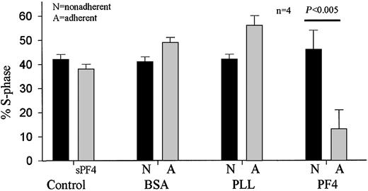
To determine whether adherence to immobilized PF4 modulates proliferation of hematopoietic progenitors, as does adherence to fibronectin,31 we performed thymidine suicide S-phase analysis of both nonadherent and adherent CD34+ cells in plastic culture wells coated with bovine serum albumin, PLL, or PF4 (Figure 5). A control shows the effect of adding 10 μg/mL PF4 in solution on proliferation of CD34+ cells cultured in suspension (left pair of bars). Neither presence of PF4 in solution nor adhesion to BSA or PLL caused any change in the percent of cells in S-phase. In striking contrast, cells adherent to immobilized PF4 had a fraction of cells in S-phase one third that of the nonadherent fraction. Thus, adhesion to immobilized PF4, but not PF4 in solution, inhibits progenitor proliferation.
Adhesion to immobilized PF4 decreases proliferation of cells giving rise to colony-forming units. Left pair of bars: CD34+ cells in suspension were incubated 16 hours either in the absence (▪) or presence (▦) of 10 μg/mL PF4 in solution (sPF4) and percent S-phase of total colony-forming cells (BFU-E, CFU-GM, and CFU-GEMM) analyzed by thymidine suicide analysis. Right 3 pairs of bars: CD34+ cells were cultured 16 hours in wells whose bottoms were coated with immobilized bovine serum albumin (BSA), poly-L-lysine (PLL), or platelet factor 4 (PF4). Adherent (A) and nonadherent (N) cells were then separated, and the fraction of total colony-forming cells in S-phase was quantified. Cells adherent to PF4 had significantly decreased proportion of cells in S-phase when compared with nonadherent cells (P < .005). Error bars represent SEMs.
Adhesion to immobilized PF4 decreases proliferation of cells giving rise to colony-forming units. Left pair of bars: CD34+ cells in suspension were incubated 16 hours either in the absence (▪) or presence (▦) of 10 μg/mL PF4 in solution (sPF4) and percent S-phase of total colony-forming cells (BFU-E, CFU-GM, and CFU-GEMM) analyzed by thymidine suicide analysis. Right 3 pairs of bars: CD34+ cells were cultured 16 hours in wells whose bottoms were coated with immobilized bovine serum albumin (BSA), poly-L-lysine (PLL), or platelet factor 4 (PF4). Adherent (A) and nonadherent (N) cells were then separated, and the fraction of total colony-forming cells in S-phase was quantified. Cells adherent to PF4 had significantly decreased proportion of cells in S-phase when compared with nonadherent cells (P < .005). Error bars represent SEMs.
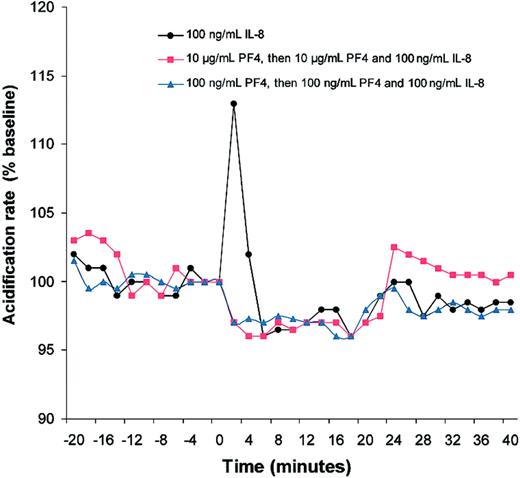
Seeking indirect evidence for a PF4 high-affinity receptor, we performed experiments using a microphysiometer (Molecular Devices). This device detects and monitors the response of cells to ligands for specific membrane receptors by assaying changes in extracellular acidification rate, an extremely sensitive though nonspecific concomitant of cellular signaling through a variety of signaling pathways.23 As a positive control we used IL-8, a chemokine from the same C-X-C family as PF4, which has well-characterized receptors (CXCR1 and CXCR2) with 7 transmembrane helices, occupation of which generates an acidification response in human CD4+ lymphocytes, monocytes, and neutrophils.13,32 IL-8 plays a role in the regulation of normal hematopoiesis in vivo as well as in vitro because mice lacking the murine IL-8 receptor homologue have greatly increased numbers of myeloid progenitors compared with normal mice.11 IL-8 also stimulates a rise in intracellular calcium in a myeloid progenitor cell line,33 a type of signal typically associated with an increase in extracellular acidification rate. Figure 6 shows that PF4 infusion, at the concentrations tested (100 ng/mL [triangles] and 10 μg/mL [squares]), failed to cause an increase in the acidification response of CD34+ cells. As predicted, IL-8 infusion resulted in a sharp and transient increase in response of CD34+ cells measured by extracellular acidification rate (solid circles). Unexpectedly, this IL-8 response was abrogated upon coinfusion with PF4 (squares and triangles).
Metabolic response of CD34+ cells to interleukin-8 in the presence of PF4. The metabolic response of CD34+ cells to PF4 and IL-8 was assessed by measurement of extracellular proton excretion (acidification rate) using a Cytosensor microphysiometer. After establishing a stable baseline, interleukin-8 (100 ng/mL) was infused over 20 minutes starting at time = 0 (closed circles). A brisk signaling response (increase in extracellular acidification over baseline) is evident. Squares: starting at t = — 16 minutes, PF4 (10 μg/mL) was infused through t = 20. At t = 0, the ongoing PF4 infusion was supplemented with IL-8 (100 ng/mL). Triangles: identical to squares except PF4 concentration is 100 ng/mL.
Metabolic response of CD34+ cells to interleukin-8 in the presence of PF4. The metabolic response of CD34+ cells to PF4 and IL-8 was assessed by measurement of extracellular proton excretion (acidification rate) using a Cytosensor microphysiometer. After establishing a stable baseline, interleukin-8 (100 ng/mL) was infused over 20 minutes starting at time = 0 (closed circles). A brisk signaling response (increase in extracellular acidification over baseline) is evident. Squares: starting at t = — 16 minutes, PF4 (10 μg/mL) was infused through t = 20. At t = 0, the ongoing PF4 infusion was supplemented with IL-8 (100 ng/mL). Triangles: identical to squares except PF4 concentration is 100 ng/mL.
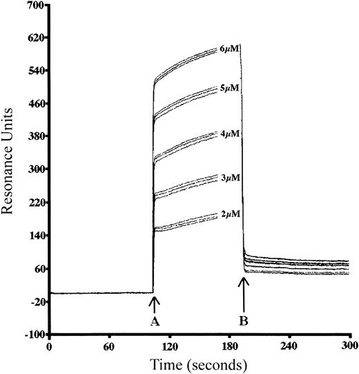
The lack of CD34+ response to IL-8 when IL-8 was coinfused with PF4 led us to hypothesize that PF4 may bind directly to IL-8 in solution, as PF4 does to VEGF.16 To test this hypothesis we performed kinetic affinity analysis of PF4 and IL-8 using surface plasmon resonance-based technology (Biacore). bPF4 was first immobilized upon a streptavidin sensor chip, and then IL-8 at different concentrations was perfused into the chamber above the chip, and potential binding interactions were monitored in real time. Figure 7 shows a series of sensorgrams resulting when increasing IL-8 concentrations (2, 3, 4, 5, and 6 μM) in triplicate were perfused over a chip upon which bPF4 had previously been immobilized.24 After establishing a stable baseline at point A, buffer containing various concentrations of IL-8 starts to be perfused and association phase begins. At point B, ligand perfusion stops and only buffer is perfused, which begins the dissociation phase. Increasing concentrations (2-6 μM) of IL-8 yield increasingly steep binding curves as well as increased amount of ligand bound seen during dissociation phase. By separate analysis of association phase and dissociation phase as described in “Materials and methods,” we determined a kassoc of 2.39 × 10⁴, and a kdiss of 9.96 × 10—4 yielding an equilibrium dissociation constant (Kd) of 42 nM, indicative of a strong binding interaction.
Sensorgram of surface plasmon resonance-based analysis of interleukin-8 binding to bPF4. Biotinylated PF4 was immobilized on SA5 streptavidin-coated dextran chip and perfused with equilibrium buffer at 20 μL/min. At time A (start of association phase), equilibrium buffer containing the indicated concentrations (2, 3, 4, 5, and 6 μM; each in triplicate) of IL-8 was perfused over the chip. At point B (start of dissociation phase), perfusion with unsupplemented equilibrium buffer is resumed. From an average kdiss/kassoc derived from a ks plot, an equilibrium dissociation constant (Kd) for the interaction of PF4 with IL-8 of 42 nM was calculated.
Sensorgram of surface plasmon resonance-based analysis of interleukin-8 binding to bPF4. Biotinylated PF4 was immobilized on SA5 streptavidin-coated dextran chip and perfused with equilibrium buffer at 20 μL/min. At time A (start of association phase), equilibrium buffer containing the indicated concentrations (2, 3, 4, 5, and 6 μM; each in triplicate) of IL-8 was perfused over the chip. At point B (start of dissociation phase), perfusion with unsupplemented equilibrium buffer is resumed. From an average kdiss/kassoc derived from a ks plot, an equilibrium dissociation constant (Kd) for the interaction of PF4 with IL-8 of 42 nM was calculated.
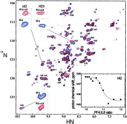
To confirm the Biacore results, interactions between IL-8 and PF4 were investigated in solution using NMR spectroscopy. Nonisotopically-enriched IL-8 was titrated into a solution of uniformly 15N-enriched native PF4. Figure 8 shows the superposition of 2 1H-15N HSQC spectra, one run with 15N-PF4 alone (blue) and the other run with 15N-PF4 to which unlabeled IL-8 (1:1 molar ratio; red) was added. The addition of IL-8 to 15N-PF4 clearly induced chemical-shift changes in many resonances. In general, the chemical-shift changes up to 0.06 ppm for proton and 0.5 ppm for nitrogen are observed. This observation alone indicates that PF4 and IL-8 indeed interact in solution. Some PF4 resonances are affected more than others and some are not affected at all, suggestive of a specific interaction. The assymetric tetramer structure of PF4 along with possible aggregate exchange effects result in the presence of multiple cross-peaks for many resonances, particularly along intersubunit interfaces.34,35 Because of this, most 15N and 1H resonance assignments could not be made, and detailed information as to how PF4 and IL-8 interact could not be definitively derived. Nonetheless, some resonances could be tentatively assigned based on analogy to NMR structural studies on the PF4-M2 mutant, which forms symmetric aggregates.34,35 Tentatively assigned cross-peaks have been labeled in Figure 8. Of the 4 inserts in Figure 8, 3 show expansions of specific HSQC cross-peaks to demonstrate more clearly that native PF4 cross-peaks shift significantly and form a more simplified pattern when IL-8 is present. In the fourth insert, chemical-shift changes for one of these cross-peaks are plotted versus the PF4/IL-8 ratio. Here, the effect is nearly saturable at a molar ratio of 1:1, suggesting that one IL-8 subunit interacts with one PF4 subunit. Although only one major cross-peak was observed for each residue at the 1:1 ratio, minor cross-peaks are still present. Observed chemical-shift changes suggest that PF4/IL-8 aggregate exchange occurs on the same time scale as PF4 monomer-dimer-tetramer association/dissociation.36 Therefore, the binding constant for heterologous association of IL-8 and PF4 falls in the inverse micromolar range. This value places a lower limit on the actual PF4/IL-8 binding constant, whereas that found using the BIAcore method provides an upper limit.
Nuclear magnetic resonance (NMR) spectroscopy of PF4 and IL-8 interaction.1H-15N HSQC spectra of 15N-labeled PF4 in the absence (blue) and presence (red, 1:1 ratio) of unlabeled IL-8 are shown. The spectra were collected at 40°C (pH = 5.0). Insets show an expansion of 3 regions (A43, I42, and H23) to exemplify the chemical-shift changes occurring upon addition of IL-8 to PF4 solution. Also, the proton chemical-shift dependence versus PF4/IL-8 ratio is presented for I42 as an example. Dashed line is plotted to visualize the trend of the chemical-shift dependence.
Nuclear magnetic resonance (NMR) spectroscopy of PF4 and IL-8 interaction.1H-15N HSQC spectra of 15N-labeled PF4 in the absence (blue) and presence (red, 1:1 ratio) of unlabeled IL-8 are shown. The spectra were collected at 40°C (pH = 5.0). Insets show an expansion of 3 regions (A43, I42, and H23) to exemplify the chemical-shift changes occurring upon addition of IL-8 to PF4 solution. Also, the proton chemical-shift dependence versus PF4/IL-8 ratio is presented for I42 as an example. Dashed line is plotted to visualize the trend of the chemical-shift dependence.
Discussion
The amino acid sequence for PF4 was determined more than 20 years ago.37 Despite this, a G protein–coupled receptor for PF4, a founding member of the C-X-C chemokine family, has yet to be identified. Unlike the other chemokines, which are active at picomolar to nanomolar concentrations, PF4 binding to CD34+ cell surface occurs at a concentration of 1 to 10 μg/mL, orders of magnitude higher. PF4 has been shown to activate neutrophils with a similar dose-response through binding to a chondroitin sulfate glycosaminoglycan moiety.15 The weak neutrophil degranulating activity of PF4 has been shown5 to be significantly increased by preincubation with TNF-α. Neutrophil adhesion to endothelial cells is enhanced by PF4 with an EC50 of approximately 1 μM by a mechanism requiring CD11a/CD18 and CD62L.38 In contrast, the prototypic C-X-C cytokine IL-8 also induced adhesion (but with an EC50 nearly 1000-fold lower), required CD11b/CD18, and was unaffected by the presence of anti-CD11a or anti-CD62 antibodies. Therefore, with regard to granulocyte function, PF4 seems to function as an “adhesogenic” protein generating adhesion-dependent signals rather than binding to a specific high affinity G protein–coupled receptor. Recently 2 other chemokines, hemofiltrate C-C chemokine-1 and regakine, have been found39 to exhibit properties similar to PF4 (ie, both are present in plasma at concentrations orders of magnitude higher than that of other more classically up-regulated chemokines; are relatively impotent at inducing chemotactic activity; and synergize with other chemokines such as IL-8 and granulocyte chemotactic protein-2). As is the case for PF4, G protein–coupled specific receptors have yet to be defined for these other chemokines.
In contrast to its mechanism for neutrophil activation, the mechanism for PF4 modulation of hematopoiesis is less clear. PF4 depresses colony formation of myeloid progenitors stimulated by GM-CSF plus steel factor at concentrations of 10 to 50 ng/mL.6,7 Inhibition of megakaryopoiesis, on the other hand, requires concentrations of PF4 in the same range we describe here for inhibition of CD34+ cell cycling (ie, 1-10 μg/mL).8,28 Of note, another biologic activity ascribed to PF4, the inhibition of angiogenesis and endothelial cell proliferation, also occurs with a similarly high ED50.16,40 Injection of high concentrations of PF4 and subsequent harvesting of bone marrow precursors from mice have demonstrated marked inhibition of hematopoietic progenitors in S-phase, a phenomenon associated with decreased sensitivity to chemotherapeutic agents subsequently administered.9 Our current findings define a potential role for PF4 in regulation of hematopoiesis that requires concentrations similar to that previously described to influence phagocyte function rather than the lower concentrations reported to inhibit colony formation in response to GM-CSF and steel factor. These findings raise the possibility that PF4 influences hematopoiesis by 2 separate mechanisms: 1 through an as-yet-unidentified high-affinity, 7-transmembrane receptor, and another depending upon a moderate-affinity interaction with a cell-surface chondroitin sulfate–containing moiety.
CD34+ progenitor adhesion to immobilized PF4 requires intact cellular metabolism and results in cytoskeletal rearrangement, implicating some sort of intracellular signaling event. The functional consequence of such adhesion is inhibition of cell cycle. In the case of neutrophils, the receptor for PF4 has been found to be a membrane chondroitin sulfate–containing moiety.13-15 Membrane-associated chondroitin sulfate also facilitates PF4 blockade of low-density lipoprotein (LDL) uptake by CHO cells through a specific binding interaction with the LDL receptor, suggesting an important role for this glycosaminoglycan in mediating the biologic effects of PF4.41 We have previously shown that the most abundant chondroitin sulfate–containing protein moiety on the surface of CD34+ hematopoietic progenitors is CD44.30 Therefore, CD44-associated chondroitin sulfate is a candidate receptor for PF4. Further studies will be required to identify the precise molecular composition of counterligand(s) on CD34+ hematopoietic progenitors that mediate interaction with PF4.
In addition to modulating hematopoiesis directly by promoting adhesion of CD34+ progenitors, PF4 may have additional, indirect effects on hematopoiesis based upon its ability to bind to chemokines such as IL-8 with high affinity (Figures 7,8) and abrogate their capacity to induce signaling (Figure 6). PF4 blockade of IL-8–induced extracellular acidification rate signaling in CD34+ progenitors is not likely due to direct competition of PF4 for IL-8 receptors CXCR1 or CXCR2, because PF4 does not displace IL-8 bound to neutrophils,5 and prior exposure of neutrophils to PF4 does not down-regulate CXCR1 and CXCR2 expression.42 A second possibility is that PF4 binds to an as-yet-uncharacterized high-affinity C-X-C receptor and generates an intracellular signal that, in turn, blocks IL-8–dependent signaling downstream from receptor occupancy. Indeed, such a mechanism is supported by the recent finding that PF4 blocks fibroblast growth factor 2 (FGF2)–dependent external signal-regulated kinase (ERK) phosphorylation but not FGF2-dependent AKT phosphorylation in endothelial cells at PF4 concentrations that do not impair FGF2 binding to cell receptors.43
Athird possibility, suggested by our current findings (Figures 6,7,8), is that PF4 may bind to IL-8 and thereby impair IL-8–dependent signaling in CD34+ human hematopoietic progenitors. This likely occurs through the formation of IL-8/PF4 heterodimers or heterotetramers. IL-8, like most chemokines, is most active in the monomeric state that predominates at physiologic (ie, nanomolar) concentrations, but at higher concentrations (ie, micromolar) forms less active homodimers.44 PF4 is unique in that even at nanomolar concentrations it is present as a mixture of monomers and dimers but at micromolar concentrations, where suppression of angiogenesis,18 activation of neutrophils,14 suppression of megakaryopoiesis,28 and binding to CD34+ hematopoietic progenitors occurs (not shown), PF4 exists as an asymmetric tetrameric aggregate.14,36,45 Based on the marked NMR-determined structural similarity of soluble PF4,45 and IL-846 and the demonstration that the principal contact points between monomers of these 2 chemokines in homodimers are through 3-stranded antiparallel, internal β-pleated sheet domains,45,46 we hypothesize that similar domains are involved in the formation of PF4/IL-8 heterodimers. We are pursuing such studies using M2-PF4, an N-terminal chimeric PF4 construct that forms symmetric dimers and tetramers that, in turn, allow unambiguous assignment of all amino acid residue NMR resonances.45 Given the strong conservation of C-X-C chemokine internal β-pleated sheet domain structure,47 demonstrating these domains to be the principal contact points in the PF4/IL-8 heterodimer would, in turn, suggest that similar interactions could occur between PF4 and other members of the C-X-C chemokine family. Although the high nanomolar Kd for the IL-8/PF4 interaction suggested by our Biacore (Figure 7) and NMR (Figure 8) studies might not account for 100 ng/mL PF4 blocking signaling by 100 ng/mL IL-8 (Figure 6), we note that in both of these methods PF4 concentrations are orders of magnitude higher, at which PF4 is present exclusively in the tetrameric state. The monomer/monomer or dimer/monomer affinities of PF4/IL-8 complexes have yet to be determined and could conceivably be higher. In any case, given the large amount of PF4 present within and released by platelets and proliferating megakaryocytes,48 in physiologic circumstances PF4 would always be present at concentrations orders of magnitude higher than that of IL-8 (or other C-X-C chemokines), a situation in which the proportion of IL-8 in the IL-8/PF4 heterodimeric complex would be high.
Our finding that PF4 blocks IL-8–dependent signaling in CD34+ human hematopoietic progenitors apparently contradicts the previous demonstration that PF4 synergizes with IL-8 to suppress myeloid, erythroid, and mixed colony progenitor outgrowth.7 However, these and the present studies were performed with different culture conditions and growth factors. We assayed the effect of PF4 only upon hematopoietic progenitor adhesion to stromal layer and immobilized PF4 and in solution in short-term experiments (Figure 5), not in methylcellulose suspension in the continued presence of PF4. The PF4 concentrations (0.1-1 ng/mL) at which synergism of IL-8 with PF4 were shown are 1000- to 10 000-fold lower than those we used in our studies, in which heterodimer formation would be highly unlikely. There is also a precedent for dose-dependent but opposing effects of PF4 upon the activity of another C-X-C chemokine, where nanogram per milliliter concentrations of PF4 inhibit, but microgram per milliliter concentrations enhance, connective tissue–activating peptide (CTAP)–III–dependent adhesion of neutrophils to endothelium.42 It is also possible that an as-yet-unidentified high-affinity PF4 chemokine receptor on CD34+ cells mediates effects at low concentrations of PF4 that are contravened by effects occurring at higher PF4 concentrations through a different mechanism involving a chondroitin sulfate–associated membrane moiety.
To our knowledge the existence of chemokine/chemokine heterodimers has previously been demonstrated only for MIP-1α and MIP-1β.49 As well as binding to IL-8 as we here show, PF4 has previously been shown to bind to (and block the biologic activities of) 2 cytokines, VEGF16516 and FGF-2.40 Thus, PF4 can participate in both chemokine/chemokine and chemokine/cytokine interactions. Further investigation will be needed to ascertain how formation of such complexes influences the biologic activity of either partner. In aggregate, however, these findings raise the possibility that PF4, especially at high (ie, micromolar) concentrations might influence the presentation and binding of numerous cytokines and chemokines in the complex milieu of the bone marrow microenvironment. Through this mechanism, as well by inducing adhesion of hematopoietic progenitors to stroma, PF4 might influence normal and pathologic hematopoiesis.
Our in vitro findings may have relevance for the pathogenesis of myelofibrotic states, which are commonly associated with hyperproliferation of the bone marrow, including megakaryocytes. Megakaryocyte proliferation has been implicated in the pathogenesis of myelofibrosis because thrombopoietin (TPO) transgenic mice develop chronic megakaryocyte hyperproliferation and myelofibrosis.50 This abnormality is corrected by transplanting the marrow from wild-type mice into the TPO transgenic mice, demonstrating that myelofibrosis results from the presence in the marrow cavity of hyperproliferating megakaryocytes. A number of potential mechanisms potentially link hyperproliferation of megakaryocytes with development of myelofibrosis, including intramedullary release of the potent mitogen platelet-derived growth factor. However, megakaryocytes and their progenitors contain and release into culture supernatant fluid high concentrations of PF4.48 It is therefore possible that, in the presence of chronic hyperproliferation and turnover of megakaryocytes, there is intramedullary accumulation of PF4 that perturbs the marrow microenvironment. This might inhibit hematopoietic progenitor proliferation and promote displacement of hematopoiesis to other organs such as the liver and spleen. Preliminary description of a PF4 knock-out mouse supports the idea that PF4 influences hematopoiesis in vivo as well as in vitro.51 We predict that the mechanisms of action of PF4 in hematopoiesis will be the subject of ongoing interest.
Prepublished online as Blood First Edition Paper, February 13, 2003; DOI 10.1182/blood-2002-08-2363.
The publication costs of this article were defrayed in part by page charge payment. Therefore, and solely to indicate this fact, this article is hereby marked “advertisement” in accordance with 18 U.S.C. section 1734.
We are grateful to Karen De Moor for her excellent technical assistance during experiments with the microphysiometer (Cytosensor, Molecular Devices, Sunnyvale, CA).


This feature is available to Subscribers Only
Sign In or Create an Account Close Modal