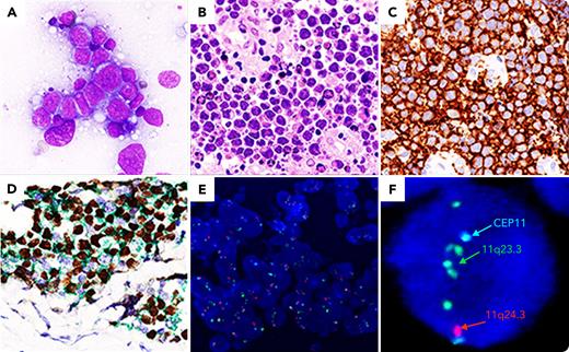
A 11-year-old boy presented with a large left neck mass. Histological examination showed diffuse proliferation of mostly large-size tumor cells (panels A-B, original magnification x400) CD20+ (panel C, original magnification x400), BCL2− and coexpressing at double immunoperoxidase staining CD10 (green)/BCL6 (brown) (panel D, original magnification x400). Fluorescence in situ hybridization (FISH) showed proximal gain in 11q23 region (green signal) and telomeric loss in 11q24 region (red signal), consistent with the typical gain-loss pattern for 11q aberration (panels E-F, E: original magnification x400, F: original magnification x1000). The MYC break-apart FISH was negative. A positron emission tomography/computed tomography was diagnostic of stage II disease.
In the World Health Organization (WHO) classification of lymphohematopoietic neoplasms, this case is regarded as a provisional entity named “Burkitt-like lymphoma with 11q aberration,” because of its resemblance to Burkitt lymphoma but lack of MYC rearrangement. This provisional entity has been now revised as “large B-cell lymphoma with 11q aberration” in the International Consensus Classification of Mature Lymphoid Neoplasms and as “high-grade B-cell lymphoma with 11q aberration” in the 5th WHO classification of lymphoid neoplasms. As compared with MYC breakpoint-positive Burkitt lymphoma, large B-cell lymphoma with 11q aberration in pediatric patients is less frequent, occurs at an older age, shows less male predominance as well as lower stage and lactate dehydrogenase and less abdominal involvement. This entity is usually characterized by an excellent outcome. Our patient was treated with a regimen as for diffuse large B-cell lymphoma, achieving a complete remission.
Author notes
For additional images, visit the ASH Image Bank, a reference and teaching tool that is continually updated with new atlas and case study images. For more information, visit http://imagebank.hematology.org.


