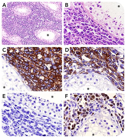
A 68-year-old man presented with a 4.5-cm painless mass in the left testicle. Orchiectomy of the involved testis was performed, and the histological examination showed diffuse proliferation of large-sized tumor cells (panels A and B; hematoxylin and eosin staining, original magnification: ×100 [A], ×400 [B]) and CD20+ (panel C; immunoperoxidase staining, original magnification ×400), CD79b+ (panel D; immunoperoxidase staining, original magnification ×400), CD10− (panel E; immunoperoxidase staining, original magnification ×400), and MUM1/IRF4+ (panel F; immunoperoxidase staining, original magnification ×400) expression. The asterisks indicate the lumen of seminiferous tubules. A diagnosis of diffuse large B-cell lymphoma (DLBCL) with activated B-cell (ABC) phenotype was made. Computed tomography was diagnostic of stage IE disease.
The International Consensus Classification regards testicular DLBCL as a new entity. Similarly, the fifth edition of the World Health Organization classification of lymphohematopoietic neoplasms has grouped DLBCL of testis under the category of large B-cell lymphomas arising in immune-privileged sites, that is, sanctuaries defined by the blood-testicular, blood-brain, and blood-retinal barriers. In the testis, mechanisms defending the developing gametocytes may provide DLBCL with an ideal milieu for acquiring an immune-escape phenotype. Testicular DLBCL shows an aggressive course and frequently relapse in the controlateral testis and the central nervous system (CNS). It shares many features with primary CNS lymphoma, including ABC cell-of-origin and mutations of NF-κB signaling pathway. Our patient was treated with radiotherapy of the controlateral testis and systemic chemotherapy (R-CHOP [rituximab plus cyclophosphamide, doxorubicin, vincristine, and prednisone]) plus CNS prophylaxis, and he is now in complete remission 23 years after the initial diagnosis.
Author notes
For additional images, visit the ASH Image Bank, a reference and teaching tool that is continually updated with new atlas and case study images. For more information, visit http://imagebank.hematology.org.


This feature is available to Subscribers Only
Sign In or Create an Account Close Modal