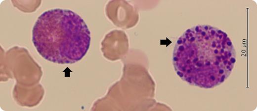
A 66-year-old man was referred to our hospital for fatigue and pancytopenia. His hemoglobin level was 7.4 g/dL, with a white cell count of 2.6 × 10⁹/L and platelet count of 39 × 109/L. To evaluate his pancytopenia, a bone marrow examination was performed. May-Giemsa staining of bone marrow aspiration showed 6% blasts and dysplastic eosinophils with large purple-violet granules resembling basophilic granules (arrows). Dysplastic eosinophils with metachromatic granules are often referred to as harlequin cells, which are commonly observed in acute myeloid leukemia (AML) with inv(16)(p13.1q22) or t(16;16)(p13.1;q22) and chronic myeloid leukemia.
A conventional cytogenetic analysis of his excess blasts revealed an inversion of chromosome 16. Moreover, the detection of CBFB-MYH11 fusion transcripts using reverse transcription polymerase chain reaction confirmed the diagnosis of AML with inv(16)(p13.1q22). In the World Health Organization classification, neoplasms with the following specific recurrent cytogenetic abnormalities are regarded as acute leukemias, irrespective of the blast percentages: t(8;21)(q22;q22.1), inv(16)(p13.1q22) or t(16;16)(p13.1;q22), and t(15;17)(q24.1;q21.2). After chemotherapy with cytarabine and anthracyclines, the patient’s pancytopenia and dysplastic eosinophils resolved. He remains in remission 6 months later. Harlequin cells imply a myeloid neoplasm regardless of the blast percentage.
For additional images, visit the ASH Image Bank, a reference and teaching tool that is continually updated with new atlas and case study images. For more information, visit http://imagebank.hematology.org.


