Abstract
Background
Primary mediastinal large B-cell lymphoma (PM LBCL) is putatively derived from thymic B cells. PM LBCL is clinically and morphologically distinct from diffuse large B-cell lymphoma (DLBCL), but shares certain phenotypic characteristics with nodular sclerosing classical Hodgkin's lymphoma. The expression and function of an intact B-cell receptor (BCR) in PM LBCL is controversial. In addition, the pathological mechanisms that drive lymphomagenesis in PM DLBCL are largely unknown. Here, we investigate the expression of functional BCR in PM LBCL and explore the hypothesis that autonomous signaling of the BCR as observed in CLL (Dühren-von Minden, Nature 2012) plays a key role in PM DLBCL pathogenesis.
Methods
The PM LBCL cell lines Farage, MedB-1, Karpas 1106P, and U2940 were tested in vitro by comparing calcium flux with and without inhibition of BCR signaling by blockade of Syk with the tyrosine kinase inhibitor R406.
BCR transcripts were identified from archived fresh-frozen biopsies from two histologically confirmed PM LBCL by ARTISAN PCR, a novel anchored RT-PCR for unbiased amplification of BCR transcripts facilitated by template switching (Koning, BJH 2016). Clonal full-length BCR sequences were identified by PacBio next-generation sequencing. To test for autonomous BCR signaling, murine triple KO (TKO) cells were transduced with functional BCR of PM LBCL (Dühren-von Minden, Nature 2012). TKO cells lack the rag2 and lambda5 genes and are therefore developmentally arrested at the pre-B-cell stage. In addition, TKO cells have their wild-type SLP65 adaptor replaced with a tamoxifen-dependent SLP65 variant. When transduced with a functional BCR, autonomous or antigen-induced BCR signaling can be measured upon induction with tamoxifen as calcium flux by flow cytometry or as robust cellular proliferation.
Results
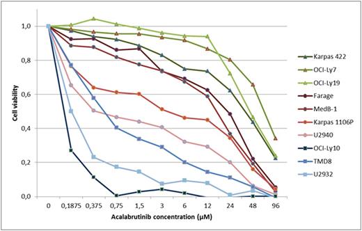
All four PM-LBCL lines had higher calcium flux in the absence of R406, suggesting autonomous BCR signaling activity without antigenic stimulation or artificial BCR crosslinking. Functional BCR transcripts could be readily identified in both primary PM LBCL biopsies by ARTISAN PCR. In accordance with our hypothesis, TKO cells transduced with the BCR of both primary PM LBCL cases showed robust calcium flux and proliferation upon activation of SLP65 by tamoxifen without additional BCR crosslinking. To test whether this autonomous BCR signal would represent a druggable therapeutic target, the PM DLBCL cell lines were cultured in the presence of increasing doses of the second generation, highly BTK-specific kinase inhibitor acalabrutinib. The ABC DLBCL cell lines OCI-Ly10, TMD8, and U2932, and the GCB cell lines Karpas 422, OCI-Ly7, and OCI-Ly19 were tested in parallel as controls. Cell viability decreased in all cell lines with increasing concentrations of acalabrutinib (Figure). Consistent with their known dependency on active BCR signalling (Young, PNAS 2015; Koning, ASH 2016), ABC DLBCL cell lines were very sensitive to acalabrutinib. As suggested by the experimental evidence for autonomous BCR signalling, PM LBCL cell lines also responded to acalabrutinib, albeit to a lesser degree than ABC DLBCL. Growth inhibition of GCB DLBCL cell lines generally required excessively high doses of acalabrutinib, consistent with the notion that this DLBCL subtype depends on tonic BCR signaling only.
Conclusion
Consistent with our hypothesis, our data demonstrate expression of an intact BCR with autonomous signaling activity in all investigated cases of PM LBCL. These results point to an oncogenic role of structurally normal BCR in PM LBCL akin to CLL. Similarly to CLL and ABC DLBCL, PM DLBCL cell lines respond to BTK inhibitors, albeit less strongly than the ABC DLBCL cell lines tested. The results identify BCR signaling as a novel therapeutic target in PM LBCL and suggest the initiation of clinical trials with BTK inhibitors in PM LBCL.
No relevant conflicts of interest to declare.
Author notes
Asterisk with author names denotes non-ASH members.


