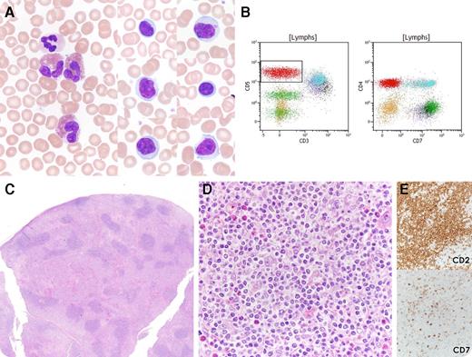
A 25-year-old woman presented with diffuse “lumps” under the skin and was found to have peripheral eosinophilia and an atypical circulating lymphocyte population (white blood cells, 10.8 × 10⁹/L; 22% lymphocytes; and 12% eosinophils) (panel A). No history of atopy was noted. Peripheral blood (PB) flow cytometry demonstrated an abnormal T-cell population with loss of surface CD3; expression of CD2, CD5, CD4; and loss of CD7 (panel B). Biopsy of one of the lesions on the chest wall revealed a paracortical infiltrate of small, atypical lymphocytes with irregular nuclei and pale cytoplasm, admixed with eosinophils (panels C-D). The immunophenotype of these lymphocytes matched the atypical T cells in the PB (panel E) (cytoplasmic CD3 is positive by immunohistochemistry) and additionally lacked staining for TFH markers CD279 and CD10. T-cell receptor gene rearrangement analysis revealed a clonal rearrangement.
The findings in this case are typical for the lymphocytic variant of hypereosinophilic syndrome, which is now regarded as a T-cell lymphoproliferative disorder characterized by eosinophilia and sCD3-negative T cells in the blood. Although rare, the lymphocytic variant of hypereosinophilic syndrome is in the differential diagnosis for a patient with isolated eosinophilia and highlights the need for PB flow cytometry to evaluate this atypical T-cell population. Recently, involvement of lymph nodes and other tissue sites has been recognized and could easily be mistaken for peripheral T-cell lymphoma and treated aggressively without the appropriate clinical history.
For additional images, visit the ASH IMAGE BANK, a reference and teaching tool that is continually updated with new atlas and case study images. For more information visit http://imagebank.hematology.org.


