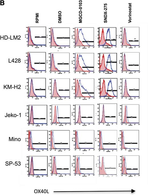
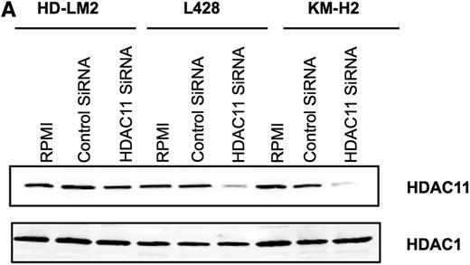
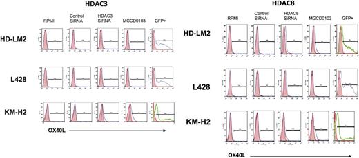
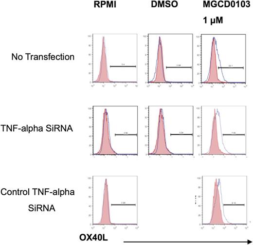
On page 2912 in the 10 March 2011 issue, Figure 1B showed a duplicate panel in the SP53 cells treated with MGCD-0103 and SNDX-275. On page 2913, Figure 2A showed a duplicate panel that was labeled “Beta actin.” This panel was inadvertently included in the figure, because HDAC1 was used as a control for loading and the siRNA specificity. On page 2914, the HDAC3 experiment in Figure 3 showed a duplicate panel for HDL-M2 and L428 cells. The KM-H2 HDAC3 panels are duplicates of the KMH2 HDAC8 panels. On page 2915, Figure 4B showed duplicate DMSO panels for TNF-alfa SiRNA and control TNF-alfa siRNA. The TNF-alfa SiRNA panel is the correct one and was used as a second control in addition to the RPMI control experiment. These errors occurred during the assembly of the figures and have no bearing on the results or conclusions of the study. The corrected Figures 1B, 2A, 3 (the HDAC3 and HDAC8 experiments), and 4B are shown. The authors apologize for these mistakes.
HDACis upregulate the expression of OX40L in HL. (A) Baseline expression of OX40L in lymphoid cell lines by western blot analysis. (B) OX40L expression in HL and MCL cells. Cells were incubated with DMSO, MGCD0103, vorinostat, and SNDX-275 (1 μM) for 24 hours before OX40L levels were analyzed by FACS. MGCD0103 and SNDX-275 increased OX40L expression in HL cells but not in MCL cells. (C) CD40 expression in HL cells. (D) OX40L expression in HL cells. Cells were incubated with DMSO, MGCD0103, and vorinostat (0.1-2.0 μM) for 48 hours and collected, and their OX40L expression was analyzed by FACS. MGCD0103 increased OX40L expression in a dose-dependent manner. Each value is the mean of 3 independent experiments (±SEM). *P < .05, **P < .005. (E) Effect of HDACis on OX40L mRNA levels in HL cells. Results of qRT-PCR after 48 hours of incubation in the absence or presence of MGCD0103 (0.5-1.0 μM) or SNDX-275 (0.5-1.0 μM). Data are presented as mRNArelative units (RU) ± SEM; n = 3 for each condition and kinetic point. *P < .05, **P < .005. (F) Effect of MGCD0103 on OX40L expression. HL cells were incubated with MGCD0103 (0.1-2.0 μM) for 48 hours. Western blot analysis was performed on whole-cell lysates. The effect was more evident with higher doses of MGCD0103 in all HL cell lines.
HDACis upregulate the expression of OX40L in HL. (A) Baseline expression of OX40L in lymphoid cell lines by western blot analysis. (B) OX40L expression in HL and MCL cells. Cells were incubated with DMSO, MGCD0103, vorinostat, and SNDX-275 (1 μM) for 24 hours before OX40L levels were analyzed by FACS. MGCD0103 and SNDX-275 increased OX40L expression in HL cells but not in MCL cells. (C) CD40 expression in HL cells. (D) OX40L expression in HL cells. Cells were incubated with DMSO, MGCD0103, and vorinostat (0.1-2.0 μM) for 48 hours and collected, and their OX40L expression was analyzed by FACS. MGCD0103 increased OX40L expression in a dose-dependent manner. Each value is the mean of 3 independent experiments (±SEM). *P < .05, **P < .005. (E) Effect of HDACis on OX40L mRNA levels in HL cells. Results of qRT-PCR after 48 hours of incubation in the absence or presence of MGCD0103 (0.5-1.0 μM) or SNDX-275 (0.5-1.0 μM). Data are presented as mRNArelative units (RU) ± SEM; n = 3 for each condition and kinetic point. *P < .05, **P < .005. (F) Effect of MGCD0103 on OX40L expression. HL cells were incubated with MGCD0103 (0.1-2.0 μM) for 48 hours. Western blot analysis was performed on whole-cell lysates. The effect was more evident with higher doses of MGCD0103 in all HL cell lines.
Silencing of HDAC11 gene expression by siRNA upregulates OX40L in HL cell lines. (A) HL cell lines were transfected with HDAC11 or control siRNA (3 μg), and after 48 hours the cellular level of HDAC11 was determined by western blot. Results represent 3 independent experiments showing the efficacy of HDAC11 siRNA in downregulating HDAC11 expression. (B) HL cell lines were analyzed by FACS after 48 hours of HDAC11 siRNA transfection. Blocking HDAC11 increased OX40L expression. (C) Mean fluorescence intensity of 3 independent experiments (±SEM). *P < .05, **P < .005. (D) The Jeko-1 cell line was analyzed by FACS after 24 hours of HDAC11 siRNA transfection. Downregulating HDAC11 did not have any effect on OX40L expression. (E) HDAC11 siRNA induced apoptosis in HL cell lines. (F) Representative experiment demonstrating the effect of downregulating HDAC11 on induction of apoptosis in 3 HL cell lines as determined by propidium iodide and Annexin V staining and FACS analysis. Results are shown after 48 hours of incubation. Each value is the mean of 3 independent experiments (±SEM). *P < .05, **P < .005. (G) Effect of HDAC11 siRNA on the caspase pathway. Downregulation of HDAC11 decreased expression of caspase 9 and caspase 8 in HL cells, as determined after 24 hours of incubation.
Silencing of HDAC11 gene expression by siRNA upregulates OX40L in HL cell lines. (A) HL cell lines were transfected with HDAC11 or control siRNA (3 μg), and after 48 hours the cellular level of HDAC11 was determined by western blot. Results represent 3 independent experiments showing the efficacy of HDAC11 siRNA in downregulating HDAC11 expression. (B) HL cell lines were analyzed by FACS after 48 hours of HDAC11 siRNA transfection. Blocking HDAC11 increased OX40L expression. (C) Mean fluorescence intensity of 3 independent experiments (±SEM). *P < .05, **P < .005. (D) The Jeko-1 cell line was analyzed by FACS after 24 hours of HDAC11 siRNA transfection. Downregulating HDAC11 did not have any effect on OX40L expression. (E) HDAC11 siRNA induced apoptosis in HL cell lines. (F) Representative experiment demonstrating the effect of downregulating HDAC11 on induction of apoptosis in 3 HL cell lines as determined by propidium iodide and Annexin V staining and FACS analysis. Results are shown after 48 hours of incubation. Each value is the mean of 3 independent experiments (±SEM). *P < .05, **P < .005. (G) Effect of HDAC11 siRNA on the caspase pathway. Downregulation of HDAC11 decreased expression of caspase 9 and caspase 8 in HL cells, as determined after 24 hours of incubation.
Effect of HDAC1, -2, -3, and -8 siRNA transfection on OX40L expression. HL cell lines were transfected with HDAC1, -2, -3, and -8 siRNA and cells were analyzed for OX40L expression after 48 hours of incubation. Inhibition of expression of HDAC1, -2, -3, or -8 had little or no effect on OX40L expression in HL cells.
Effect of HDAC1, -2, -3, and -8 siRNA transfection on OX40L expression. HL cell lines were transfected with HDAC1, -2, -3, and -8 siRNA and cells were analyzed for OX40L expression after 48 hours of incubation. Inhibition of expression of HDAC1, -2, -3, or -8 had little or no effect on OX40L expression in HL cells.
HDAC11 regulates the production of cytokines and chemokines in HL. (A) We analyzed 4 cell lines: HD-LM2, L428, KM-H2, and Mino. Cell culture supernatants were collected after 24 hours of HDAC11 siRNA transfection. HDAC11 siRNA increased the production of IL-13, TNF-α, interferon-γ, IL-6, and IL-17. Each value is the mean of 3 independent experiments (±SEM). *P < .05, **P < .005. (B) Expression of OX40L after TNF-α siRNA transfection. The L428 cell line was analyzed by FACS after TNF-α siRNA transfection and after 48 hours of treatment with MGCD0103 (1.0 μM). MGCD0103 increased OX40L expression. (C) OX40L expression in HD-LM2 cells. HD-LM2 cells were incubated with recombinant TNF-α (1-10 ng) for 48 hours and then analyzed by FACS. Recombinant TNF-α did not have a significant effect on OX40L expression in these cells.
HDAC11 regulates the production of cytokines and chemokines in HL. (A) We analyzed 4 cell lines: HD-LM2, L428, KM-H2, and Mino. Cell culture supernatants were collected after 24 hours of HDAC11 siRNA transfection. HDAC11 siRNA increased the production of IL-13, TNF-α, interferon-γ, IL-6, and IL-17. Each value is the mean of 3 independent experiments (±SEM). *P < .05, **P < .005. (B) Expression of OX40L after TNF-α siRNA transfection. The L428 cell line was analyzed by FACS after TNF-α siRNA transfection and after 48 hours of treatment with MGCD0103 (1.0 μM). MGCD0103 increased OX40L expression. (C) OX40L expression in HD-LM2 cells. HD-LM2 cells were incubated with recombinant TNF-α (1-10 ng) for 48 hours and then analyzed by FACS. Recombinant TNF-α did not have a significant effect on OX40L expression in these cells.


