Abstract
Interleukin-21 (IL-21) enhances T helper 1 (Th1) and Th17 differentiation while inhibiting the conversion of inducible regulatory T cells (Tregs) from naive T cells. To determine the role of IL-21 in graft-versus-host disease (GVHD), anti–IL-21 antibody (Ab) was given to recipients of CD25−CD4+ or CD4+ and CD8+ T-effectors. IL-21 neutralization attenuated GVHD-related weight loss and prolonged survival. Likewise, a majority of mice receiving IL-21−/− CD25− T-effectors survived long term, whereas those receiving wild-type T cells died. The latter recipients had higher grades of GVHD in the ileum and colon. Surprisingly, disruption of IL-21 signaling did not affect IL-17 production, although colon-infiltrating T-effector cells had decreased interferon γ (IFNγ) and increased IL-4 production. FoxP3+ Tregs were increased in colons of anti–IL-21 Ab-treated recipients of FoxP3− IL-21−/− T cells, indicating Treg conversion. Recipients of FoxP3-deficient T-effectors isolated from chimeras were resistant to the GVHD protective effects of IL-21 blockade. Whereas graft-versus-leukemia (GVL) can occur in the absence of IL-21, loss of both IL-21 and perforin expression abrogated GVL. Together, these data indicate that IL-21 suppresses inducible Treg conversion and further suggest that IL-21 blockade is an attractive strategy to reduce GVHD-induced injury.
Introduction
Graft-versus-host disease (GVHD) remains an important complication after allogeneic bone marrow transplantation (BMT). Despite broadly reactive pharmacologic agents, GVHD is not uniformly avoided and immunosuppression may cause malignancy recurrence or immunodeficiency. Selective GVHD preventive approaches retaining a graft-versus-leukemia (GVL) effect are needed.
Interleukin-21 (IL-21) is produced by CD4+ T cells (especially T helper 17 [Th17]–producing cells) and natural killer T (NKT) cells1 and signals through the IL-2γc and IL-21R complex. IL-21R is expressed on hematopoietic and epithelial cells and promotes the activation, differentiation, maturation, or expansion of NK cells, B cells, CD8+ and CD4+ T cells, dendritic cells, and macrophages, resulting in anticancer activity.2-5 IL-21 facilitates autoimmunity in some6-8 but not all9,10 experimental models by supporting immunoglobulin production and Th17 cell–mediated pathogenesis.
Because IL-21 augments Th17 cell differentiation, indirect evidence for the role of IL-21 in GVHD pathogenesis may be derived from such GVHD studies. Whereas IL-17 and Th17 cells reduce GVHD mediated by CD4+ and CD8+ donor T cells,11 Th17 cells accelerated GVHD mediated exclusively by CD4+ T cells.12 Naive CD4+ T cells skewed toward a Th17 phenotype in vitro have been used to demonstrate that Th17 cells contribute to GVHD pathogenesis, especially involving the skin and lung.13 IL-21 has been described variably as an inhibitor14 or enhancer15 of Th1 differentiation. IL-21 supports Th17 cell survival at the expense of regulatory T cells (Tregs), which are reciprocally controlled by Th17 cells.16 By inhibiting naive T-cell conversion into CD4+25+FoxP3+ regulatory T cells (termed inducible Tregs, iTregs),17,18 limiting the suppression of T-effectors (Teffs) by Tregs, and augmenting Th17 responses,19,20 IL-21 may increase GVHD lethality.
The present studies were conducted to delineate the influence of IL-21 on GVHD and GVL and to elucidate the mechanisms associated with the observed biologic effects. We show that blocking or abrogating the IL-21 signaling pathway reduced acute GVHD mortality and tissue damage in the small intestine and the colon associated with decreased frequencies of interferon γ (IFNγ)–producing tissue-resident donor T cells in the colonic lamina propria (LP). At the same time FoxP3-expressing Tregs, which were virtually absent in the presence of IL-21, were found at relatively high frequencies at the site of inflammation in the colon and the small intestine in the absence of IL-21. These data, which are the first to demonstrate an in vivo role for IL-21 in iTreg generation, suggested a causative role of iTregs in GVHD attenuation. This was confirmed using Teffs incapable of generating iTregs. Despite acute GVHD attenuation, we show that GVL can occur in the absence of IL-21. Lastly, we show that the perforin and IL-21 pathways are nonredundant in the context of both the GVHD and GVL settings.
Methods
Mice
C57BL/6 (H-2b, termed B6) mice were purchased from the National Institutes of Health. B10.BR (H2k), BALB/c (H2d), B6.SJL-Ptprca Pep3b/BoyJ congenic mice (H2b, termed CD45.1), C57BL/6-Prf1tm1Sdz/J (termed perforin−/−), male B.Cg-Foxp3sf (scurfy) mice lacking the Treg transcription factor FoxP3 (H2b, CD45.2), and B6 IL-2Rγc–deficient mice were purchased from The Jackson Laboratory. B6-FoxP3-GFP knock-in breeders that express green fluorescent protein (GFP) (Memorial Sloan-Kettering Cancer Institute, New York, NY) under the control of the FoxP3 promoter were provided by A. Rudensky (Memorial Sloan-Kettering Cancer Institute, New York, NY). Heterozygote IL-21−/− and IL-21R−/− mice generated in the 129S7/SvEvBrd-Hprtb-m2 strain by Lexicon Genetics were backcrossed 7 generations to B6 mice by ZymoGenetics. Mice were used at 6 to 14 weeks of age, kept under specific pathogen-free conditions, and used according to University of Minnesota Institutional Animal Care and Use Committee–approved protocols.
GVHD induction and IL-21 neutralization
Recipients received total body irradiation at a lethal dose (B10.BR 11 Gy; Balb/c 8.75 Gy) by a 137Cesium source 24 hours before cell infusion. Unless otherwise specified, B10.BR mice were used as recipients. CD4+ and CD8+ T cell–depleted bone marrow (BM) cells (107) from B6 mice or unmanipulated BM from IL-2Rγc−/− mice were injected intravenously on day 0. Teffs were purified from splenocytes shipped overnight on ice. After red cell lysis, cells were incubated with phycoerythrin (PE)–labeled antibodies (Abs) with specificities for CD19, CD11b, NK1.1 (eBioscience), and T-cell receptor γδ (BD) and, where indicated, also for CD8 or/and CD25 and incubated with anti-PE microbeads followed by passage over LD columns (Miltenyi Biotech). Purity was more than 95%. For some experiments, FoxP3−CD4+ or FoxP3− T cells were sorted based on the absence of GFP expression using a FACSDiva sorter (BD). GFP+ cells after sorting were less than 0.01%. Naive scurfy Teffs were generated by injecting scurfy (CD45.2) BM cells (107) into lethally irradiated congenic (CD45.1) mice. After 5 weeks, C45.1−CD25−CD4+scurfy cells were purified from splenocytes using a FACSDiva sorter. All recipients were monitored daily for survival, weighed twice weekly for 30 days, and weekly thereafter. Where indicated, irrelevant rat immunoglobulin G (IgG; Rockland) or chimeric mouse/rat anti–IL-21 Ab (mIgG1, clone Ch268.5.1; ZymoGenetics) was administered at a dose of 200 μg twice weekly for either 5 or 6 weeks, as indicated.
Tissue analysis
Representative mice were killed on indicated days and spleens, livers, lungs, small intestine, colon, kidney, and skin specimens were embedded in OCT compound and snap frozen, and 6-μm sections were cut, fixed in acetone for 5 minutes, and stained with hematoxylin and eosin. Scoring was done as previously published with an Olympus BX51 microscope using a 10× lens (aperture 0.4).21 In some experiments, sections were air dried overnight, blocked with 100% horse serum, and incubated with biotinylated anti-FoxP3 (FJK16s; eBioscience) overnight. Secondary incubation was done with streptavidin cyanin 3 (Jackson ImmunoResearch). CD4 staining was done with anti–CD4–fluorescein isothiocyanate. Confocal images were acquired on an Olympus FluoView 500 confocal laser scanning microscope using a 10× lens (aperture 0.7). Images shown as acquired.
Flow cytometry
Abs with specificity for CD4, CD8, CD25, tumor necrosis factor-α (TNFα), granzyme B, IFNγ, IL-4, IL-10, and IL-17, were purchased from eBioscience. Antibodies for Fas ligand (FasL) and Kits containing Abs binding to Ki-67 and allophycocyanin-labeled annexin-V were purchased from BD Biosciences. Colon and liver cell purification was done as described.22 Some cells were stained with Abs to CD4 and CD8, Ki-67, and annexin-V without restimulation. For cell number quantification, PE-labeled counting beads (Sigma) were used. For intracellular cytokine staining, cells were incubated for 4 hours in αCD3ϵ Ab-coated plates (5 mg/mL) at 37°C. Cells were stained for CD4 and CD8, fixed, and permeabilized with a commercial kit (BD) and stained according to manufacturer's protocol with antibodies to intracellular antigens. Cells were acquired on a 4-color FACSCalibur instrument (BD). Analysis was done with FlowJo8.5 (TreeStar).
Assessment of GVL activity
A20 lymphoma cells were purchased from ATTC and nucleoporated with a Sleeping Beauty transposon construct to permit the coexpression of dsred2 and renilla luciferase (termed A20luc) as described.23 Lethally irradiated Balb/c recipients undergoing BMT received A20luc (105). Groups received 2 × 10⁶ CD25− Teffs from wild type (wt), IL-21−/−, IL-21R−/−, or perforin−/− mice in the presence or absence of anti–IL-21 mAb, as indicated, for a period of 6 weeks. For A20luc cell imaging, mice were anesthetized with pentobarbital. EnduRen live cell substrate (final volume, 0.1 mL; Promega) at a final concentration of 60mM was injected intraperitoneally into the mice 5 minutes before imaging. Images were acquired after exposure for 1 minute with an in vivo imaging system (IVIS100; Xenogen).
Statistical analysis
Survival was analyzed by life-table methods and actuarial survival data are shown. Comparison between groups was done by log-rank test. Group comparisons of GVHD scores were done using the Mann-Whitney test. Comparisons were performed using an unpaired 2-sided Student t test. P values of .05 or less were considered significant. Box plots show mean plus or minus SE of the mean.
Results
IL-21 signaling is critical for acute GVHD-induced lethality and target organ injury
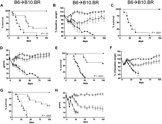
To investigate the role of IL-21 in GVHD, lethally irradiated B10.BR mice were given B6 BM with or without 106 B6 CD4+25− T cells. Cohorts were treated with irrelevant IgG or neutralizing anti–IL-21 Ab for a period of 5 weeks after BMT. Whereas all controls died of GVHD lethality, anti–IL-21 Ab-treated recipients survived significantly longer and 31% survived beyond 100 days after BMT (Figure 1A). Mean weights reflected survival differences (Figure 1B). In other studies in which 106 CD4+CD25− cells from wt or IL-21−/− T cells were infused, the latter had 100% day-100 survival versus 0% in recipients of wt CD4+25− T cells (Figure 1C-D). Recipients of IL-21−/− versus wt CD4+CD25− T cells had minimal GVHD-induced weight loss, in contrast to controls (Figure 1D).
Reduced GVHD lethality as a consequence of inhibiting IL-21/IL-21R signaling on donor T cells. Survival and weight curves of lethally irradiated B10.BR recipients of T cell–depleted BM and Teffs from B6 mice. * indicates recipients of wt BM if not otherwise indicated. Recipients of 106 wt CD4+CD25− T cells treated with irrelevant IgG (▾) or anti–IL-21Ab (▿; P = .01; A-B). Recipients of CD4+CD25− T cells from wt (▾) and IL-21−/− (▿) donors (P < .001; C-D). Recipients of CD25− T cells from wt (▾) or IL-21−/− (▿) donors (P < .001; E-F). Recipients of IL-2γcR−/− BM (*) plus wt (▾) or IL-21−/− (▿) CD25− T cells (P < .001; C-D). Data from 2, 1, 4, and 1 experiments with 16, 8, 22, and 12 mice per group for panels A and B, C and D, E and F, and G and H, respectively.
Reduced GVHD lethality as a consequence of inhibiting IL-21/IL-21R signaling on donor T cells. Survival and weight curves of lethally irradiated B10.BR recipients of T cell–depleted BM and Teffs from B6 mice. * indicates recipients of wt BM if not otherwise indicated. Recipients of 106 wt CD4+CD25− T cells treated with irrelevant IgG (▾) or anti–IL-21Ab (▿; P = .01; A-B). Recipients of CD4+CD25− T cells from wt (▾) and IL-21−/− (▿) donors (P < .001; C-D). Recipients of CD25− T cells from wt (▾) or IL-21−/− (▿) donors (P < .001; E-F). Recipients of IL-2γcR−/− BM (*) plus wt (▾) or IL-21−/− (▿) CD25− T cells (P < .001; C-D). Data from 2, 1, 4, and 1 experiments with 16, 8, 22, and 12 mice per group for panels A and B, C and D, E and F, and G and H, respectively.
To determine whether the GVHD-protective effect of IL-21−/− T cells was restricted to the CD4+25− T-cell subset, CD25− T cells, containing both CD4+ and CD8+ T cells from wt or IL-21−/− mice, were used as donor Teffs. Survival was significantly prolonged in recipients of IL-21−/− versus wt CD25− T cells, with 59% versus 0% surviving long term (P < .001, Figure 1E). Recipients of IL-21−/− CD25− T cells had reduced weights compared with BM-only recipients (Figure 1F). We considered the possibility that donor IL-21 produced by NKT cells or residual BM T cells remaining after pan-T-cell depletion may contribute to GVHD-induced lethality even when donor Teffs are obtained from IL-21−/− mice. To determine whether the complete absence of donor IL-21–producing cells would lessen GVHD-induced mortality, B10.BR recipients were reconstituted with BM cells from T-, B-, and NK-deficient IL-2Rγc−/− mice with or without CD25− T cells (2 × 10⁶/recipient) from wt or IL-21−/− mice (Figure 1G-H). The superior survival of recipients of IL-21−/− T cells confirmed results of the initial experiments.
To study the impact of IL-21 on GVHD target tissues, tissue sections from mice electively killed on day 7 and day 14 after BMT were scored for the histologic grade of GVHD. Although no differences were noted in mean GVHD scores on day 7 (data not shown), recipients of IL-21−/− versus wt CD25− T cells had significantly less injury and lower mean GVHD scores in the colon and ileum (Figure 2Av-vi vs iii-iv). No significant differences were found in the tissue scores in the liver, spleen, lung, skin, or kidney (Figure 2B). The effects of IL-21 on intestinal GVHD pathology are associated with differences in the mean weight loss curves (Figure 1F,H).
Reduced GVHD histopathology in recipients of IL-21−/− CD25− T cells. (A) Lethally irradiated recipients were injected with BM only, BM + 2 × 10⁶ wt CD25− T cells, or BM + IL-21−/− CD25− T cells, killed on day 14 after BMT, and hematoxylin and eosin–stained tissue sections were scored for GVHD: Colon (i,iii,v) and ileum (ii,iv,vi) from recipients of BM (i-ii), BM + wild-type T-effector cells (iii-iv) or BM + IL-21−/− cells (v-vi). Mean ± SEM of histologic grade of graft-versus-host disease in target organs in mice injected with BM only (□), BM + 2 × 10⁶ wt CD25− T cells (■), or BM + IL-21−/− CD25− T cells ( ; wt vs IL-21−/−, P = .020 [colon] and P = .005 [small intestine], all others not significant [ns]; B).
Reduced GVHD histopathology in recipients of IL-21−/− CD25− T cells. (A) Lethally irradiated recipients were injected with BM only, BM + 2 × 10⁶ wt CD25− T cells, or BM + IL-21−/− CD25− T cells, killed on day 14 after BMT, and hematoxylin and eosin–stained tissue sections were scored for GVHD: Colon (i,iii,v) and ileum (ii,iv,vi) from recipients of BM (i-ii), BM + wild-type T-effector cells (iii-iv) or BM + IL-21−/− cells (v-vi). Mean ± SEM of histologic grade of graft-versus-host disease in target organs in mice injected with BM only (□), BM + 2 × 10⁶ wt CD25− T cells (■), or BM + IL-21−/− CD25− T cells ( ; wt vs IL-21−/−, P = .020 [colon] and P = .005 [small intestine], all others not significant [ns]; B).
GVHD-induced lethality requires donor T-cell IL-21R signaling that augments donor Teff-mediated intestinal injury
To begin to discern the mechanism(s) responsible for the acceleration of GVHD by IL-21, studies were performed to determine whether IL-21 signaling was dependent on IL-21R expression in donor T cells that, we hypothesized, would support Teff differentiation or expansion. Recipients were given BM with or without IL-21R−/− versus wt CD25− T cells. Recipients of IL-21R−/− CD25− T cells had reduced GVHD-induced mortality and weight loss (supplemental Figure 1A-D, available on the Blood website; see the Supplemental Materials link at the top of the online article), suggesting that IL-21 produced by donor T cells functions in an autocrine loop to drive pathogenic donor Teff generation.
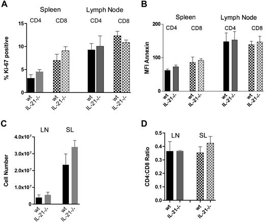
Increased GVHD-induced mortality conferred by an intact IL-21/IL-21R signaling axis on donor T cells could result from an increased expansion of Teffs as a consequence of higher proliferative or lower apoptosis rates. However, no significant differences were seen in the frequency of proliferating (Ki-67+) or apoptotic (annexin-V+) IL-21−/− versus wt CD4+ or CD8+ T cells isolated from the lymph nodes (LNs) or spleen on day 7 after BMT (Figure 3A-B). Absolute LNs and splenocyte numbers and CD4/CD8 ratios were unchanged (Figure 3C-D). These data indicate that IL-21 is not an important regulator of the early post-BMT splenic T-cell proliferation and apoptosis in this model.
Comparative T-cell proliferation and apoptosis of IL-21−/− versus wt CD25− T cells during a GVHD response. Frequency of Ki-67–positive CD4+ and CD8+ spleen and lymph node T cells (A); mean fluorescence intensity (MFI) of annexin-V–allophycocyanin on CD4+ and CD8+ spleen and lymph node T cells (B). Total cell numbers in spleen and lymph nodes; (C) CD4/CD8 ratio in spleens and lymph nodes (D). All data at day 7 after BMT of lethally irradiated recipients injected with 107 T cell–depleted BM cells and 5 × 10⁶ CD25− wt or IL-21−/− CD25− T cells. P = ns for all wt versus IL-21−/− comparisons. Data from 1 experiment with 3 mice per group.
Comparative T-cell proliferation and apoptosis of IL-21−/− versus wt CD25− T cells during a GVHD response. Frequency of Ki-67–positive CD4+ and CD8+ spleen and lymph node T cells (A); mean fluorescence intensity (MFI) of annexin-V–allophycocyanin on CD4+ and CD8+ spleen and lymph node T cells (B). Total cell numbers in spleen and lymph nodes; (C) CD4/CD8 ratio in spleens and lymph nodes (D). All data at day 7 after BMT of lethally irradiated recipients injected with 107 T cell–depleted BM cells and 5 × 10⁶ CD25− wt or IL-21−/− CD25− T cells. P = ns for all wt versus IL-21−/− comparisons. Data from 1 experiment with 3 mice per group.
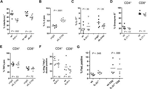
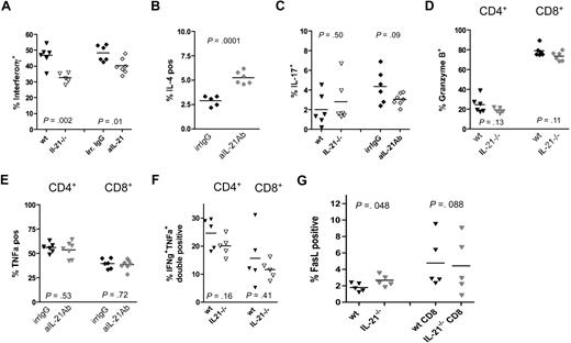
The relative importance of IL-21 in Th generation in vivo has not been reported in the context of GVHD-induced alloresponses. Published literature indicate that IFN-γ (Th1) but neither IL-4 (Th2) nor IL-17 (Th17) can increase GVHD-related colon damage.12,13,24-26 To determine whether IL-21 signaling blockade is associated with the reduced differentiation of IFN-γ–secreting cells in situ, at day 14 after BMT splenocytes and colon LP T cells from recipients of wt or IL-21−/− CD25− T cells were analyzed for the frequency of IFN-γ–secreting T cells after in vitro polyclonal stimulation. Mice receiving IL-21−/− T cells had a significantly lower frequency of IFN-γ–producing CD4+ and CD8+ T cells in both the spleen (not shown) and colonic LP (CD4+ T cells: Figure 4A; CD8+ T cells: data not shown). A similar reduction of IFN-γ–producing T cells was observed when analyzing LP T cells isolated from recipients of wt T cells treated with anti–IL-21 Ab versus irrelevant IgG (Figure 4A). Conversely, a significant increase of IL-4–producing LP T cells was found in recipients treated with anti–IL-21 Ab versus irrelevant IgG (Figure 4B). The frequency of IL-17–expressing LP T cells was not affected by IL-21 (Figure 4C). Although anti–IL-21 Ab decreased the numbers of IL-17–expressing cells, this was not statistically significant compared with control animals (Figure 4C).
Effects of IL-21−/− CD25− T cells on cytokine and cytolytic Teff molecule production. Lethally irradiated recipients were injected with 2 × 10⁶ wt of IL-21−/− CD25− T cells (triangles) or injected with 2 × 10⁶ FoxP3− T cells and treated with irrelevant IgG or anti–IL-21 (diamonds). Colon lamina propria cells were isolated on day 14 and analyzed by FACS and the frequency of CD4+ cells expressing IFNγ (A), IL-4 (B), and IL-17 (C) is shown. The frequency of granzyme B (D) or TNFα (E) was determined on CD4+ (triangles) and CD8+ (diamonds) T cells. The frequency of CD4+ and CD8+ T cells coexpressing IFNγ/TNFα (F) or expressing FasL (G) is shown. P values are indicated. Data from 2 experiments (A,C) or 1 experiment (B,D-G) with 6 mice per group total.
Effects of IL-21−/− CD25− T cells on cytokine and cytolytic Teff molecule production. Lethally irradiated recipients were injected with 2 × 10⁶ wt of IL-21−/− CD25− T cells (triangles) or injected with 2 × 10⁶ FoxP3− T cells and treated with irrelevant IgG or anti–IL-21 (diamonds). Colon lamina propria cells were isolated on day 14 and analyzed by FACS and the frequency of CD4+ cells expressing IFNγ (A), IL-4 (B), and IL-17 (C) is shown. The frequency of granzyme B (D) or TNFα (E) was determined on CD4+ (triangles) and CD8+ (diamonds) T cells. The frequency of CD4+ and CD8+ T cells coexpressing IFNγ/TNFα (F) or expressing FasL (G) is shown. P values are indicated. Data from 2 experiments (A,C) or 1 experiment (B,D-G) with 6 mice per group total.
In addition to IFN-γ secretion, GVHD-induced colon injury can be generated by the infiltration of donor T cells that express the cytolytic effector molecules granzyme B27-29 or TNF-α.30 Granzyme B expression was quantified in LP CD4+ or CD8+ T cells isolated from mice injected with wt or IL-21−/− CD25− T cells during BMT. No significant differences were seen between groups (Figure 4D). In other studies, recipients of wt CD25− T cells were treated with anti–IL-21 or irrelevant Ab and the frequency of TNF-α–expressing T cells was quantified. No significant differences between groups (Figure 4E) were found. The frequency of cells producing both IFN-γ/TNF-α dual-expressing cells also was similar in the presence or absence of IL-21 (Figure 4F). In contrast, FasL was expressed on a significantly higher proportion of CD4+ T cells isolated from the LP (Figure 4G) and liver but not spleen (data not shown) of recipients of IL-21−/− versus wt T cells. The frequency of CD8+ T cells expressing FasL from all 3 tissues was not significantly different between groups (Figure 4G and data not shown). Of the molecules associated with GVHD-induced gastrointestinal injury, our studies indicate that reduced GVHD-induced gastrointestinal injury seen when IL-21/IL-21R signaling was blocked is not due to a decrease in granzyme B–, TNF-α–, or FasL-expressing donor T cells but is associated with a reduction in IFN-γ–secreting T cells.
IL-21 blockade induces FoxP3+ Tregs that reside within the LP and inhibit GVHD lethality
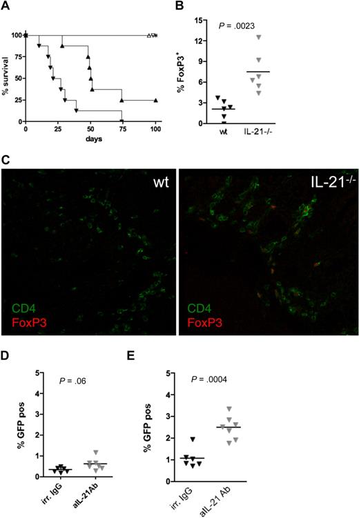
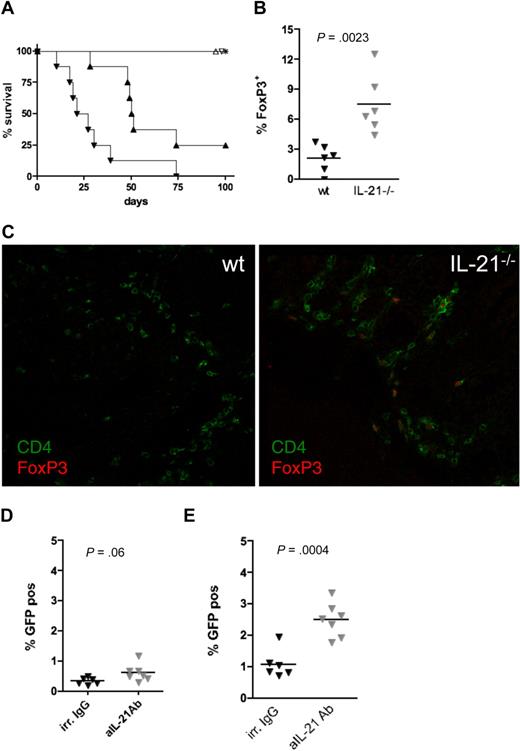
Treg depletion of the T-cell inoculum for BMT accelerates GVHD-induced mortality (Figure 5A and Taylor et al31 ), whereas the adoptive cotransfer of high numbers of Tregs inhibits the generation of GVHD-causing Th1 effector cells via suppression of T-cell activation in LNs.31-34 To determine whether an increase in LP Tregs might be observed under IL-21 blockade, we quantified the frequency of FoxP3-expressing CD4+ T cells in the LP after BMT. Recipients of wt versus IL-21−/− CD25− T cells had a significantly lower frequency of FoxP3+ cells (2.1% ± 0.6% for wt versus 7.2% ± 1.2% for IL-21−/−; Figure 5B). This finding was even more pronounced when tissue sections from mice at day 30 after BMT were analyzed by confocal microscopy. Whereas only occasional T cells were seen in the single surviving recipient of wt T cells, FoxP3+ Tregs constituted a large fraction of CD4+ T cells infiltrating the LP in all surviving mice analyzed at this time point (Figure 5C). Interestingly, Tregs were rarely found in other organs studied either on day 14 after BMT by fluorescence-activated cell sorting (FACS) (liver and spleen: data not shown) or on day 30 after BMT by confocal microscopy (spleen, liver, skin, lung, and kidney: data not shown), regardless of the presence or absence of donor T-cell IL-21 production.
Effects of IL-21 on FoxP3+ T-cell generation. Lethally irradiated B10.BR recipients were injected with 107 T cell–depleted bone marrow and (106 CD4+ (▴, ▵) or CD4+25− (▾, ▿) T cells from wt (▴,▾) or IL-21−/− (▵, ▿) mice and monitored for survival (A; P = .001 for wt vs IL-21−/− groups). Lethally irradiated recipients were injected with 2 × 10⁶ wt of IL-21−/− CD25− T cells and lamina propria (LP) cells were isolated from the colon on day 14, stained with anti-CD4 and anti-FoxP3, and analyzed by FACS. The frequency of FoxP3-expressing CD4+ cells was determined (B; P = .002). Subgroups of mice described in panel A were killed on day 30 and sections from frozen tissue blocks were analyzed for expression of CD4 (green) and FoxP3 (red) by confocal microscopy (C). Lethally irradiated recipients were injected with 2 × 10⁶ FoxP3− T cells from FoxP3-GFP knock-in mice. Subgroups were treated with irrelevant IgG or anti–IL-21 Ab. The frequency of FoxP3-expressing CD4+ spleen cells (D; P = .06) or CD4+LP T cells (E; P = .004) was determined. Data were obtained from 1 experiment each with 8 (A), 6 (B), 4 (C), and 6 (D-E) mice per group.
Effects of IL-21 on FoxP3+ T-cell generation. Lethally irradiated B10.BR recipients were injected with 107 T cell–depleted bone marrow and (106 CD4+ (▴, ▵) or CD4+25− (▾, ▿) T cells from wt (▴,▾) or IL-21−/− (▵, ▿) mice and monitored for survival (A; P = .001 for wt vs IL-21−/− groups). Lethally irradiated recipients were injected with 2 × 10⁶ wt of IL-21−/− CD25− T cells and lamina propria (LP) cells were isolated from the colon on day 14, stained with anti-CD4 and anti-FoxP3, and analyzed by FACS. The frequency of FoxP3-expressing CD4+ cells was determined (B; P = .002). Subgroups of mice described in panel A were killed on day 30 and sections from frozen tissue blocks were analyzed for expression of CD4 (green) and FoxP3 (red) by confocal microscopy (C). Lethally irradiated recipients were injected with 2 × 10⁶ FoxP3− T cells from FoxP3-GFP knock-in mice. Subgroups were treated with irrelevant IgG or anti–IL-21 Ab. The frequency of FoxP3-expressing CD4+ spleen cells (D; P = .06) or CD4+LP T cells (E; P = .004) was determined. Data were obtained from 1 experiment each with 8 (A), 6 (B), 4 (C), and 6 (D-E) mice per group.
Although the frequency of natural Tregs in the adoptively transferred, CD25−-depleted T cells was below 0.5% in both groups, it was possible that this low level of residual cotransferred natural Tregs may have preferentially expanded in recipients of IL-21−/− versus wt CD25− T cells. To address this possibility, FoxP3− T cells were sorted from FoxP3-GFP knock-in donors and injected into recipients treated with irrelevant IgG or anti–IL-21 Ab. Splenocytes and LP T cells were isolated on day 14 after BMT and the frequency of induced FoxP3+ T cells was determined. Only very low frequencies of FoxP3+ Tregs (mean < 1% of CD4 T cells) were found in the spleen of mice in both treatment groups (Figure 5D). A significant increase in FoxP3-expressing Tregs was observed in the LP of anti–IL-21 versus irrelevant Ab-treated mice (2.5% vs 1.1%; Figure 5E). These data indicate that the loss of IL-21 favors the development and infiltration of FoxP3+ Tregs into the LP during GVHD generation.
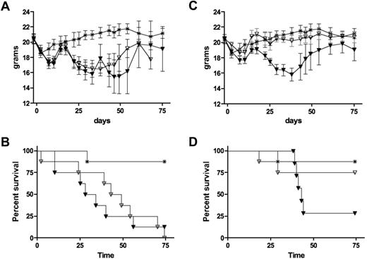
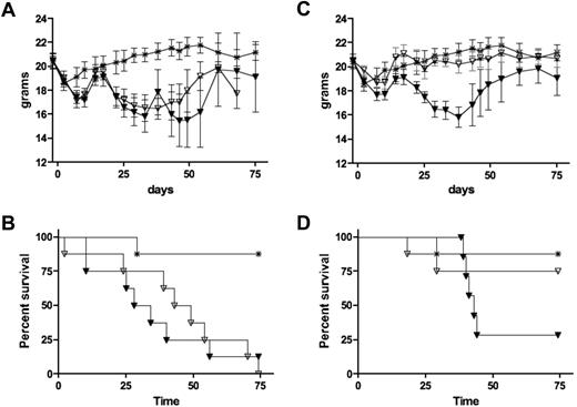
To determine whether the increased frequency of LP Tregs in anti–IL-21 Ab-treated mice per se was responsible for the decrease in GVHD lethality, we generated chimeras containing FoxP3-deficient cells by infusing BM from 20-day-old scurfy mice into lethally irradiated congenic donors to generate scurfy chimeras. The presence of congenic, radioresistant host Tregs in these chimeras prevents the unrestrained activation of scurfy T cells, permitting the isolation of nonactivated scurfy CD4+25− T cells. Scurfy CD4+CD25− cells from chimeric donors or wt CD4+CD25− cells from wt donors were injected into secondary lethally irradiated major histocompatibility complex–disparate recipients treated with anti–IL-21 Ab or irrelevant IgG. All mice receiving scurfy cells died within 79 days and no difference in weight or survival curves was observed between groups that received scurfy cells and anti–IL-21 Ab versus irrelevant mAb (Figure 6A-B). In contrast, mice receiving CD4+CD25− wt cells treated with anti–IL-21 Ab had significantly less weight loss (Figure 6C) and survived longer than mice treated with irrelevant IgG (75% vs 25%, respectively; Figure 6D). Thus, the attenuation of GVHD by IL-21 blockade requires the induction of FoxP3+ Tregs.
Effect of donor IL-21 expression on the in vivo conversion of CD4+25− T cells into FoxP3+ cells. Weights (A,C) and survival (B,D) of lethally irradiated recipients injected with 107 T cell–depleted bone marrow and 0.5 × 10⁶ flow-sorted CD45.1−CD4+CD25− T cells isolated from scurfy chimeric (A-B) or wt (C-D) donors. Subgroups were treated with irrelevant IgG (▾) or aIL-21 (▿). P values between treated and untreated groups for survival were .90 (B) and .12 (D). Data from 1 experiment with 8 mice per group.
Effect of donor IL-21 expression on the in vivo conversion of CD4+25− T cells into FoxP3+ cells. Weights (A,C) and survival (B,D) of lethally irradiated recipients injected with 107 T cell–depleted bone marrow and 0.5 × 10⁶ flow-sorted CD45.1−CD4+CD25− T cells isolated from scurfy chimeric (A-B) or wt (C-D) donors. Subgroups were treated with irrelevant IgG (▾) or aIL-21 (▿). P values between treated and untreated groups for survival were .90 (B) and .12 (D). Data from 1 experiment with 8 mice per group.
IL-21 signaling is not required for GVL generation
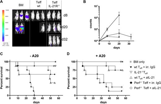
IL-21 can augment antitumor-directed cytotoxic T lymphocyte activity and has direct antitumor effects.2 To determine whether the lack of donor T-cell IL-21 signaling, which reduces GVHD lethality, would also interfere with GVL activity, lethally irradiated BALB/c recipients were given B6 BM and A20luc (H2d) lymphoma cells intravenously on day 0. Groups were given CD25− T cells from wt or IL-21-R−/− mice. All mice receiving A20luc and BMT without supplemental T cells died within 25 days due to tumor burden, as evidenced by hind leg paralysis and high levels of photon emission (Figure 7A-B). Mice receiving wt T cells showed no or minimal signs of tumor growth, but all mice died of GVHD by day 32. All mice that received A20luc and IL-21R−/− T cells showed minimal initial tumor growth followed by regression with minimal evidence of tumor or GVHD by day 32 after BMT (Figure 7A-B).
Loss of IL-21 signaling on donor T cells does not eliminate GVL activity. Lethally irradiated Balb/c recipients were injected with T cell–depleted bone marrow and 105 A20 lymphoma cells on day 0. Subgroups received 2 × 10⁶ CD25− T cells from wt or IL-21R−/− mice also on day 0. (A) Tumor growth was monitored by luciferase imaging on days 8, 20, and 32 after BMT. (B) Photon counts of BM-only recipients (*), recipients of BM + wt T-effector cells (▾), and BM + IL-21−/− cells (▿) on days 8, 20, and 32. (C) Survival of lethally irradiated Balb/c recipients injected with T cell–depleted bone marrow alone (*) or together with 2 × 10⁶ CD25− T cells from wt B6 mice treated with irrelevant (irr) IgG (▾) or aIL-21 Ab (▵), or with 2 × 10⁶ CD25− T cells from IL-21−/− (▿) or perforin−/− mice treated with irr IgG (♦) or aIL-21 Ab (◇; P < .001 for ▾ versus ▿, ▾ versus ♦, and ▾ versus ◇; P < .05 for ▾ versus ▿, ▿ versus ◇; P = ns for all others). (D) Survival of the same recipients as in panel C, but coinjected with 105 A20 lymphoma cells (P ≤ .001 for ▾ versus ▿; P < .05 for ▾ versus ▵, ♦ versus ◇, and ▾ versus ♦; P = ns for all others). Data from 2 experiments with a total of 8 (A-B) and 7 (C-D) mice per group.
Loss of IL-21 signaling on donor T cells does not eliminate GVL activity. Lethally irradiated Balb/c recipients were injected with T cell–depleted bone marrow and 105 A20 lymphoma cells on day 0. Subgroups received 2 × 10⁶ CD25− T cells from wt or IL-21R−/− mice also on day 0. (A) Tumor growth was monitored by luciferase imaging on days 8, 20, and 32 after BMT. (B) Photon counts of BM-only recipients (*), recipients of BM + wt T-effector cells (▾), and BM + IL-21−/− cells (▿) on days 8, 20, and 32. (C) Survival of lethally irradiated Balb/c recipients injected with T cell–depleted bone marrow alone (*) or together with 2 × 10⁶ CD25− T cells from wt B6 mice treated with irrelevant (irr) IgG (▾) or aIL-21 Ab (▵), or with 2 × 10⁶ CD25− T cells from IL-21−/− (▿) or perforin−/− mice treated with irr IgG (♦) or aIL-21 Ab (◇; P < .001 for ▾ versus ▿, ▾ versus ♦, and ▾ versus ◇; P < .05 for ▾ versus ▿, ▿ versus ◇; P = ns for all others). (D) Survival of the same recipients as in panel C, but coinjected with 105 A20 lymphoma cells (P ≤ .001 for ▾ versus ▿; P < .05 for ▾ versus ▵, ♦ versus ◇, and ▾ versus ♦; P = ns for all others). Data from 2 experiments with a total of 8 (A-B) and 7 (C-D) mice per group.
To begin to elucidate the potential mechanisms for the preserved GVL effect seen in recipients given A20luc cells and IL-21R−/− T cells, recombinant murine IL-21 or TNFα (10-100 pg/mL) was added to A20 cells in vitro. No differences were seen in the expansion or viability of annexin-V staining over 4 days of analysis (data not shown). These data excluded a direct effect of IL-21 or TNFα on A20 tumor killing. A recent study showed no protection against GVHD lethality and a complete loss of GVL against A20 cells when perforin−/− T cells were infused into BALB/c recipients.35 The lack of GVHD inhibition contrasted with our studies in a different fully allogeneic strain combination.36 We hypothesized that IL-21 blockade should not be fully redundant with perforin pathway blockade because a GVL effect was seen. B6 BM with or without wt versus IL-21−/−, perforin−/− CD25− T cells was given to BALB/c recipients. For GVHD studies, no A20 cells were given. For GVL studies, recipients of wt versus perforin−/− CD25− T cells were given A20 cells (105) at the time of BMT. Recipients of wt and perforin−/− Teffs received irrelevant or anti–IL-21 Ab. Non–tumor-bearing recipients of IL-21−/− CD25− T cells had an 88% survival at 2 months versus 0% survival in recipients given wt CD25− T cells and irrelevant Ab (Figure 7C). Anti–IL-21 Ab resulted in 63% survival until after the time of discontinuation of the Ab at 6 weeks after BMT when survival diminished to 25%, significantly better than controls. Recipients of perforin−/− donor CD25− T cells had 50% survival, significantly better than recipients of wt CD25− T cells and comparable with anti–IL-21 Ab. Adding anti–IL-21 Ab to recipients of perforin−/− CD25− T cells significantly improved survival versus Ab alone, suggesting that the perforin and IL-21 pathways are nonredundant for inhibiting GVHD lethality. In recipients challenged with a lethal dose of A20 cells at the time of BMT, infusing IL-21−/−, wt + anti–IL-21 Ab or perforin−/− CD25− T cells and irrelevantIgG significantly improved survival compared with those given wild-type CD25− T cells (Figure 7D). Combined anti–IL-21 Ab and perforin−/− T cells resulted in the loss of any survival advantage by either pathway alone. These data indicate that effective GVL responses that occur in the absence of IL-21 signaling in donor T cells is dependent upon the perforin pathway, suggesting that IL-21 signaling blockade does not interfere with perforin expression.
Discussion
Here we demonstrate an important role of IL-21 in GVHD. Donor T-cell production of IL-21 and its signaling via the IL-21R expressed on donor T cells were essential for optimal GVHD-induced gastrointestinal tract injury and subsequent lethality. Blockade of IL-21/IL-21R signaling was associated with a decrease of IFN-γ–secreting T cells infiltrating the LP of the colon. Moreover, these data demonstrate for the first time that blockade of IL-21 signaling increases FoxP3+ iTregs in vivo. A direct cause-and-effect relationship between the capacity for iTreg generation by donor T cells and the amelioration of GVHD lethality by IL-21/R blockade was firmly established. Because GVL activity was retained by IL-21/R blockade, these data suggest that anti–IL-21 Abs could be useful for GVHD prevention in the clinic.
IL-21 blockade by genetic or pharmacologic means led to a reduced GVHD-induced lethality. The greater magnitude of GVHD inhibition using genetic compared with pharmacologic approaches to block IL-21 signaling may be due to inadequate blockade at the tissue level or the relatively shorter duration of blockade that accompanied a 5- to 6-week Ab course versus the permanent blockade with IL-21 or IL-21R gene deletion. In both instances, the frequency of IFNγ-secreting CD4+ and CD8+ LPs was significantly reduced on day 14 after BMT (Figure 4A). Although no reduction in the mean GVHD scores was noted in the spleen, the frequency CD4+ and CD8+ T cells producing IFNγ was significantly reduced in the absence of IL-21 signaling. In the liver, there was a reduction in the frequency of IFNγ-expressing CD8+ and to a lesser extent CD4+ T cells. These data pointed to the possibility of a more local (gastrointestinal) influence of IL-21 on GVHD lethality. IFN-γ–producing Th1 cells are known to cause gastrointestinal injury during GVHD.24,25 Seemingly conflicting data exist for the relationship between IL-21 and IFN-γ production. Although one report suggested an inhibitory effect of IL-21 on Th1 differentiation and IFN-γ production in vitro and in vivo,14 another describes a facilitating effect of IL-21 on Th1 differentiation.37 Our data indicate that during GVHD, IL-21 blockade reduced the generation of IFN-γ–secreting cells, consistent with findings by Strengell et al.37 We observed an increase in the frequency of IL-4–producing cells in the colon. Consistent with the limited role of Th17 cells in gastrointestinal GVHD,12,13 we did not observe a difference in the frequency of Th17 cells relative to the presence of IL-21. The reduced frequency of IFN-γ–secreting LP T cells alone would not seem to explain the reduced GVHD lethality observed because our prior studies have indicated that IFN-γ−/− T cells accelerated whereas IL-4−/− T cells reduced GVHD lethality. In some settings in which GVHD colon injury is attenuated due to indoleamine 2,3-dioxygenase, a tryptophan catabolic enzyme, only the number but not the frequency of IFN-γ–secreting cells in the colon was affected by this pathway. We favor the explanation that the balance between cytokine-secreting cells in a given site is more important than the frequency of a single cytokine. An alternative and not a mutually exclusive explanation is that the reduced frequency of IFN-γ–secreting cells may be reflective of a more immune suppressive environment that is not conducive to Teff generation. Consistent with this hypothesis, the in vivo administration of ex vivo–expanded natural Tregs reduces Teff generation and IFN-γ secretion.38
Because GVHD typically is initiated in secondary lymphoid organs, splenic T cells were analyzed for proliferation and apoptosis in recipients of IL-21−/− versus wt CD25− T cells. No differences were seen on day 7 after BMT, suggesting that the major effects of IL-21 blockade may be found in GVHD parenchymal organs and especially the gastrointestinal tract, consistent with our pathologic GVHD scores. In that regard, we found an increase in percentage of annexin-V staining of CD4+ T cells (data not shown) along with increased Tregs in the colon at 2 to 3 weeks after BMT. Our initial studies indicated that the increased frequency of Tregs observed in the colon of recipients of IL-21−/− versus wt T cells could be compatible with preferential expansion of natural Tregs that were cotransferred with donor CD25− T cells or iTreg generation. Intriguingly, although ex vivo IFNγ has been shown to promote iTreg generation of CD4+ T cells in response to allogeneic immature dendritic cells39 and we observed a significantly lower frequency of day 14 post-BMT IFNγ–expressing T cells isolated from 3 distinct GVHD target organs, serum IFNγ levels were similar in recipients of IL-21−/− versus wt T cells when assayed on day 8 (mean levels, ∼ 125 pg/mL on day 8; data not shown). By day 14, mean IFNγ levels had fallen to 10 pg/mL or less in both groups. Thus, we could not find evidence for a direct link between IL-21 production and iTreg generation that functioned via IFNγ regulation.
Using CD4+FoxP3− donor T cells, we were able to demonstrate that IL-21 production by donor T cells impedes iTreg generation. This has been previously reported only in vitro.17 In recipients given wt T cells with irrelevant Ig versus anti–IL-21 Ab or IL-21−/− T cells, a significant decrease in Treg frequency was seen in LP (2.5- to 3-fold; Figure 5B-E) and hepatic (1.6-fold; data not shown) T cells. The overall higher Treg frequencies in the LP versus the liver may account for the greater reduction in pathologic scores of the gastrointestinal tract seen in the context of IL-21 blockade. Alternatively, in recipients of IL-21−/− versus wt T cells, the 1.5-fold increased level of FasL expression (2.67% vs 1.78%, respectively; P = .048) and significant increase in hepatic CD8+ T-cell granzyme B expression may have contributed to liver GVHD,27 offsetting other benefits of IL-21 blockade.
The requirement for FoxP3 expression and function in donor T cells for ameliorating GVHD in recipients of IL-21−/− versus wt T cells was best demonstrated by studies in which FoxP3-deficient scurfy T cells isolated from chimeras that also contained congenic FoxP3+ wt T cells were unable to reduce GVHD lethality after adoptive transfer into major histocompatibility complex–disparate recipients treated with anti–IL-21 Ab. The lack of a beneficial effect of IL-21 blockade in mice receiving scurfy cells supports our contention that FoxP3+ Tregs are necessary for optimal GVHD inhibition and suggests that other Treg-independent mechanisms such as the direct effects of IL-21 on epithelial tissue injury or cytokine skewing are unlikely explanations for the beneficial effects of IL-21 blockade on GVHD.
Because NKT cells were depleted from the transferred donor T cells, the source of IL-21 in our model is CD4+ T cells. The influence of IL-21 on T-cell function has been most intensively studied in CD8+ T cells where it has been shown to augment CD8+ cytotoxic T-cell function. However, morbidity and mortality in our model is not dependent on CD8+ T cells, suggesting that IL-21 inhibits GVHD via direct effects on CD4+ T-cell differentiation or function. In models of autoimmunity, IL-21 releases Teffs from Treg-induced suppression, resulting in augmented effector function.19,20 However, we were unable to identify an influence of IL-21 on the ability of LP T cells to differentiate into cytolytic Teffs as we found no difference in the frequency of granzyme B– and/or TNF-α–expressing T cells in the LP. Suppression by Tregs has been shown to have no effect on the expression of cytolytic molecules, but rather on their release.40
Although we did not formally test the functional significance of IL-21R expression by gastrointestinal epithelial cells in this model, the lack of a significant difference using donor IL-21−/− versus IL-21R−/− and the lack of any effect of IL-21 blockade in the absence of donor T cells that have lost the capacity to acquire FoxP3 expression and Treg function indicate that IL-21R expression on gastrointestinal epithelial cells likely plays a limited role, if any, in GVHD. Although the IL-21R is abundantly expressed on B cells, NK cells, and epithelial tissues, the results obtained from experiments using IL-2Rγc−/− BM donors and the lack of any effect on GVHD mortality using scurfy donor T cells suggest that at least in this model of acute GVHD, only the IL-21R expression on T cells is responsible for maximal disease.
IL-21 blockade, although allowing significant GVHD attenuation, does not preclude effective GVL responses. A20luc clearance in GVHD has been shown to partially depend on the presence of perforin35 on CD4+ T cells.41 Under the conditions here, perforin−/− T cells reduced GVHD, consistent with studies by us36 and others,27,28,42 although this is not a uniform finding.35 In contrast to a recent report, no significant survival effect of perforin deficiency was noted for A20 clearance.35 Differences in GVL responses may be explained by the coexpression of dsred2, which may serve as a helper epitope for A20luc elimination. The frequency of granzyme B–expressing splenic CD4+ T cells, the dominant GVL Teffs present in the site of A20 lymphoma seeding and metastases, was significantly lower in recipients of IL-21−/− versus wt T cells (7.6% vs 11.6%, respectively), although the physiologic significance is unknown because survival was comparable in recipients of A20 cells and wt versus perforin−/− T cells. Because no changes in FasL expression were observed in splenic T cells and FasL deficiency does not abrogate a GVL effect,35 this pathway is unlikely involved in retaining GVL responses in recipients of IL-21−/− T cells.
In summary we have observed that IL-21 production by donor T cells and signaling of donor T cells induced by IL-21 is an essential driving force for optimal GVHD generation. We have shown for the first time that IL-21/R signaling of donor T cells impedes the generation of iTregs, which then fail to restrain GVHD-induced colon injury and lethality. Our studies demonstrate GVHD attenuation and GVL preservation with anti–IL-21 Ab and provide an attractive strategy for GVHD prevention that should be explored for clinical application.
The online version of this article contains a data supplement.
The publication costs of this article were defrayed in part by page charge payment. Therefore, and solely to indicate this fact, this article is hereby marked “advertisement” in accordance with 18 USC section 1734.
Acknowledgments
We thank Bartosz Grzywacz and Johan Dirks for helpful discussions, and Chris Lees and Luna Liu as well as the Flow Cytometry Core Facility of the Masonic Cancer Center for technical assistance. We also acknowledge Harald Haugen and Wendy Curtis for breeding and maintenance of the IL-21−/− and IL-21R−/− mice at Zymogenetics.
This work was supported in part by the Children's Cancer Research Fund and the National Institutes of Health R01 AI34495, HL56067, and CA72669. The confocal microscope was made available through a National Center for Research Resources Shared Instrumentation Grant (1 S10 RR16851). C.B. was in part funded by the Swiss National Science Foundation (PBBSB-108600) and the Swiss Cancer League Oncosuisse (BIL KLS 01617-12-2004).
National Institutes of Health
Authorship
Contribution: C.B. designed and performed experiments, analyzed data, and wrote the paper; L.K., C.V., and M.M. performed experiments and analyzed data; E.G. performed experiments; A.P-M. performed experiments and edited the paper; and P.S. and B.R.B. designed experiments, analyzed data, and edited the paper.
Conflict-of-interest disclosure: P.S. is a paid employee of Zymogenetics Inc. The remaining authors declare no competing financial interests.
Correspondence: Bruce R. Blazar, MMC 109, University of Minnesota Hospital, 420 Delaware St SE, Minneapolis, MN 55455; e-mail: blaza001@umn.edu.

![Figure 2. Reduced GVHD histopathology in recipients of IL-21−/− CD25− T cells. (A) Lethally irradiated recipients were injected with BM only, BM + 2 × 10⁶ wt CD25− T cells, or BM + IL-21−/− CD25− T cells, killed on day 14 after BMT, and hematoxylin and eosin–stained tissue sections were scored for GVHD: Colon (i,iii,v) and ileum (ii,iv,vi) from recipients of BM (i-ii), BM + wild-type T-effector cells (iii-iv) or BM + IL-21−/− cells (v-vi). Mean ± SEM of histologic grade of graft-versus-host disease in target organs in mice injected with BM only (□), BM + 2 × 10⁶ wt CD25− T cells (■), or BM + IL-21−/− CD25− T cells (; wt vs IL-21−/−, P = .020 [colon] and P = .005 [small intestine], all others not significant [ns]; B).](https://ash.silverchair-cdn.com/ash/content_public/journal/blood/114/26/10.1182_blood-2009-05-221135/4/m_zh89990945750002.jpeg?Expires=1769532395&Signature=zusBa6u3MtfrJG5DaSKM7nRyYr6FV3h3x3FEkZreBIipyh8CrK-uKWBF0EyBmYQ1a~xR21pLynaghE7q-YzJv4DAGQdrGojffJ641w7xxrzylOwGy7IJvu-EDZYlWv441S3PRVEg8bBNQWFfqRsvcTRHWAGu1gXgxPF5CxHvAzvd3dfzG1Jzdd59lDkNMc1Iabdb2rDemvS5KkrWJTyxmNivIG8suVObSOSYVTbpu6QjQuRvEkYAMFrShMz7nFGZa5DkwxWeLI6bDgPihI0bBnr1chei2eIpi8JfqJIDVwB85bfDWQ85kB-rmcY-rnPlzVERktcjVydgOJta912BVQ__&Key-Pair-Id=APKAIE5G5CRDK6RD3PGA)


![Figure 2. Reduced GVHD histopathology in recipients of IL-21−/− CD25− T cells. (A) Lethally irradiated recipients were injected with BM only, BM + 2 × 10⁶ wt CD25− T cells, or BM + IL-21−/− CD25− T cells, killed on day 14 after BMT, and hematoxylin and eosin–stained tissue sections were scored for GVHD: Colon (i,iii,v) and ileum (ii,iv,vi) from recipients of BM (i-ii), BM + wild-type T-effector cells (iii-iv) or BM + IL-21−/− cells (v-vi). Mean ± SEM of histologic grade of graft-versus-host disease in target organs in mice injected with BM only (□), BM + 2 × 10⁶ wt CD25− T cells (■), or BM + IL-21−/− CD25− T cells (; wt vs IL-21−/−, P = .020 [colon] and P = .005 [small intestine], all others not significant [ns]; B).](https://ash.silverchair-cdn.com/ash/content_public/journal/blood/114/26/10.1182_blood-2009-05-221135/4/m_zh89990945750002.jpeg?Expires=1769532396&Signature=mFN0yhfFpaZU-cJ51T8BD9G-9g5zWuSD8n~PKfMg1ku552iiH0toU3frPPH7nS7HIqKebFGVeAofi1KRAH8~RL7VAsTj~ihR3upLCm936Eneo8eYcy78djiUNZ9fB65fYSDBJtIjkMnqb1~kBiyXUj--NRbbovDXuJ0pVxgMX4k84a74H~sCORT2RgYbE7qDH03sS6sKB0HK-NND2R~mFXebHSfFDuTEk~OXwOakjo-OV6YTNALn84MN6XEaprCk57Z55TmBJUX65FC8c1BThEtAuRUAYRyI7W6nfwQ9attHVd7NrrvU8VrvFPbBvz5a311nIaMRSifV~2Y1RPXrqA__&Key-Pair-Id=APKAIE5G5CRDK6RD3PGA)
 ; wt vs IL-21−/−, P = .020 [colon] and P = .005 [small intestine], all others not significant [ns]; B).
