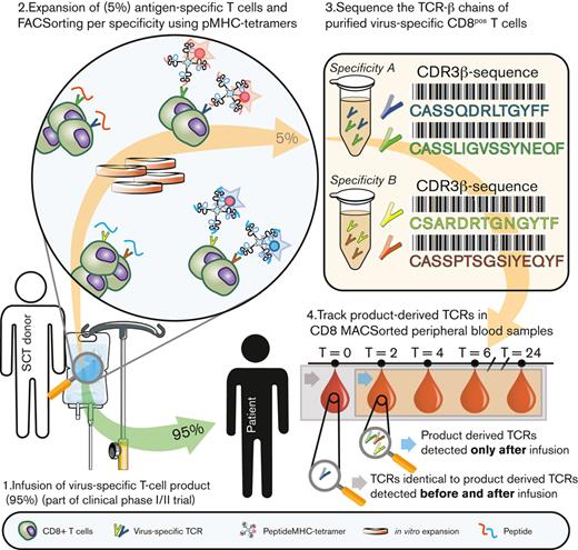
Key Points
In vivo expansion of infused multiantigen-specific T-cell products in response to antigen encounter.
In vivo persistence of infused multiantigen-specific T-cell products in absence of viral reactivation.
Abstract
Adoptive cellular therapies with T cells are increasingly used to treat a variety of conditions. For instance, in a recent phase 1/2 trial, we prophylactically administered multivirus-specific T-cell products to protect recipients of T-cell–depleted allogeneic stem cell grafts against viral reactivation. To establish treatment efficacy, it is important to determine the fate of the individual transferred T-cell populations. However, it is difficult to unequivocally distinguish progeny of the transferred T-cell products from recipient- or stem cell graft–derived T cells that survived T-cell depletion during conditioning or stem cell graft manipulation. Using messenger RNA sequencing of the T-cell receptor β-chains of the individual virus-specific T-cell populations within these T-cell products, we were able to track the multiple clonal virus-specific subpopulations in peripheral blood and distinguish recipient- and stem cell graft–derived virus-specific T cells from the progeny of the infused T-cell products. We observed in vivo expansion of virus-specific T cells that were exclusively derived from the T-cell products with similar kinetics as the expansion of virus-specific T cells that could also be detected before the T-cell product infusion. In addition, we demonstrated persistence of virus-specific T cells derived from the T-cell products in most patients who did not show viral reactivation. This study demonstrates that virus-specific T cells from prophylactically infused multiantigen-specific T-cell products can expand in response to antigen encounter in vivo and even persist in the absence of early viral reactivation.
Introduction
The use of adoptive T-cell therapies has increased in frequency and options in the last few decades.1 Although these strategies show promising results, it is currently difficult to adequately follow the fate of these products in the patient. In vivo tracking and tracing of the progeny from the infused T-cell products is vital in the assessment of treatment efficacy. For instance, when the products administered fail to achieve expected results, it is important to determine whether this might be explained by the lack of persistence. Likewise, selective in vivo expansion of the transferred T cells in response to antigen encounter would be an argument to support the case that engrafted T cells helped to elicit the clinical effect. Chapuis et al recently tackled this by tracking adoptively transferred T-cell products using T-cell receptor (TCR) deep sequencing.2
Patients that undergo allogeneic stem cell transplantation (alloSCT) are (temporarily) immunocompromised and reactivations of cytomegalovirus (CMV), Epstein-Barr virus (EBV), and adenovirus (AdV) are frequently observed. The adoptive transfer of HLA-matched virus-specific T cells from the original stem cell donor can reduce viral infection and reactivation risk in these patients.3 Safety and feasibility of adoptive transfer of donor-derived virus-specific memory T-cell products have been demonstrated in multiple phase 1/2 clinical studies by different groups, including ours.4-12 However, persistence of the virus-specific T cells could not be attributed unequivocally to the transferred T-cell product, because T cells with the same antigen specificity might already have been present in the patient or derived from the stem cell graft. Furthermore, the currently used techniques (ie, peptide major histocompatibility complex [pMHC] tetramer staining, marker-gene analysis, and/or enzyme-linked immunospot) allow only for the detection of frequencies higher than ∼0.1% within total peripheral blood mononuclear cells (PBMCs).13-18 More sensitive and specific detection methods are required to track individual T-cell populations derived from the T-cell products in vivo.
In a recent phase 1/2 trial in the department of Hematology (LUMC), 24 patients who received T-cell–depleted alloSCT were treated posttransplant with a prophylactic infusion of a stem cell donor–derived multiantigen (virus)–specific T-cell product, containing CD8+ T cells directed against CMV, EBV, and/or AdV antigens.19,20 The aim was to prevent uncontrolled viral reactivation in these patients. The T cells from the products had to persist long enough without the presence of viral antigen to be able to prevent or control the viral reactivation. Safety and feasibility of this approach and appearance in peripheral blood (PB) of virus-specific T cells as detected by conventional pMHC tetramer staining were demonstrated. However, we could not determine whether these virus-specific T cells were derived from the infused product, and whether virus-specific T cells persisted in patients whose frequencies of such cells were below the detection threshold of conventional pMHC tetramer staining.
This study aimed to track prophylactically administered virus-specific T cells by using high-throughput TCR sequencing of the infused T-cell products and PBMC samples of the patients after administration. We analyzed in detail how many and which T cells persisted and expanded after administration of the products. Using the TCR sequencing technology, we were able to distinguish the virus-specific T cells that were already present before infusion of the product from persisting or expanding T cells that were exclusively derived from the product.
Materials and methods
Collection of patient and donor material
After receiving informed consent according to the Declaration of Helsinki, PBMCs were isolated from patients who underwent alloSCT and their stem cell donors by standard Ficoll-isopaque separation and stored in the vapor phase of liquid nitrogen.20 The patients and donors had been included in a previous single-center, phase 1/2 study exploring the safety, feasibility, and first evidence of efficacy of prophylactic infusion of multiantigen-specific T cells to prevent complications early after T-cell–depleted alloSCT20 (T Control, EudraCT-number 2014-003171-39). Clinical results can be found in the original article.20 In the current analysis, patients and donor-derived T-cell products were numbered identically to the phase 1/2 study.20 See supplemental Table 1 for relevant patient and donor characteristics.
Generation of multiantigen-specific T-cell products and isolation of single-antigen–specific T-cell populations
T-cell products were generated using the MHC I streptamer isolation technology, as previously described.19,20 In brief, isolation complexes (MHC I streptamers) were generated per target antigen T-cell specificity. For every patient, MHC I streptamers for 4 HLA∗02:01-restricted viral antigens (2 CMV, 1 EBV, and 1 AdV) were pooled. In addition; depending on HLA type of patient/donor, and regardless of donor CMV, EBV, and AdV serostatus; MHC I streptamers for peptides presented in HLA-A∗01:01, HLA-A∗24:02, HLA-B∗07:02, and/or HLA-B∗08:01 were added to this pool (Table 1). The pool of MHC I streptamers was incubated with 2 × 10⁹ donor-derived PBMCs and MHC I streptamer–bound cells were isolated using a CliniMACS Plus Instrument (Miltenyi Biotec, Bergisch Gladbach, Germany) under Good Manufacturing Practices (GMP) conditions. Five percent of the cells from all products, except products U (3%) and Y (4%), were nonspecifically expanded using 800 ng/mL phytohemagglutinin with autologous PBMCs as the feeder mixture, as previously described.20 Products were cryopreserved and used for in-depth analysis in this study. To isolate single-antigen–specific T-cell populations from the products, cells were first incubated with pMHC tetramer complexes for 30 minutes at 4°C followed by incubation with fluorescein isothiocyanate–labeled CD8 (BD Biosciences) antibodies at 4°C for 30 minutes. pMHC tetramer–positive virus-specific T cells were bulk sorted by fluorescence-activated cell sorting (FACS) for each specificity and directly lysed. Sorting was performed on a FACSAria (BD Biosciences) using Diva software (BD Biosciences). For the generation of pMHC I tetramers, see supplemental Material and methods.
pMHC streptamers used for the generation of multivirus-specific T-cell products
| Virus . | Protein . | Peptide . | HLA-restriction . | Specificity included in T-cell product manufacturing for these patients, n . |
|---|---|---|---|---|
| CMV | pp50 | VTEHDTLLY | HLA-A∗01:01 | 3 |
| pp65 | NLVPMVATV | HLA-A∗02:01 | 20 | |
| IE1 | VLEETSVML | HLA-A∗02:01 | 20 | |
| pp65 | QYDPVAALF | HLA-A∗24:02 | 6 | |
| pp65 | TPRVTGGGAM | HLA-B∗07:02 | 7 | |
| IE1 | QIKVRVDMV | HLA-B∗08:01 | 5 | |
| EBV | BMLF1 | GLCTLVAML | HLA-A∗02:01 | 20 |
| EBNA3A | RPPIFIRRL | HLA-B∗07:02 | 7 | |
| BZLF1 | RAKFKQLL | HLA-B∗08:01 | 5 | |
| AdV | HEXON | TDLGQNLLY | HLA-A∗01:01 | 3 |
| E1A | LLDQLIEEV | HLA-A∗02:01 | 20 | |
| HEXON | TYFSLNNKF | HLA-A∗24:02 | 6 | |
| HEXON | KPYSGTAYNAL | HLA-B∗07:02 | 7 |
TCRβ library preparation
TCRβ sequences were identified using ARTISAN polymerase chain reaction (PCR) adapted for TCR PCR as previously described.21-23 See supplemental Material and methods for TCR PCR procedure. In short, total messenger RNA (mRNA) (10 μL) was extracted from T cells from 20 unsorted products and 81 pMHC tetramer–positive CD8+ T-cell populations directly after FACS using magnetic beads (Dynabead mRNA DIRECT Kit, Invitrogen, ThermoFisher Scientific; supplemental Figure 1A). To determine whether T cells with TCRβ sequences detected in the T-cell products were present in PB of the patients after administration, we isolated primary CD8+ T cells from follow-up PB samples with magnetic-activated cell sorting (MACS) using CD8 T-cell isolation kits with LS Columns (Miltenyi Biotec). PB samples at the moment of product infusion contained a median of 110 × 106 CD8+ T cells per liter (interquartile range [IQR], 47-509; supplemental Figure 2A). After CD8 enrichment, all PB samples contained a median of 81% CD8+ cells (IQR, 61%-92%; supplemental Figure 2B). PB samples for follow-up were taken every 2 weeks until 8 weeks after T-cell product infusion and every 4 weeks thereafter until 6 months after undergoing alloSCT.20 Total mRNA (10 μL) was extracted from 109 CD8+ populations isolated from follow-up PB samples from the patients (supplemental Figure 1B), containing a median of 0.3 × 10⁶ cells (IQR, 0.16 × 10⁶ to 0.66 × 10⁶ cells; supplemental Figure 2C). The samples from each patient were sequenced separately. Unique identifiers were used for the PCR products of each virus-specific T-cell population, unsorted T-cell product, and monitoring sample (see supplemental Tables 2 and 3 for primer and identifier sequences, respectively). These amplicons with identifiers were purified, quantified, and pooled into 1 library per patient for paired-end sequencing of 125 base pairs on an Illumina HiSeq 4000 or NovaSeq 6000. The samples of each patient were separately sequenced on different chips to obtain enough reads per sample (oversampling). Each CD8+ PB follow-up sample contained a median of 8.5 × 10⁶ reads (IQR, 5.5 × 10⁶ to 12.8 × 10⁶ reads; supplemental Figure 2D). Oversampling resulted in a median of 28 reads per cell per sample (IQR, 14-46 reads per cell; supplemental Figure 2E). Deep sequencing was performed at GenomeScan (Leiden, The Netherlands) and almost all reads contained a Phred quality score above Q30 (median, 92%; IQR, 90%-93%). One-third of a lane (∼100 000 000 reads) was used per library and the other lanes were used for projects that did not contain TCR sequences. Raw data were demultiplexed and aligned to the matching TCRβ variable diversity joining and constant genes. In total, a median of 33% (IQR, 22%-49%) of all reads were successfully aligned (supplemental Figure 2F). Complementary determining region 3 β (CDR3β) sequences were built using MIXCR software using a bidirectional approach (5′-3′ and 3′-5′ read).24 MIXCR corrects for PCR and sequencing errors (https://mixcr.readthedocs.io/en/master/). CDR3β sequences that were present in multiple T-cell populations with different specificities as a result of FACS contamination were annotated to the T-cell population that contained 10-fold more of this sequence than the other T-cell populations with different specificities (see representative example in supplemental Figure 3). An exception was made for dominant T-cell populations that contained 1 or 2 CDR3β sequences that were present at high frequencies (>40%) within this T-cell population and within the total product. Such sequences contaminated T-cell populations that were of low frequency within the product. This was only the case for CDR3β sequences specific for CMV-pp65TPR, CMV-pp50VTE, EBV-EBNA3ARPP, and EBV-BZLF1RAK from products F, T, Y, and V, respectively. In these cases, a fivefold difference in frequency compared with T-cell populations with other specificities was used for annotation.
Target antigen–specific immune reconstitution
Absolute numbers of circulating CD3+/CD8+ T cells per liter of blood were determined on fresh blood by flow cytometry (see supplemental Material and methods). Frequencies of target antigen–specific T cells were determined based on the percentages of target antigen–specific TCR nucleotide sequences from each CD8+ population isolated from a follow-up sample. Absolute numbers of target antigen–specific T cells per liter of blood were calculated by multiplying the percentages of target antigen–specific TCR nucleotide sequences with the absolute numbers of CD3+CD8+ T cells per liter of blood.
Detection limit and cutoff value
A median of 3.7 × 10⁵ (range, 0.6 × 10⁵ to 74 × 105) and a median of 3.6 × 10⁵ (range, 1.2× 10⁵ to 47 × 105) CD8+ T cells were MACS isolated from PB from each patient before infusion of the T-cell product and from subsequent monitoring samples, respectively. In theory, we could detect the amplified reads from 1 T cell. One T cell would correspond with a median frequency of 0.0037% and 0.0036% for samples before and after infusion of the product, respectively. Based on this detection limit, we used a cutoff of 0.001%; sequences that occurred at frequencies <0.001% were not analyzed.
Results
Annotation and quantification of virus-specific TCR sequences from the T-cell products
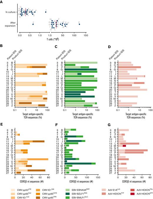
In a recent phase 1/2 trial, 24 stem cell donor–derived multiantigen (virus)–specific T-cell products, each containing CD8+ T-cell populations specific for several CMV, EBV, and AdV antigens, were generated and prophylactically administered to the respective patients early after T-cell–depleted alloSCT.20 All donors were seropositive for EBV and 13 donors were seropositive for CMV, whereas the serostatus for AdV was not determined (supplemental Table 1). All products contained EBV-specific T cells and all but 1 contained AdV-specific T cells, whereas CMV-specific T cells were only detectable in products from CMV-seropositive donors. We aimed to track the in vivo fate of the transferred virus-specific T cells using high-throughput TCR sequencing of virus-specific T cells from the products and of CD8+ T cells in PB samples collected from patients at different times after infusion. To track these T cells, TCR sequences had to be correctly allocated to the virus-specific T-cell populations that were present in each product. To this end, T-cell products were first polyclonally expanded in vitro to have sufficient T cells to sort each virus-specific T-cell population and to allocate the TCR sequences to the specific populations (see supplemental Figure 1A for a schematic overview). Insufficient PB samples were available from 2 patients who were, therefore, excluded from analysis. Furthermore, T cells of 2 T-cell products could not be expanded in vitro, thus, the patients that had received the corresponding T-cell products were also excluded from analysis. From 20 out of the initial 24 products, a median of 0.29 × 10⁶ T cells were expanded to a median of 21.9 × 10⁶ T cells (Figure 1A). From these expanded fractions, we separately isolated all the different virus-specific T-cell populations that were present in each product20 by FACS using pMHC tetramers and performed TCR sequencing of the CDR3β regions of each isolated virus-specific T-cell population. Unsorted fractions of the expanded T-cell products were also sequenced in parallel to quantify the different virus-specific TCRs present in the products (supplemental Figure 1A). In total, 97.5% (range, 81%-100%) of the TCR sequences from the sorted virus-specific T-cell populations could be detected in the unsorted bulk products (data not shown). To investigate the distribution of the different specificities in each product, we analyzed the frequencies of the TCR sequences for each target antigen of each virus. Overall, from the 13 products that were generated from CMV-seropositive donors, TCRs of 32 CMV-specific T-cell populations were annotated (Figure 1B). From the 20 products (all of which were generated from EBV-seropositive donors), TCRs of 31 EBV-specific T-cell populations were annotated (Figure 1C). Finally, TCRs from a total of 22 AdV-specific T-cell populations were annotated from 17 expanded products (Figure 1D), whereas 2 expanded products yielded insufficient numbers of AdV-specific cells for analysis.
Annotation and quantification of virus-specific TCR sequences from the T-cell products. T cells from 20 T-cell products were successfully expanded. The different virus-specific T-cell populations were sorted by FACS from these expanded products into separate pure populations using pMHC tetramers followed by direct mRNA isolation and TCR sequencing of the CDR3β regions. The remaining unsorted T cells from the T-cell products were sequenced in parallel to quantify the TCRs that were present in the products. (A) The numbers of T cells from the products that were put in culture and the cell numbers after expansion are shown. The red lines represent medians. Shown are the frequencies of CMV- (B), EBV- (C), and AdV- (D) specific TCR nucleotide sequences (CDR3β sequences) that were present in the target antigen–specific T-cell products. The sum of all target antigen–specific TCR nucleotide sequences were set to 100%. The different virus specificities are shown as stacked columns for each product. The number of different CDR3β sequences are shown that were specific for CMV- (E), EBV- (F) and AdV- (G) derived antigens for each product. nt, nucleotide; ID, identification.
Annotation and quantification of virus-specific TCR sequences from the T-cell products. T cells from 20 T-cell products were successfully expanded. The different virus-specific T-cell populations were sorted by FACS from these expanded products into separate pure populations using pMHC tetramers followed by direct mRNA isolation and TCR sequencing of the CDR3β regions. The remaining unsorted T cells from the T-cell products were sequenced in parallel to quantify the TCRs that were present in the products. (A) The numbers of T cells from the products that were put in culture and the cell numbers after expansion are shown. The red lines represent medians. Shown are the frequencies of CMV- (B), EBV- (C), and AdV- (D) specific TCR nucleotide sequences (CDR3β sequences) that were present in the target antigen–specific T-cell products. The sum of all target antigen–specific TCR nucleotide sequences were set to 100%. The different virus specificities are shown as stacked columns for each product. The number of different CDR3β sequences are shown that were specific for CMV- (E), EBV- (F) and AdV- (G) derived antigens for each product. nt, nucleotide; ID, identification.
To investigate how many different clonal virus-specific T-cell populations from each product we could potentially track in vivo, we quantified the different TCR nucleotide sequences for each virus-specific T-cell population within the T-cell products. The CMV-, EBV-, or AdV-specific T-cell populations from the products contained a median of 30 (range, 1-79), 34 (range, 5-140), and 20 (range, 2-74) different TCR nucleotide sequences, respectively (Figure 1E-G). Most of the virus-specific TCR nucleotide sequences were found at low frequencies (between 0.001% and 0.1%) in these expanded T-cell products (supplemental Figure 4).
T cells with TCR nucleotide sequences found in the T-cell products could be identified in patients with and without detectable viral loads
To investigate whether virus-specific T cells from the infused T-cell products could be found back in PB samples of the patients, we sequenced the TCRs of CD8+ T-cell fractions from PB samples taken at various time points after infusion of the T-cell products (see supplemental Figure 2 for sample quality assessments). These TCR nucleotide sequences were then compared with those that were present in the T-cell products (see supplemental Figure 1B for a schematic overview).
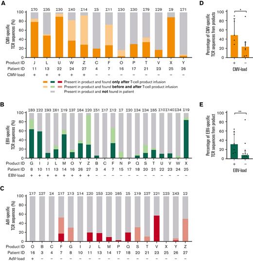
We previously illustrated that despite T-cell depletion of the graft, part of the donor-derived T-cell compartment can survive this procedure.25,26 This implies that TCRs detected in postinfusion samples may not necessarily be derived from the infused products, but may have already been introduced in the patients with the stem cell grafts. Therefore, we also analyzed PB samples that were obtained from patients before infusion of the products. In Figure 2, we separately depict the virus-specific TCR nucleotide sequences present in the products that could be found in patient PB samples only after infusion (orange: CMV, green: EBV, and red: AdV), both before and after infusion (light orange: CMV, light green: EBV, and light red: AdV), or could not be found after infusion (white) (Figure 2A-C). In addition, we determined whether the diversity (number of different TCR nucleotide sequences) correlated with viral reactivation as determined by presence of viral load in PB.
Virus-specific T cells with TCR nucleotide sequences found in the T-cell products could be identified in patients with and without detectable viral loads. The total numbers (Σ) of different virus-specific TCR nucleotide sequences are shown for each product (above each bar). Patients are grouped according to detectable viral loads after infusion of the T-cell product. Shown are the percentages of different CMV- (A), EBV- (B), and AdV- (C) specific TCR nucleotide sequences that could be detected only after infusion of the products (color scale), before and after infusion (light color scale), or that could not be detected (white). The numbers of different TCR nucleotide sequences that were found only after infusion, shown as percentages in bar plots with the median and iIQR, were compared between patients with and without CMV viral load (D) and with and without EBV viral load (E). Statistical differences were assessed with the Mann-Whitney t test (D-E). ∗P < .05; ∗∗P < .01.
Virus-specific T cells with TCR nucleotide sequences found in the T-cell products could be identified in patients with and without detectable viral loads. The total numbers (Σ) of different virus-specific TCR nucleotide sequences are shown for each product (above each bar). Patients are grouped according to detectable viral loads after infusion of the T-cell product. Shown are the percentages of different CMV- (A), EBV- (B), and AdV- (C) specific TCR nucleotide sequences that could be detected only after infusion of the products (color scale), before and after infusion (light color scale), or that could not be detected (white). The numbers of different TCR nucleotide sequences that were found only after infusion, shown as percentages in bar plots with the median and iIQR, were compared between patients with and without CMV viral load (D) and with and without EBV viral load (E). Statistical differences were assessed with the Mann-Whitney t test (D-E). ∗P < .05; ∗∗P < .01.
In 9 patients, the presence of some CMV-specific T-cell populations could not unequivocally be ascribed to the infused T-cell product, because the same sequences were also found in PB samples taken before infusion (Figure 2A light orange bars). However, many CMV-specific TCR nucleotide sequences were only found in PB samples of the 13 patients after infusion, strongly indicating that they originated from the infused products (Figure 2A orange bars). Such TCRs were found in patients with and without viral load after infusion, and a higher number (diversity) of different TCRs were found in samples from patients with CMV reactivation than in samples from patients without CMV reactivations following infusions, indicating in vivo expansion in response to the virus (Figure 2D). Similar results were obtained for the EBV-specific T-cell populations (Figure 2B). In 13 patients, a number of EBV-specific TCR nucleotide sequences were found both before and after infusion. Conclusions about their origin from the infused products (light green bars), therefore, could not be made. However, in 16 patients, TCR nucleotide sequences of EBV-specific T cells could be tracked in PB samples only after infusion of the products (green bars) and these EBV-specific TCRs were more abundant in patients with EBV reactivation than in those without (Figure 2E). In 2 patients without EBV reactivation, EBV-specific TCR nucleotide sequences from the infused products could not be detected in the PB samples. Finally, although PB samples from 6 patients contained AdV-specific TCR nucleotide sequences that were also present before infusion, samples from 8 patients contained AdV-specific TCR nucleotide sequences only after infusion of the products (Figure 2C red bars). In PB samples from the patient with a single positive AdV viral load, no AdV-specific TCRs could be tracked back.
These data show that in most patients after infusion of the multiantigen-specific T-cell products, high frequencies of virus-specific TCRs that were derived from the infused T-cell products could be identified. As measured by frequencies of virus-specific TCRs, significantly larger proportions of the T-cell products were found back in patients with viral reactivation compared with those without, indicating that these virus-specific T cells from the T-cell products contributed to the antiviral immune response.
Kinetics of virus-specific T cells with TCR nucleotide sequences from the infused products in patients with and without viral loads after infusion
For a number of T-cell populations, it was impossible to determine on the basis of TCR sequence detection alone, to what extent their presence was explained by infusion of the T-cell product, given that they were already present in the patients before infusion. However, we reasoned that we might still be able to assess the contribution of transfused T cells to viral control by examining their expansion kinetics after infusion of the T-cell product. If T cells derived from the product would significantly contribute to controlling the virus, we hypothesized that irrespective of their presence or absence in the patient before infusion, similar kinetics of the progeny of the virus-specific T-cell populations from the products should be found. Virus-specific T cells would show expansion in case of reactivation and only persistence in the absence of viral load after infusion. Therefore, we investigated the kinetics of virus-specific T-cell populations that appeared after infusion of T-cell products in patients with and without viral reactivations (colored) and compared these with the kinetics of virus-specific T-cell populations that were present before product infusion (gray). In Figures 3, 4, and 5, we separately depicted the kinetics of CMV-, EBV-, and AdV-specific T-cell populations (with TCR nucleotide sequences that were found in the products) in the presence and absence of viral loads after infusion.
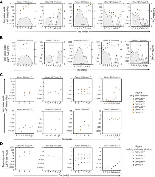
Kinetics of CMV-specific T cells with TCR nucleotide sequences also present in the infused T-cell products in patients with and without viral loads after infusion. Positive CMV viral loads were detected in 5 out of 13 patients that received a T-cell product containing CMV-specific T cells. PB samples that were obtained before and after infusion of the products were sorted by MACS for CD8+ T cells followed by mRNA isolation and sequencing of the TCRs. The numbers of target antigen–specific CD8+ T cells per liter blood were calculated by multiplying the frequencies of CMV-specific TCR nucleotide sequences with the absolute numbers of CD3+/CD8+ T cells per liter. CMV viral loads (dashed lines with gray area under the curve) and absolute numbers of CMV target antigen–specific T cells in PB samples are illustrated from the moment just before product infusion (day 0) until the end of follow-up. Shown are the kinetics of CMV target antigen–specific T cells in patients with CM -reactivations in whom TCR nucleotide sequences were found that only appeared after infusion of the products (A) and appeared before and after infusion of the products (B). Shown are the kinetics of CMV target antigen–specific T cells in patients without detectable CMV viral loads in whom TCR nucleotide sequences that were identical to the products were found that only appeared after infusion of the products (C) and appeared before and after infusion of the products (D). NP, not performed.
Kinetics of CMV-specific T cells with TCR nucleotide sequences also present in the infused T-cell products in patients with and without viral loads after infusion. Positive CMV viral loads were detected in 5 out of 13 patients that received a T-cell product containing CMV-specific T cells. PB samples that were obtained before and after infusion of the products were sorted by MACS for CD8+ T cells followed by mRNA isolation and sequencing of the TCRs. The numbers of target antigen–specific CD8+ T cells per liter blood were calculated by multiplying the frequencies of CMV-specific TCR nucleotide sequences with the absolute numbers of CD3+/CD8+ T cells per liter. CMV viral loads (dashed lines with gray area under the curve) and absolute numbers of CMV target antigen–specific T cells in PB samples are illustrated from the moment just before product infusion (day 0) until the end of follow-up. Shown are the kinetics of CMV target antigen–specific T cells in patients with CM -reactivations in whom TCR nucleotide sequences were found that only appeared after infusion of the products (A) and appeared before and after infusion of the products (B). Shown are the kinetics of CMV target antigen–specific T cells in patients without detectable CMV viral loads in whom TCR nucleotide sequences that were identical to the products were found that only appeared after infusion of the products (C) and appeared before and after infusion of the products (D). NP, not performed.
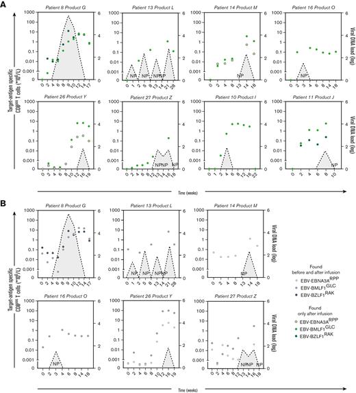
Kinetics of EBV-specific T cells with TCR nucleotide sequences present in the infused T-cell products in patients with and without viral loads after infusion. Positive EBV viral loads were detected in 8 out of 20 patients that received a T-cell product containing EBV-specific T cells. PB samples that were obtained before and after infusion of the product were sorted by MACS for CD8+ T cells followed by mRNA isolation and sequencing of the TCRs. The numbers of target antigen–specific CD8+ T cells per liter blood were calculated by multiplying the frequencies of EBV-specific TCR nucleotide sequences with the absolute numbers of CD3+CD8+ T cells per liter of blood. EBV viral loads (dashed lines with gray area under the curve) and absolute numbers of EBV target antigen–specific T cells in PB samples are illustrated from the moment just before product infusion (day 0) until the end of follow-up. Shown are the kinetics of EBV target antigen–specific T cells in patients with EBV reactivations where TCR nucleotide sequences were found that only appeared after infusion of the products (A) and appeared before and after infusion of the products (B). Shown are the kinetics of EBV target antigen–specific T cells in patients without detectable EBV viral loads where TCR nucleotide sequences that were identical to the products were found that only appeared after infusion of the products (C) and appeared before and after infusion of the products (D).
Kinetics of EBV-specific T cells with TCR nucleotide sequences present in the infused T-cell products in patients with and without viral loads after infusion. Positive EBV viral loads were detected in 8 out of 20 patients that received a T-cell product containing EBV-specific T cells. PB samples that were obtained before and after infusion of the product were sorted by MACS for CD8+ T cells followed by mRNA isolation and sequencing of the TCRs. The numbers of target antigen–specific CD8+ T cells per liter blood were calculated by multiplying the frequencies of EBV-specific TCR nucleotide sequences with the absolute numbers of CD3+CD8+ T cells per liter of blood. EBV viral loads (dashed lines with gray area under the curve) and absolute numbers of EBV target antigen–specific T cells in PB samples are illustrated from the moment just before product infusion (day 0) until the end of follow-up. Shown are the kinetics of EBV target antigen–specific T cells in patients with EBV reactivations where TCR nucleotide sequences were found that only appeared after infusion of the products (A) and appeared before and after infusion of the products (B). Shown are the kinetics of EBV target antigen–specific T cells in patients without detectable EBV viral loads where TCR nucleotide sequences that were identical to the products were found that only appeared after infusion of the products (C) and appeared before and after infusion of the products (D).
Kinetics of AdV-specific T cells with TCR nucleotide sequences present in the infused T-cell products in patients without viral loads after infusion. In 16 out of 17 patients that received a product that contained AdV-specific T cells, no AdV viral load was detected after infusion of the product. PB samples of these 16 patients that were obtained before and after infusion of the product were sorted by MACS for CD8+ T cells followed by mRNA isolation and sequencing of the TCRs. The numbers of target antigen–specific CD8+ T cells per liter were calculated by multiplying the frequencies of AdV-specific TCR nucleotide sequences with the absolute numbers of CD3+CD8+ T cells per liter of blood. Absolute numbers of AdV target antigen–specific T cells in PB samples are illustrated from the moment just before product infusion (day 0) until the end of follow-up. Shown are the kinetics of AdV target antigen–specific T cells in patients without detectable AdV viral loads in whom TCR nucleotide sequences that were identical to the products were found that only appeared after infusion of the products (A) and appeared before and after infusion of the products (B).
Kinetics of AdV-specific T cells with TCR nucleotide sequences present in the infused T-cell products in patients without viral loads after infusion. In 16 out of 17 patients that received a product that contained AdV-specific T cells, no AdV viral load was detected after infusion of the product. PB samples of these 16 patients that were obtained before and after infusion of the product were sorted by MACS for CD8+ T cells followed by mRNA isolation and sequencing of the TCRs. The numbers of target antigen–specific CD8+ T cells per liter were calculated by multiplying the frequencies of AdV-specific TCR nucleotide sequences with the absolute numbers of CD3+CD8+ T cells per liter of blood. Absolute numbers of AdV target antigen–specific T cells in PB samples are illustrated from the moment just before product infusion (day 0) until the end of follow-up. Shown are the kinetics of AdV target antigen–specific T cells in patients without detectable AdV viral loads in whom TCR nucleotide sequences that were identical to the products were found that only appeared after infusion of the products (A) and appeared before and after infusion of the products (B).
CMV
As illustrated in Figure 3A-B, positive CMV viral loads were detected in 5 out of 13 patients before and after infusion of the T-cell products. In 4 of these 5 patients, CMV viral loads were also detected at the moment of infusion of the T-cell products. In all 5 patients, CMV-specific TCR nucleotide sequences that only appeared after and those that were already present before infusion of the T-cell products exhibited similar expansion and contraction that correlated with the increase and decrease of viral loads (Figure 3A-B). The responding individual CMV-specific T-cell clones all showed similar expansion kinetics, including T-cell clones that contained public TCRs (TCR amino acid sequences that are identical in different individuals) and T-cell clones containing private TCRs (supplemental Figure 5A-B). During follow-up of the other 8 patients, no CMV viral loads were detected after infusion. Two patients (patients 7 and 16) had positive CMV viral loads, which had been cleared before infusion of the products (data not shown). In all 8 patients, we detected T cells with CMV-specific TCR nucleotide sequences from the products that only appeared after infusion, and these persisted without clear expansion in 6 out of 8 patients (Figure 3C). In 4 of these patients, we also detected CMV-specific TCR nucleotide sequences that were present before and after infusion of the T-cell products, showing similar kinetics (Figure 3D).
EBV
As shown in Figure 4A-B, reactivations as reflected by positive EBV viral loads, were detected in 8 out of 20 patients after infusion of the T-cell products. Detectable EBV viral loads were absent in all 8 patients at the time of T-cell product infusions, but EBV viral loads had been detected before infusion in 3 out of 8 patients (patients 11, 13, and 26; data not shown). In all 8 patients, EBV-specific TCR nucleotide sequences that only appeared after infusion and EBV-specific TCR nucleotide sequences that were present before and after infusion of the T-cell products showed similar expansion and contraction that correlated with the increase and decrease of viral loads (Figure 4A-B). Individual EBV-specific T-cell clones all showed similar expansion kinetics, including T-cell clones that contained public and private TCRs (supplemental Figure 5C-D). During follow-up of the other 12 patients, no EBV viral loads were detected after infusion, but EBV viral loads had been detected before infusion in 2 of the 12 patients (patients 3 and 23; data not shown). In 8 of the 12 patients, we detected EBV-specific TCR nucleotide sequences from the products that only appeared after infusion, showing persistence without clear expansion in 5 out of 8 patients (Figure 4C). In 5 patients, we also detected T cells with EBV-specific TCR nucleotide sequences that were present before and after infusion of the T-cell products, showing similar kinetics in all patients, except for patient 25 (Figure 4D).
AdV
One patient out of 17 (patient 16) who was prophylactically infused with a product that contained AdV-specific T cells, showed a detectable AdV viral load at week 16 after infusion. AdV-E1ALLD– and AdV-HEXONTYF–specific T cells were present in T-cell product P, but no AdV-specific TCR nucleotide sequences could be detected in the PB samples before or after infusion of the product. During follow-up of the other 16 patients, AdV viral loads were undetectable after infusion. In 8 out of 16 patients, we detected T cells with AdV-specific TCR nucleotide sequences from the products that only appeared after infusion, showing persistence without clear expansion in 7 out of 8 patients (Figure 5A). In 2 of these patients, we also detected T cells with AdV-specific TCR nucleotide sequences that were present before and after infusion of the T-cell products, showing similar kinetics, except for patient 26 (Figure 5B).
Based on these results, we conclude that in 5 out of 5 patients with a detectable CMV viral load, and in 8 out of 8 patients with a detectable EBV viral load, T cells with CMV- and EBV-specific TCR nucleotide sequences that were only found after infusion of the products displayed similar kinetics to those that were found before and after infusion of the products. One patient had a detectable AdV viral load during follow-up, but no T cells with AdV-specific TCR nucleotide sequences could be detected. In 8 out of 8 patients without detectable CMV viral loads, 8 out of 12 patients without detectable EBV viral loads, and 8 out of 16 patients without AdV viral loads, persistence of CMV-, EBV-, and AdV-specific T cells, respectively, with TCR nucleotide sequences from the products were observed, which were not detected before infusion.
Longitudinal analysis of total numbers of product-derived CMV- and EBV-specific T cells
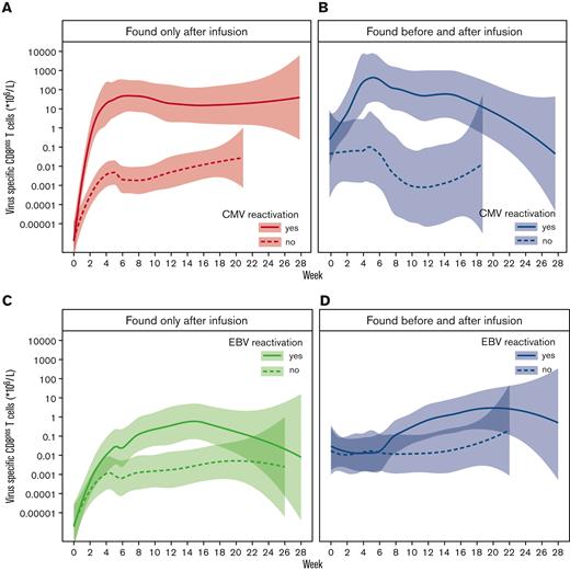
To study the association between viremia and the kinetics of expansion/persistence of adoptively transferred CMV- and EBV-specific T cells, we performed statistical modeling. First, we compared the smoothed Loess curves of the kinetics of all product-derived CMV- and EBV-specific T cells for patients with and without detectable viral loads during follow-up after product infusion. As expected, product-derived CMV-specific T cells that were only detected after product infusion showed more vigorous expansion in patients with CMV viral loads during follow-up (orange solid line) compared with that in patients who never had detectable CMV viral loads (Figure 6A orange dashed line). Product-derived CMV-specific T cells that were detected before and after infusion followed a similar pattern, with more expansion in patients with CMV viral loads (gray solid line) than in patients without (Figure 6B gray dashed line). Product-derived EBV-specific T cells that were only detected after product infusion showed the same pattern as CMV-specific T cells, with more expansion in patients with EBV viral loads (green solid line) than in patients without (Figure 6C gray dashed line). Similar trends were found for EBV-specific T cells that were detected before and after infusion (Figure 6D). To investigate whether the increases in numbers of CMV- and EBV-specific T cells were indeed significantly associated with viremia, we constructed 4-linear mixed models with the presence of viral load as a time-dependent covariate. The models contained 2 fixed effects, including time since infusion (in weeks), and whether or not the measurement was taken after the first appearance of viral load (per patient). For the modeling of product-derived T cells that were found only after infusion, a patient-specific, random slope effect for time was included to account for the heterogeneity in the trajectories between patients. For the modeling of product-derived T cells that were found before and after infusion, a random intercept effect was added, because the T cells were already detectable at time of infusion. For CMV, the start of detectable viral loads were significantly associated with higher T-cell numbers (P ≤ .0001 and P = .0001 for T cells found only after infusion and T cells found before and after infusion, respectively). Similarly, the appearance of EBV viral loads was significantly associated with higher numbers of EBV-specific T cells that were found only after infusion (P ≤ .0001), but a nonsignificant association (P = .1771) with viral load was observed for EBV-specific T cells that were found before and after infusion. These data show that the in vivo expansion/persistence kinetics of adoptively transferred CMV- and EBV-specific T cells that were only detected after infusion were significantly different for patients with viral loads (strongly expanding/proliferating) than for patients that did not develop detectable viral loads in the follow-up period after T-cell product infusion (persisting/maintenance).
Statistical modeling of the expansion/persistence kinetics of adoptively transferred CMV- and EBV-specific T cells and the presence of viral loads in the follow-up period after T-cell product infusion. Positive CMV or EBV viral loads were detected after infusion of T-cell products in 5 out of 13 patients and 8 out of 20 patients that received a T-cell product containing CMV- or EBV-specific T cells, respectively. Smoothed Loess curves were plotted to study the association between viremia and expansion/persistence kinetics of adoptively transferred CMV- and EBV-specific T cells. Kinetics of the numbers of product–derived CMV-specific T cells that were only detected after T-cell product infusion (A) or both before and after infusion (B) are shown for patients with a positive CMV viral load during follow-up (solid line) and without detectable viral loads in the follow-up period (dashed line). Product–derived EBV-specific T cells that were only detected after infusion (C) or both before and after infusion (D) are shown for patients with a positive EBV viral load during follow-up (solid line) and without detectable viral loads (dashed line).
Statistical modeling of the expansion/persistence kinetics of adoptively transferred CMV- and EBV-specific T cells and the presence of viral loads in the follow-up period after T-cell product infusion. Positive CMV or EBV viral loads were detected after infusion of T-cell products in 5 out of 13 patients and 8 out of 20 patients that received a T-cell product containing CMV- or EBV-specific T cells, respectively. Smoothed Loess curves were plotted to study the association between viremia and expansion/persistence kinetics of adoptively transferred CMV- and EBV-specific T cells. Kinetics of the numbers of product–derived CMV-specific T cells that were only detected after T-cell product infusion (A) or both before and after infusion (B) are shown for patients with a positive CMV viral load during follow-up (solid line) and without detectable viral loads in the follow-up period (dashed line). Product–derived EBV-specific T cells that were only detected after infusion (C) or both before and after infusion (D) are shown for patients with a positive EBV viral load during follow-up (solid line) and without detectable viral loads (dashed line).
Discussion
This study investigated the persistence and expansion in patients of in vitro–isolated and prophylactically infused multiantigen virus-specific T-cell products in the presence or absence of viral reactivation by in vivo tracking of individual T-cell populations. In contrast with the pMHC tetramer technology that we previously used,20 TCR sequencing of purified viral antigen–specific T-cell populations allowed us to identify multiple different clonal T-cell populations within the antigen–specific T-cell compartments. This permitted their tracking with high sensitivity and specificity in PB of patients after infusion of the virus-specific T-cell products. TCR mapping of the donor-derived CMV-, EBV-, and AdV-specific T cells in the products revealed the presence of medians of 30, 34, and 20 different TCR sequences per product, respectively. This technology allowed us to follow the presence and kinetics of the virus-specific T cells after infusion into patients after undergoing alloSCT. It also made it possible to distinguish donor-derived virus-specific T cells that were already present in the patient before infusion from those exclusively derived from the infused T-cell products. TCR sequences from the products that were exclusively found in PB after infusion were documented in all patients infused with CMV-specific T cells, in 80% of patients infused with EBV-specific T cells, and in 47% of patients infused with AdV-specific T cells. As expected, higher frequencies of TCRs identical to the T-cell products could be tracked in PB of patients with CMV or EBV reactivations, compared with patients without reactivations. Since only 1 patient experienced AdV reactivation, no conclusions could be drawn about the T-cell kinetics in the presence of this virus. All patients with CMV or EBV reactivations showed expansion of virus-specific TCRs with similar kinetics, irrespective of the presence of some of these T cells in the patients before infusion, suggesting that the virus-specific T cells from the T-cell products contributed to the antiviral immune response. Statistical modeling of the expansion/persistence kinetics of the adoptively transferred virus-specific T cells showed a significant correlation between the vigorous increase in the numbers of circulating product-derived CMV- and EBV-specific T cells and viral loads. In 100%, 67%, and 50% of the patients infused with CMV-, EBV-, and AdV-specific T cells, respectively, T cells with TCRs identical to those in infusion products could be tracked for extended periods, even in the absence of viral reactivation, indicating T-cell persistence and a potential role of the adoptively transferred T-cell products in long-term protection against viral reactivation.
Several phase 1 and 2 clinical studies have been performed, exploring safety, feasibility, and potential efficacy of adoptive transfer of in vitro–selected virus-specific T-cell products derived from the original stem cell donors to control refractory viral reactivation in patients following alloSCT.20,27-31 Presence of virus-specific T cells in patients, in some of these studies, was demonstrated using pMHC tetramer–based flow cytometry assays or enzyme-linked immunospot analysis. However, the origin of the detected virus-specific T cells could not be demonstrated using these assays. Similar studies have been performed after solid organ transplantation in patients who received autologous virus-specific T-cell products derived from the naïve T-cell repertoire.32 In this case, the origin of the virus-specific T cells that were observed in the patients was clear, but it remained difficult to unequivocally link the clinical effects to the infused T-cell product. In many cases, control of reactivation or even disappearance of clinical symptoms was documented after infusion of the virus-specific T cells.9,33 However, even in case of (partial) T-cell depletion of the stem cell grafts, virus-specific T cells that survive the depletion are coadministered with the graft and already present at the time of infusion of the T-cell product.25,26 This makes it difficult to address whether the infused T-cell products were actually responsible for the clinical effects, despite a clear correlation between infusion of the cells and clinical benefit.29 With next-generation TCR sequencing techniques, adoptively transferred donor-derived T cells (eg, virus-specific, tumor-specific, or regulatory T cells) could be more efficiently tracked in PB samples of patients with virus reactivations,34 organ transplantations,35 cancer,36 or autoimmune diseases.37 However, donor-derived virus-specific T cells have not yet been tracked using TCR sequences in patients who prophylactically received them. By tracking many individual clonal virus-specific T-cell populations from the T-cell products and doing so in patients clearing viral reactivations, we could demonstrate that most T cells expanding in response to in vivo appearance of the virus were derived from the infused T-cell products. However, de novo generation of public TCRs in the recipient that originate from the donor-derived stem cells/naïve precursor T cells cannot be unequivocally excluded. Therefore, T cells expressing public TCRs are difficult to link to the product. However, from previous studies we know that memory CD8+ T cells dominate the immune reconstitution at week 6 after alloSCT compared with naïve T cells. Also, naïve T cells are more efficiently depleted following alemtuzumab-based T-cell depletion,25 making it unlikely that such T cells expressing public TCRs emerging early after alloSCT would be derived from the naïve donor T-cell repertoire. In addition, patient-derived virus-specific T cells can also contribute to the control of viral reactivation after alloSCT, especially after NMA TCD alloSCT, as described previously.38 Because the focus within the current study was on the in vivo fate of donor-derived virus-specific T cells on adoptive transfer, rather than on the entire virus-specific immunity posttransplant, we did not quantify and annotate patient-derived virus-specific T cells and therefore, cannot elaborate on the role of host immunity in these specific patients. Gene-edited donor-derived T cells could circumvent part of this problem by tracking T cells with the inserted gene in vivo, thereby limiting the influence of autologous-derived TCR sequences.15,39,40 Long-term persistence was shown for adoptively transferred gene-marked EBV-specific T cells using this approach.15 It has been suggested that coinfusion of virus-specific CD4+ and CD8+ T cells may be beneficial.41-43 Our study also strongly supports previous indications that in vitro selection based on purification of virus-specific CD8+ T cells by pMHC complexes using the streptamer technology does not hamper the in vivo functionality of these cells after infusion,20 resulting in expansion and long-term persistence without the coinfusion of virus-specific CD4+ T cells. It was reported that the diversity of TCRs of CMV-specific T-cell populations decreases during viral reactivation.44 However, in our analyses we did not observe any reduction in diversity of the TCR repertoire, even in those patients where clonal expansion of virus-specific T-cell populations was overt. The high sequencing depth of our strategy allowed us to also detect persisting virus-specific T-cell clones that were not expanding, but that contributed to the diversity of the TCR repertoire. Because our clinical phase 1/2 study20 did not include a control arm without T-cell product infusion or placebo, we cannot demonstrate the reconstitution and expansion kinetics in patients who did not receive adoptively transferred virus-specific T cells.
The strategy we applied to perform bulk TCRβ sequencing of thousands of cells in parallel is a powerful tool to dissect the diversity of the TCR repertoire of multiple T-cell populations. However, the main limitation is the inability to pair the information regarding TCRα and TCRβ sequences of individual T cells. Paired single-cell sequencing of TCRα and TCRβ chains would have allowed us to provide information on the TCRα usage of product-derived virus-specific T cells. However, single-cell sequencing is more limited in the number of cells that can be sequenced simultaneously, thereby losing resolution required for the detection of T-cell populations that are present at low frequencies in the PB samples of patients. This can potentially result in undetected donor-derived virus-specific T cells before infusion or undetected persisting virus-specific T cells when viral loads are absent.
Although TCRβ sequencing of product-derived virus-specific T-cell populations allowed for tracking of individual T-cell clones on adoptive transfer to patients, it remains possible that TCRs of T cells from the patient contain exactly the same nucleotide sequence as the donor/product–derived TCRs. We recently demonstrated that the magnitude, defined as frequency and occurrence, of such public TCRs is high, but that this was only on the amino acid level,23 illustrating that most TCRs contained different nucleotide sequences as a result of convergent recombination and random nucleotide insertion between variable diversity joining regions. Therefore, the chance of shared TCR nucleotide sequences between patient and donor has been estimated to be relatively low.
Most of the studies exploring the potential benefit of in vitro–selected virus-specific T cells has been performed in a pre-emptive or therapeutic setting. In these cases, the cells are infused when viral reactivation has already occurred. This does not allow evaluation of survival/persistence of the T-cell products when they do not immediately encounter their antigen in vivo. It has been suggested that under those circumstances, survival of the adoptively transferred T cells may be poor. Obviously, in the absence of antigen and expansion, the contribution of the infused T cells to the total peripheral T-cell repertoire in the patient is generally too low to allow detection using the pMHC tetramer technology.45 However, our approach allowed us to determine the in vivo persistence of prophylactically infused T-cell products even in the absence of viral reactivation. We found evidence that infused virus-specific T cells persisted at very low frequencies without clear expansion. In a few patients, we could also detect late expansion of these infused T cells, supporting the persistent functionality following in vitro selection and infusion of the virus-specific T-cell products, even in the absence of direct in vivo antigen encounter. Statistical modeling of the expansion/persistence kinetics of the adoptively transferred virus-specific T cells showed a significant correlation between the vigorous increase in the numbers of circulating product-derived CMV- and EBV-specific T cells and viral loads. It could be assumed that different virus-specific T-cell populations targeting different antigens can have different efficacies, but modeling of the different CMV/EBV-specificities separately was not possible in our study because of the limited sample numbers and resulting insufficient power. However, as shown in Figures 3 and 4, different virus-specific T-cell populations showed very similar expansion kinetics within each patient, assuming no large differences in efficacies between specificities.
In conclusion, our study shows that TCR sequencing allows highly sensitive and specific tracking and tracing of multiple clonal T-cell populations from in vitro–selected donor-derived virus-specific CD8+ T-cell products, following infusion in patients who underwent alloSCT. Using this methodology, we were able to distinguish expansion and persistence of virus-specific T-cell populations selectively derived from the infused T-cell products from those T cells that were already present in the patients before infusion. We demonstrated after viral reactivation in vivo, expansion of multiple clonal T-cell populations from multivirus-specific CD8+ T-cell products in vitro purified by the streptamer technology. We showed persistence of prophylactically infused virus-specific T cells derived from the infused T-cell products in the absence of viral reactivation, illustrating long-term persistence. These results suggest that infusion of donor-derived multivirus-specific T cells in a prophylactic setting, early after T-cell–depleted alloSCT, may be a viable option to prevent viral complications.
Acknowledgments
This work was supported by Sanquin Research and Landsteiner Laboratory for Blood Cell research (PPO 15-37/L number 2101) and by research funding from Stichting den Brinker (Zeist, The Netherlands). This study was additionally supported by grants from the European Union’s seventh Framework Program (FP/2007-2013) under grant agreement (number 601722), by Dutch Cancer Society (grant UL 2008 to 4263), and by German Cancer Society (grant DFG-FOR 28030, project P09).
The sponsors are nonprofit organizations that support science in general. They had no role in gathering, analyzing, or interpreting the data.
Authorship
Contribution: J.H.F.F., I.J., and H.E. developed the clinical study concept; P.v.B. and J.H.F.F. enrolled study participants and monitored patients clinically; L.G. provided major histocompatibility complex I streptamer technology equipment; L.H., H.M.v.E., S.A.J.V., C.H., and M.C.J.R. generated multivirus-specific T-cell products; W.H., L.H., M.C.J.R., and C.A.M.v.B. performed in-depth analysis of the T-cell products; W.H., L.H., and C.A.M.v.B. performed in-depth T-cell receptor sequencing analysis of the follow-up peripheral blood samples; E.A.S.K. performed statistical analysis and trajectory analysis of virus-specific T cells using Loess curves; W.H., I.J., J.H.F.F., and D.A. wrote the manuscript; and all authors reviewed the manuscript.
Conflict-of-interest disclosure: L.G. is a senior vice president and managing director of Juno Therapeutics GmbH, a Celgene company (Munich, Germany). The remaining authors declare no competing financial interests.
Correspondence: W. Huisman, Department of Hematology, Leiden University Medical Center, Postal code 9600, 2300 RC Leiden, The Netherlands; e-mail: w.huisman@lumc.nl.
References
Author notes
Deidentified individual participant data that underlie the reported results will be made available after publication at the National Center for Biotechnology Information Sequence Read Archive (accession number SUB12065193, ncbi.nlm.nih.gov/sra).
Data are available on request from the corresponding author, W. Huisman (w.huisman@lumc.nl).
The full-text version of this article contains a data supplement.

