TO THE EDITOR:
Mouse models are indispensable tools to study the impact of genetic alterations in cancer. Only a few studies have thus far undertaken functional characterizations of recurrent genomic-discovered gene mutations of chronic lymphocytic leukemia (CLL)1 despite recent advances in genetic engineering that have increasingly facilitated an improved ability to assess the in vivo effects of candidate disease drivers. In this context, conditional knockin and knockout strategies have been applied to the study of individual lesions, including del(13q), modeled in the MDR mice,2 the B-cell developmental factor Ikzf3,3 or in the splicing factor Sf3b1 in combination with Atm deletion,4 all of which recapitulate indolent CLL development, consistent with the phenotype of human disease.
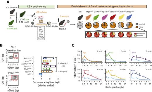
We hypothesize that the most common loss-of-function (LOF) putative drivers typical of CLL (Atm, Trp53, Chd2, Birc3, Mga, or Samhd1)1,5 (supplemental Figure 1A-B) may affect preleukemic signaling, thus dysregulating B-cell–intrinsic prosurvival pathways. To address this question, we developed a versatile platform for the introduction of individual LOF lesions in vivo in murine B cells. We generated a transplant model that efficiently delivers single guide RNAs (sgRNAs) to hematopoietic stem and progenitor cells of strains expressing B-cell–restricted Cas9 (ie, Cd19-Cas9; Figure 1A [left]), thus generating gene edits only in developing Cas9-expressing B cells and allowing the introduction of insertions-deletions (indels) at the target locus, leading to LOF of the target gene. First, we intercrossed mice carrying B-cell–restricted Cre expression (Cd19-cre) with those carrying conditional Cas9–green fluorescent protein (Cas9-GFP)6 to generate a strain expressing B-cell–restricted Cas9 (Cd19-Cas9) (Figure 1A [left]). Next, we optimized the in vitro engineering of early stem and progenitor cells (ie, Lin- Sca-1+ c-kit+ [LSK]) from Cd19-Cas9 mice using lentivirus expressing sgRNAs (mCherry+) targeting Atm, Trp53, Chd2, Birc3, Mga, or Samhd1. We chose LSKs because of their high transducibility and long-term repopulating potential and designed sgRNAs targeting early exonic regions in each of the 6 genes to allow early disruption of the protein sequence (supplemental Table 1). Lastly, we transplanted the single sgRNA-expressing LSKs into sublethally irradiated CD45.1 recipient mice. We confirmed the consistent presence of ∼45% to 85% gene-edited sequences (>70% carrying frameshift mutations, the majority resulting from 1 or 2 base pair indels; supplemental Figure 2) in edited B cells (GFP+mCherry+, isolated from the peripheral blood by cell sorting), but not in T cells or monocytes, at 4 months after transplant by polymerase chain reaction-based targeted deep sequencing (analyzed by CRISPResso2 software7; Figure 1A [right]; supplemental Figure 1C-D). A minority of lesions (<30%) led to in-frame alterations (ie, ±3 nucleotides in length). Reproducible on-target editing was detected across the 6 cohorts (with no detectable gene edits in 24 putative off-targets), per single-cell DNA-sequencing analysis (DNA-seq) of 1787 single B cells (supplemental Figure 1E). All LOF mutations were found to confer a survival advantage to B cells compared with the non-targeting scramble control when exposed to the mitogenic stimuli lipopolysaccharide (LPS) and interleukin-4 (IL-4) for 72 hours in vitro (n = 5 per group; P < .05; Figure 1B). Likewise, circulating edited B cells from animals following transplant were elevated across the mutated models compared with the non-targeting control line (Nt-1; P < .05; RM-ANOVA; Figure 1C). Despite these early changes, mostly marked at 4 months after transplant, a time point at which B cells are considered to achieve optimal host reconstitution, none of the single-edited animals (n = 10 per group) developed clonal B220+ CD5+ Igκ+ circulating B-cell expansions over 24-months of observation. Mga- and Chd2- depleted animals showed trajectories that were particularly short-lived (supplemental Figure 1F; P < .0002), in line with limited in vitro persistence observed in previous cell line studies.8 These observations collectively suggest the dual possibility of limited cell-intrinsic dysregulation of prosurvival pathways introduced by these lesions and/or insufficient cell-extrinsic contribution to leukemia phenotypes in these otherwise healthy animals.
Individual CLL LOF lesions alter mature B-cell survival but are insufficient to drive leukemia in mouse models. (A) Transplant schema and generation of singly-edited mouse lines. LSKs were lentivirally transfected in vitro with sgRNAs targeting the single LOFs (or a non-targeting scramble control, ie, Nt-1) in addition to the mCherry marker before the engraftment into sublethally irradiated CD45.1 recipient mice. PCR-based targeted deep sequencing of DNA from peripheral blood edited B cells (ie, GFP+mCherry+) was used to confirm presence of ∼45% to 85% gene edits (>70% frameshift mutations) across the 6 genes. (B) In vitro survival of normal B cells isolated from the spleen of 5 animals per group (6 LOF-expressing strains and 1 Nt-1) and cultured in vitro in presence of LPS+IL4 for 3 days. Fold increase over day 0 is displayed alongside representative flow cytometric plots from 1 Nt-1 and 1 Trp53-depleted sample. P-value lower or equal to 0.05, ANOVA with Dunnett’s correction for multiple comparisons. (C) Percent (%) GFP+mCherry+ B cells as assessed on longitudinal bleeds over the course of 24 months, in 10 animals per group. ∗P-value lower or equal to 0.05; lower or equal to 0.05; RM-ANOVA, repeated measures analysis of variance.
Individual CLL LOF lesions alter mature B-cell survival but are insufficient to drive leukemia in mouse models. (A) Transplant schema and generation of singly-edited mouse lines. LSKs were lentivirally transfected in vitro with sgRNAs targeting the single LOFs (or a non-targeting scramble control, ie, Nt-1) in addition to the mCherry marker before the engraftment into sublethally irradiated CD45.1 recipient mice. PCR-based targeted deep sequencing of DNA from peripheral blood edited B cells (ie, GFP+mCherry+) was used to confirm presence of ∼45% to 85% gene edits (>70% frameshift mutations) across the 6 genes. (B) In vitro survival of normal B cells isolated from the spleen of 5 animals per group (6 LOF-expressing strains and 1 Nt-1) and cultured in vitro in presence of LPS+IL4 for 3 days. Fold increase over day 0 is displayed alongside representative flow cytometric plots from 1 Nt-1 and 1 Trp53-depleted sample. P-value lower or equal to 0.05, ANOVA with Dunnett’s correction for multiple comparisons. (C) Percent (%) GFP+mCherry+ B cells as assessed on longitudinal bleeds over the course of 24 months, in 10 animals per group. ∗P-value lower or equal to 0.05; lower or equal to 0.05; RM-ANOVA, repeated measures analysis of variance.
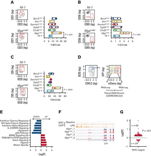
We asked whether any of the modeled lesions (also frequent drivers of other aggressive B-cell malignancies, including diffuse large B-cell lymphoma)9 would affect B-cell developmental trajectories (supplemental Figure 3A; supplemental Table 2). Although transitional B-cell population abundance was unchanged across genotype-defined groups (supplemental Figure 3B-C), Mga-depleted mice showed increased marginal zone (P = .003) (with concordantly decreased follicular B cells) and germinal center (P = .009) splenic subpopulations (Figure 2A-B; supplemental Figure 3D). We also identified an abundance of peritoneal B1a and a concordant decrease in B1b cells (P < .05; Figure 2C; supplemental Figure 3E), suggesting a role of Mga in regulating mature B-cell development and lineage-specification. In particular, the increased spontaneous germinal center formation may relate to the lymphomogenic activity of this lesion, whereas increased CD5+B1a peritoneal B cells may instead relate to CLL predisposition, as B1a cells are considered to be the cell of origin of murine CLL.10
Mga mutation alters B-cell developmental pathways. (A) Percent (%) abundance of MZ B cells, (B) GC B cells in spleen and (C) B1a cells in peritoneum preparations from 5 animals per group, including non-targeting controls. ∗∗P-value, lower or equal to 0.01, ∗∗∗P-value, lower or equal to 0.001, ANOVA with Dunnett's correction for multiple comparisons. (D) Flow-plot of edited vs unedited fractions analyzed via RNA-seq and CISTROME-GO. (E) Pathway enrichment analysis of Mga-depleted vs unedited cells, per Enrichr. (F) IGV screenshot of MYC ChIP-seq data of mouse B cells with or without LPS treatment and RNA-seq data of Lck gene expression in edited (Mgaindel) and unedited (MgaWT) fractions of 2 independent mice. (G) Change in expression (Mgaindel vs MgaWT) of genes that were direct Myc targets upon LPS stimuli, as assessed by CISTROME-GO analysis. ∗∗∗P-value, lower or equal to 0.001; χ2 test. GC, germinal center; IGV, integrative genomics viewer; MZ, marginal zone.
Mga mutation alters B-cell developmental pathways. (A) Percent (%) abundance of MZ B cells, (B) GC B cells in spleen and (C) B1a cells in peritoneum preparations from 5 animals per group, including non-targeting controls. ∗∗P-value, lower or equal to 0.01, ∗∗∗P-value, lower or equal to 0.001, ANOVA with Dunnett's correction for multiple comparisons. (D) Flow-plot of edited vs unedited fractions analyzed via RNA-seq and CISTROME-GO. (E) Pathway enrichment analysis of Mga-depleted vs unedited cells, per Enrichr. (F) IGV screenshot of MYC ChIP-seq data of mouse B cells with or without LPS treatment and RNA-seq data of Lck gene expression in edited (Mgaindel) and unedited (MgaWT) fractions of 2 independent mice. (G) Change in expression (Mgaindel vs MgaWT) of genes that were direct Myc targets upon LPS stimuli, as assessed by CISTROME-GO analysis. ∗∗∗P-value, lower or equal to 0.001; χ2 test. GC, germinal center; IGV, integrative genomics viewer; MZ, marginal zone.
Given the known role of Mga as negative regulator of MYC targets in other cellular contexts,11 we further asked whether the observed B-cell developmental phenotypes could result from aberrant MYC target activation in mature B cells. To this end, we analyzed the transcriptomes of gene-edited mature B cells (GFP+mCherry+) and compared them with those of unedited (GFP+mCherry-) B cells isolated from the same animals (2 animals per group; Figure 2D). Analyses were performed 4 months after transplant because this was observed to be the peak of edited B-cell abundance across cohorts (Figure 1C). Via Enrichr analysis, using the Molecular Signatures Database (MSigDB), we identified enrichment in PI3K/AKT/mTOR and Notch signaling and diminished interferon gamma, p53, and apoptotic gene expression in Mga-depleted cells compared with those in the control unedited fraction (false discovery rate <0.10; Figure 2E; supplemental Tables 3-4). To identify direct MYC targets affected by loss of Mga, we performed an integrated analysis of Mga-depleted RNA-seq and existing chromatin immunoprecipitation–seq (ChIP-seq) data of MYC from mouse B cells exposed to LPS stimulation for 8 hours12 using CISTROME-GO,13 which allows sorting of direct targets of specific transcription factors based on the integrative ranking of transcription factor–binding (ChIP-seq) combined with change in gene expression (RNA-seq). Our results showed that Mga depletion activated transcription of several MYC target genes with relevance to B-cell developmental biology (supplemental Table 5), including the B-cell receptor signaling kinase lymphocyte cell-specific protein-tyrosine kinase (Lck), the cell cycle and Wnt regulators Stag3 and Ptprs and the ribosomal protein Rps29 in response to LPS (Figure 2F; supplemental Figure 4). Notably, direct MYC targets were more likely to be upregulated than Mga-depleted cells, consistent with MYC activation (P = .001; χ2 test; Figure 2G). Together, these results support the notion that Mga plays a unique role in regulating MYC targets involved in B-cell proliferation and differentiation, resulting in a direct impact on cell fate determination of mature naïve B cells.
In conclusion, we demonstrate that our models represent a valuable and versatile platform for the study of individual genetic lesions in a preleukemic B-cell context. We show that introduction of individual CLL LOF mutations in maturing B cells can alter prosurvival and B-cell developmental pathways and increase cellular fitness in vitro and in vivo. We further observe that individually they are insufficient to drive leukemia and motivate studies of evaluating the effects of combinatorial assortment of lesions in B-cell leukemogenesis.
Acknowledgments: The authors thank members of the Wu lab for the valuable discussions. The authors are also grateful for excellent technical assistance from the Dana-Farber Cancer Institute Animal Research Facility and the Dana-Faber Cancer Institute Flow Cytometry Core. This study was supported by grants from the National Institutes of Health/National Cancer Institute (P01 CA206978 and R01CA216273). E.t.H. is a scholar of the American Society of Hematology. S.Y. is supported by a research fellowship from the Lauri Strauss Leukemia Foundation and National Cancer Institute (R21 CA267527-01). M.H.S. has been supported by a Sara Borrell postdoctoral contract (CD19/00222) from the Instituto de Salud Carlos III (ISCIII), cofounded by Fondo Social Europeo “El Fondo Social Europeo invierte en tu futuro.” K.C. is supported by National Human Genome Research Institute Career Development Award K99HG011658. S.L. is supported by the National Cancer Institute Research Specialist Award (R50CA251956). L.P. is partially supported by National Human Genome Research Institute Genomic Innovator Award R35HG010717.
Contribution: E.t.H., S.Y., and C.J.W. designed the study and wrote the manuscript; E.t.H. generated mouse models, performed most experiments, and analyzed data; S.Y. performed RNA-seq and ChIP-seq analyses; S.L. performed the single-cell DNA-seq experiments; R.R., M.H.S, K.C., L.P., K.J.L., and D.N. assisted with study design and data analysis; G.B.H., F.F.R., and E.W. provided technical support; and C.J.W. provided overall supervision to the study.
Conflict-of-interest disclosure: C.J.W. is an equity holder of BioNTech Inc. and receives research funding from Pharmacyclics. L.P. has financial interests in Edilytics Inc. K.C. is an employee, shareholder, and officer of Edilytics Inc. The interests of L.P. and K.C. were reviewed and are managed by Massachusetts General Hospital and Partners HealthCare in accordance with their conflict-of-interest policies. The remaining authors declare no competing financial interests.
Correspondence: Catherine J. Wu, Department of Medical Oncology, Dana-Farber Cancer Institute, 450 Brookline Ave, Dana Building, Room DA-520, Boston, MA 02115; e-mail: cwu@partners.org.
References
Author notes
∗E.H., S.Y., and C.J.W. contributed equally to this study.
Mouse RNA-seq data reported in this article have been deposited in the Gene Expression Omnibus database (accession number GSE197061).
Data are available on request from the corresponding author, Catherine J. Wu (cwu@partners.org).
The full-text version of this article contains a data supplement.


