TO THE EDITOR:
To date, little is known about what causes the breakdown of immune tolerance or the significance of increased cytokines found by some investigators in patients with warm autoimmune hemolytic anemia (wAIHA).1-6 In this retrospective study, plasma was collected from 54 direct antiglobulin test (DAT)-positive samples of patients with wAIHA, documented by the highly respected laboratory of the late George Garratty at the American Red Cross Blood Services, Southern California Region, Pomona, California. This reference laboratory specializes in confirmation of autoimmune hemolytic anemias by serological testing as having active or compensated wAIHA (supplemental Table S1 for clinical details). Plasma samples from 36 healthy controls (HC) obtained from the Canadian Blood Services, Toronto, were used for comparison. The samples were analyzed for 38 cytokines, chemokines, and growth factors as described in the supplemental Methods. Ethics approvals (CBS protocol for normal healthy donors #2009.015 and American Red Cross Protocol for patients with wAIHA #2011-007) were obtained to do the study, which was conducted according to the Declaration of Helsinki.
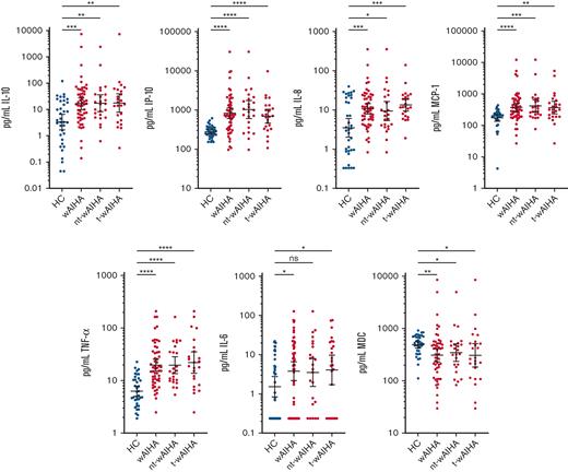
Using the samples from 54 patients, we identified a significant difference in 15 cytokines when compared with HC; an elevation of cytokines IL-1Ra, IL-6, IL-10, IL12p70, TNFα and chemokines IL-8/CXCL8, IP-10/CXCL10, GRO/CXCL1, MCP-1/CCL2 and a decrease in cytokines IFNα2, IL-3, IL-13, TGFα and chemokines fractalkine/CX3CL1, MDC/CCL22 (supplemental Table S2 in supplemental Methods and Results for statistical methods and summary statistics for all cytokines analyzed). For subsequent analysis, we considered the stability of these 15 cytokines. Although our samples were stored as ethylenediaminetetraacetic acid (EDTA) whole blood for up to 6 days before having the plasma separated and stored, we previously determined that blood collected into EDTA anticoagulant maintains stability of many cytokines, even up to 10 days of storage.7 Applying the results of that storage study, we identified the following 7 (3 cytokines and 4 chemokines) from the 15 above that were stable in stored EDTA whole blood: IL-6, IL-10, TNFα, IL-8/CXCL8, IP-10/CXCL10, MCP-1/CCL2, and MDC/CCL22 (Figure 1).
Identification of plasma cytokines altered in wAIHA. Cytokines found significantly changed between HC (n = 36) and wAIHA patients (n = 54) are shown. HC are also compared with nontransfused (nt-wAIHA, n = 24) or transfused (t-wAIHA, n = 24) patient subsets (transfusion status was not available for 6 patients). Transfusion status did not affect significance with the exception of IL-6, which was not different from HC in nt-wAIHA patients. Shown here for visual comparison are the original data plotted on log scale. The geographic mean (equivalent to mean of log-transformed data) and 95% confidence intervals of the mean is shown for each cytokine. Significance bars above data indicate the degree of difference between HC and wAIHA; ∗P <.05; ∗∗P <.01; ∗∗∗P <.001; ∗∗∗∗P <.0001 were determined using t-test for log-normal cytokines, and Mann Whitney nonparametric analysis for nonlog normal cytokines. Supplemental Methods provides the details of statistical analysis of log-transformed cytokine data.
Identification of plasma cytokines altered in wAIHA. Cytokines found significantly changed between HC (n = 36) and wAIHA patients (n = 54) are shown. HC are also compared with nontransfused (nt-wAIHA, n = 24) or transfused (t-wAIHA, n = 24) patient subsets (transfusion status was not available for 6 patients). Transfusion status did not affect significance with the exception of IL-6, which was not different from HC in nt-wAIHA patients. Shown here for visual comparison are the original data plotted on log scale. The geographic mean (equivalent to mean of log-transformed data) and 95% confidence intervals of the mean is shown for each cytokine. Significance bars above data indicate the degree of difference between HC and wAIHA; ∗P <.05; ∗∗P <.01; ∗∗∗P <.001; ∗∗∗∗P <.0001 were determined using t-test for log-normal cytokines, and Mann Whitney nonparametric analysis for nonlog normal cytokines. Supplemental Methods provides the details of statistical analysis of log-transformed cytokine data.
Approximately half of the patients with wAIHA had a history of recent transfusion (past 3 months). Transfusion-related immunomodulation is well known, and its effects on circulating cytokine levels have been described.1 To examine whether transfusion status affects the change profile of the significant cytokines, we included this variable in the scatterplot representation of the data (Figure 1).
We found IL-6, IL-8/CXCL8, IL-10, IP-10/CXCL10, MCP-1/CCL2, and TNFα significantly elevated in wAIHA compared with HC, whereas only the chemokine MDC/CCL22 was significantly decreased in our study (Figure 1). There have been 2 other studies that corroborate our findings for IL-6. One study using cultured peripheral blood mononuclear cell (PBMCs) from 28 patients with wAIHA found IL-6 to increase3, and 1 study that used serum from 35 patients with wAIHA also found IL-6 to be increased.6 We also found IL-10 to be elevated as has been described in previous studies using cultured PBMCs2 and 1 study using patient sera.6 In this study, we evaluated more cytokines than any previous study and, for the first time, included chemokines and some growth factors. Of the 38 cytokines/chemokines included in our study, we found 3, TNFα, IP-10/CXCL10, and IL-8/CXCL8, having the highest significance values in wAIHA. TNFα has been previously described as elevated in patients with autoimmunity and suggested to be a potential biomarker,8 but IP-10/CXCL10 and IL-8/CXCL8 have never been examined. Indeed, to the best of our knowledge, our study is the first to evaluate chemokine production in wAIHA, and the first to find that IP-10/CXCL10 and IL-8/CXCL8 correlate with wAIHA.
The cytokine/chemokine profile that we observed in wAIHA is based on differences with HC (univariate comparison). Using a logistic regression model to identify which of these cytokines/chemokines best predict wAIHA, we identified 4, TNFα, MDC/CCL22, MCP-1/CCL2, and IL-8/CXCL8 (supplemental Figure S1). Using this particular analysis, IP-10/CXCL10 was not significant (P = .07), which surprised us given its threefold elevation in wAIHA. This is explained by a strong correlation between IP-10/CXCL10 and TNFα (supplemental Figure S2). Our interpretation is that because of its strong correlation with TNFα, IP-10/CXCL10 does not add statistically to the predictive model, including these 4 cytokines. However, these 4 to 5 cytokines are identified here as good biomarkers of wAIHA, which should be included in future studies of cytokines important in wAIHA, and may lead to potential therapeutic targets for novel therapies.
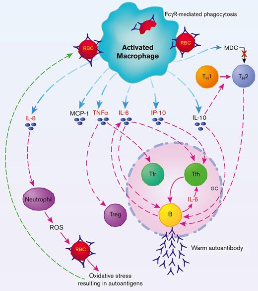
Based on our findings, and the existing knowledge of cytokine/chemokine function and cell-specific production, we hypothesize a mechanistic model of immune dysfunction in wAIHA (Figure 2). When loss of tolerance occurs causing wAIHA,9 uncontrolled activation of the mononuclear phagocyte system ensues as a consequence,10-13 and this results in high production of IL-6, IL-8/CXCL8, IL-10, TNFα, IP-10/CXCL10, MCP-1/CCL2, and MDC/CCL22.
Hypothetical schematic representation of proposed interplay between cytokine biomarkers and immune cells in wAIHA. Activated macrophages in spleen and/or liver produce wAIHA biomarkers IL-6, IL-8/CXCL8, IL-10, MCP-1/CXCL2, IP-10/CXCL10, TNFα, and MDC/CCXL22. IL-6, TNFα, and IL-10 dysregulate the humoral immune response in GC. TNFα downregulates T regulatory (Treg) cells in the periphery and Tfr cells in the GC. IL-10 is essential for generation of GC B-cell response; IL-6, produced by activated macrophages and plasmablast B cells, enhances gene transcription and function of Tfh cells that pushes and maintains autoantibody production. IL-8/CXCL8 may play a pivotal role in inducing autoantigens on the RBC because of ROS-induced oxidative stress. MCP-1 and IP-10 may indicate severity of wAIHA as MCP-1 is produced from macrophages owing to antibody-sensitized RBCs binding to FcRs, and IP-10 is necessary for effector T-cell generation and may influence Tfh effector cells as well. Down regulation of MDC may help to shift the TH2 response to a more inflammatory TH1 response and help drive the wAIHA. Cytokine/chemokines in red are potential biomarkers of wAIHA.
Hypothetical schematic representation of proposed interplay between cytokine biomarkers and immune cells in wAIHA. Activated macrophages in spleen and/or liver produce wAIHA biomarkers IL-6, IL-8/CXCL8, IL-10, MCP-1/CXCL2, IP-10/CXCL10, TNFα, and MDC/CCXL22. IL-6, TNFα, and IL-10 dysregulate the humoral immune response in GC. TNFα downregulates T regulatory (Treg) cells in the periphery and Tfr cells in the GC. IL-10 is essential for generation of GC B-cell response; IL-6, produced by activated macrophages and plasmablast B cells, enhances gene transcription and function of Tfh cells that pushes and maintains autoantibody production. IL-8/CXCL8 may play a pivotal role in inducing autoantigens on the RBC because of ROS-induced oxidative stress. MCP-1 and IP-10 may indicate severity of wAIHA as MCP-1 is produced from macrophages owing to antibody-sensitized RBCs binding to FcRs, and IP-10 is necessary for effector T-cell generation and may influence Tfh effector cells as well. Down regulation of MDC may help to shift the TH2 response to a more inflammatory TH1 response and help drive the wAIHA. Cytokine/chemokines in red are potential biomarkers of wAIHA.
IL-6, previously shown to be elevated in patients with autoimmunity, including wAIHA,3,14 is an early inducer of T-follicular helper (Tfh) differentiation,15,16 which may result in high autoantibody levels.14 Tfh cells promote B-cell activation, expansion, and differentiation in the germinal centers (GC).17,18 In addition to activated macrophages, IL-6 is also produced by plasmablasts, which contributes to an increase in Tfh cells and uncontrolled autoantibody production.16 Therapy using tocilizumab, a humanized monoclonal antibody that targets the IL-6 receptor, has been reported to be successful in rheumatoid arthritis and other autoimmune diseases and has been shown to ameliorate wAIHA in 2 reports.19,20
TNFα is commonly elevated in autoimmune diseases and has been shown to negatively regulate T-regulatory cells (Tregs).8 As TNFα downregulates Tregs, it may also downregulate T-follicular regulatory (Tfr) cells in GCs. Tfr cells are a specialized subset of Tregs, which control GC response by suppressing the activation of B cells and Tfh cells.18 Dysregulation of Tfr, leading to pathogenic Tfh and B-cell activation and development of autoimmune-mediated diseases, likely involves IL-6 in addition to TNFα.8,16 Thus, elucidation of IL-6 and TNFα biomarkers tend to support a Treg-Tfr/Tfh-centered mechanism for wAIHA.
IL-10 has previously been shown to be increased in wAIHA, in conjunction with decreased Treg levels4; thus, increased IL-10 in our study is probably not from Tregs but from activated macrophages and likely functions to favor a TH2 humoral immune response.
Our novel finding of increased IL-8/CXCL8 in patients with wAIHA is perhaps not surprising. There is increasing evidence on the role of reactive oxygen species (ROS) in AIHA pathogenesis.21 Neutrophils are one of the main producers of ROS and are recruited by IL-8/CXCL8. Studies have shown that increased oxidative stress in red blood cells (RBCs) because of neutrophil ROS production is associated with anemia and triggers autoantibody production by providing epitopes for autoantibodies on RBC membranes.22
The increased production of the chemokines IP-10/CXCL10 and MCP-1/CCL2 may simply represent the inflammatory response through leukocyte recruitment in wAIHA, with IP-10/CXCL10 and MCP-1/CCL2 contributing to the maintenance of the activated macrophages. However, IP-10 and MCP-1 have been reported as biomarkers associated with the severity of COVID-19 disease23; perhaps these 2 chemokines could be biomarkers that could be related to the severity of wAIHA. Alternatively, IP-10/CXCL10 has been reported to have a role in effector T-cell generation and trafficking,24 so it may also have a role in effector Tfh cells in the GC. MDC/CCL22 is produced by macrophages and dendritic cells and recruits TH2 cells to sites of inflammation.25 Reduced MDC/CCL22 would inhibit TH2 cells and may allow for increased TH1 cell involvement in wAIHA to further drive the inflammation.
In our study, we could not confirm all the previous work reporting on cytokines in wAIHA. We did not find elevations in IL-173,6 or IFNγ levels6 neither in IL-2 or IL-4 levels.3,6 Use of cultured PBMCs for their analysis may explain our differences.3 IL-21 and IL-23, previously reported as elevated,6 were not included in our test panel. Our results are most consistent with the study that examined serum from patients with wAIHA, where IL-6 and IL-10 levels were increased6; however, in contrast, these investigators found a decrease in TNFα and increases in IL-17 and IFNγ, which we did not find using plasma. Perhaps these differences can be explained by the stability of the cytokines in serum vs plasma. Another study using serum proposed IL-33 to be a potential wAIHA biomarker,5 which we did not examine in our study.
For the first time, we report significantly elevated chemokines IP-10/CXCL10 and IL-8/CXCL8 in patients with wAIHA. These novel chemokines may serve as biomarkers along with IL-6 and TNFα for difficult-to-diagnose cases of wAIHA, such as DAT-negative wAIHA, and provide a rationale for therapeutic targets.
Acknowledgments: The authors are most grateful to the late George Garratty for his enthusiasm and support for this work. The authors also thank David W.H. Dai for additional discussion. This work was funded in part by a Canadian Blood Services Graduate Fellowship Program award (E.M.B.) and an Ontario Graduate Scholarship (E.M.B.). R.S.M.Y. is supported by the Hak-Ming and Deborah Chiu Chair in Paediatric Translational Research. This research was also supported by the Centre for Innovation of Canadian Blood Services, using general resources provided in part by Health Canada, a department of the federal government of Canada (D.R.B.). The views expressed herein do not necessarily represent the view of the Federal Government of Canada.
Contribution: D.R.B. and E.M.B. designed the research; R.M.L. provided patient samples; T.D., B.B., and E.M.B. performed the research; D.R.B., Q.Y., T.D., B.B., and E.M.B. analyzed the data; D.R.B., B.B., and E.M.B. wrote the manuscript; and all authors edited and approved the final version of the manuscript.
Conflict-of-interest disclosure: The authors declare no competing financial interests.
Correspondence: Donald R. Branch, Senior Scientist, Centre for Innovation, Canadian Blood Services, Keenan Research Centre, Room 420, 30 Bond St, Toronto, ON, M5B 1W8, Canada; e-mail: don.branch@utoronto.ca.
References
Author notes
The raw cytokine dataset is available on Figshare: https://doi.org/10.6084/m9.figshare.20771233.v1.
Data are available on request from the corresponding author, Donald R. Branch (don.branch@utoronto.ca).
The full-text version of this article contains a data supplement.
B.B. and E.M.B. contributed equally to this work.


