Key Points
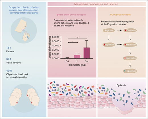
Dysbiosis of the oral microbiome with flourishment of pathobionts are accentuated in patients undergoing HSCT with oral mucositis.
Salivary microbial composition and associated metabolites, specifically the polyamine pathway, are associated with mucositis development.
Abstract
Oral mucositis (OM) is a common debilitating dose-limiting toxicity of cancer treatment, including hematopoietic stem cell transplantation (HSCT). We hypothesized that the oral microbiome is disturbed during allogeneic HSCT, partially accounting for the variability in OM severity. Using 16S ribosomal RNA gene sequence analysis, metabolomic profiling, and computational methods, we characterized the behavior of the salivary microbiome and metabolome of 184 patients pre- and post-HSCT. Transplantation was associated with a decrease in oral α diversity in all patients. In contrast to the gut microbiome, an association with overall survival was not detected. Among 135 patients given methotrexate for graft-versus-host disease prophylaxis pre-HSCT, Kingella and Atopobium abundance correlated with future development of severe OM. Posttransplant, Methylobacterium species were significantly enriched in patients with severe OM. Moreover, the oral microbiome and metabolome of severe OM patients underwent distinct changes post-HSCT, compared with patients with no or mild OM. Changes in specific metabolites were well explained by microbial composition, and the common metabolic pathway was the polyamines pathway, which is essential for epithelial homeostasis. Together, our findings suggest that salivary microbial composition and metabolites are associated with the development of OM, offering new insights on pathophysiology and potential avenues of intervention.
Introduction
Oral mucositis (OM) is a common debilitating toxicity of cancer therapy, including hematopoietic stem cell transplantation (HSCT).1-3 Chemoradiotherapy-induced damage to epithelial stem cells, release of inflammatory cytokines, and activation of the innate immune system drive OM pathophysiology.4 Since commensal bacteria facilitate immune responses,5 the oral microbiome may play a role in OM pathogenesis.6-9 Several groups have reported alterations in the oral microbiota following intensive cancer treatment, but cohorts were small and HSCT recipients were underrepresented.8-12
We aimed to characterize the salivary microbiota among allogeneic HSCT recipients and identify patterns associated with OM. In a relatively large cohort, we demonstrate that HSCT is associated with oral microbiota injury, with greater disruption among patients developing OM. Furthermore, the pretransplantation salivary bacterial composition is associated with susceptibility to OM. Finally, metabolic changes partially explained by microbial derangement accompany OM.
Study design
This was a single-center prospective study of patients who underwent allogeneic HSCT at Sheba Medical Center, Israel, between 2016 and 2018. There was no restriction on transplant indication, donor, or conditioning regimen. Saliva samples were collected weekly from hospital admission and continued after discharge. Saliva samples were also collected from 19 healthy controls. Day 0 was defined as the day of stem cell infusion.
OM was documented prospectively and graded according to the Common Terminology Criteria for Adverse Events version 4.0 criteria.
Microbial analyses and metabolomics profiling of saliva samples were done using 16 ribosomal RNA sequencing and ultrahigh performance liquid chromatography-tandem mass spectroscopy (Metabolon), respectively.
See supplemental Methods for additional details.
Results and discussion
We included 184 adult allogeneic HSCT recipients (supplemental Table 1) and 19 healthy controls. The majority of patients received high-intensity conditioning regimens (myeloablative [41.3%] and reduced toxicity [28.8%]). Methotrexate-based graft-versus-host disease prophylaxis was administered to 135 patients (73.4%). In the complete cohort, 106 (57.6%) and 79 (42.9%) of patients developed grade 2 to 4 and grade 3 to 4 OM (defined as severe), while 78 patients (42.4%) had no (grade 0) or grade 1 OM. OM appeared at a median of 7 (interquartile range, 5-9) days from stem cell infusion and continued for a median of 9 (interquartile range, 6-13) days. The distribution of antibiotic exposure from 10 days before transplantation to 6 days after was similar in patients with and without severe OM (supplemental Figure 1).
A total of 604 saliva specimens were collected, starting from hospital admission up to 34 days posttransplantation. Patients provided a median of 4 samples (range, 1-7). Samples were grouped in bins of 7 days (supplemental Figure 2).
At baseline (days −7 to −1), saliva samples had similar β and α diversity (measured by weighted UniFrac and Shannon index, respectively), compared with healthy controls (Figure 1A-B). However, bacterial composition differed markedly (supplemental Figure 3), possibly reflecting previous treatment exposure. In transplantation recipients, α diversity decreased over time, reaching a minimum on day 14 (Figure 1C). Peled et al and others have observed a similar decrease in the gut microbiota, where low periengraftment α diversity is correlated with survival.13,14 We, however, did not detect such an association in periengraftment saliva samples (Figure 1D). The periengraftment samples, collected within ±10 days of neutrophil engraftment, had a lower α diversity and higher β diversity compared with baseline samples and controls (Figure 1A-B). This is in line with findings reported by Oku et al, decreased bacterial diversity in the tongue microbiome of HSCT recipients.15
Salivary microbiome alterations in HSCT recipients. (A) Principal-coordinates analysis based on weighted UniFrac distance matrix of saliva samples from healthy individuals and HSCT recipients before transplantation and periengraftment. An increase in distance between periengraftment samples and healthy control and between periengraftment samples and pretransplantation samples was observed (false discovery rate [FDR]–corrected P = .0015). (B) Shannon index of healthy controls and pretransplant and periengraftment patients (**FDR-corrected P < .001). (C) Changes in the Shannon index over time for all HSCT patients (n = 184). Day 0 represents the day of the HSCT. (D) Kaplan-Meier estimated overall survival (OS) and progression-free survival (PFS) by pre-engraftment α diversity; high and low diversity are relative to median Shannon index. P value was calculated using the log-rank test.
Salivary microbiome alterations in HSCT recipients. (A) Principal-coordinates analysis based on weighted UniFrac distance matrix of saliva samples from healthy individuals and HSCT recipients before transplantation and periengraftment. An increase in distance between periengraftment samples and healthy control and between periengraftment samples and pretransplantation samples was observed (false discovery rate [FDR]–corrected P = .0015). (B) Shannon index of healthy controls and pretransplant and periengraftment patients (**FDR-corrected P < .001). (C) Changes in the Shannon index over time for all HSCT patients (n = 184). Day 0 represents the day of the HSCT. (D) Kaplan-Meier estimated overall survival (OS) and progression-free survival (PFS) by pre-engraftment α diversity; high and low diversity are relative to median Shannon index. P value was calculated using the log-rank test.
Rates of severe OM were 70% with myeloablative conditioning, followed by 38% with reduced-intensity conditioning and 21% with reduced-toxicity conditioning. Among patients who received methotrexate, 47% developed severe OM vs 31% with mycophenolate mofetil. Since methotrexate has been shown to alter the gastrointestinal microbiota9 and reduce the population heterogeneity, we restricted the subsequent analysis to patients receiving methotrexate.
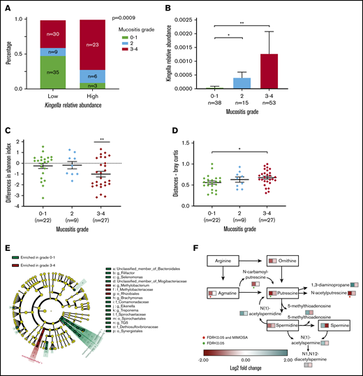
When comparing extreme grades of OM (0-1 vs 3-4), oral α and β diversity was similar at pretransplantation irrespective of future mucositis severity (supplemental Figure 4A-B). However, Kingella and Atopobium were identified as predictive of severe OM using 2 independent methods (XGBoost and linear discriminant analysis effect size analysis; supplemental Figure 4C-D). Indeed, patients with a higher pretransplantation relative abundance of Kingella were more likely to develop severe mucositis (Figure 2A-B). The same pattern was noted, irrespective of conditioning-regimen intensity (supplemental Figure 5). High and low relative abundance of Atopobium corresponded with severe OM rates of 58% vs 42% (P = .12); relative abundance of Atopobium did increase with greater mucositis severity (P < .05) (supplemental Figure 4E-F). The genera Kingella and Atopobium have been linked to infections and inflammation in the oral cavity,16,17 but an association with OM has yet to be described.
Salivary microbiota injury and oral mucositis. Pretransplantation bacteria correlate with future severe OM. (A) Proportion of OM by the relative abundance of pretransplantation Kingella. Abundance groups are relative to the median, which was 0 and shared by >50% of patients. P value was calculated using the χ2 test. (B) Pretransplantation Kingella relative abundance by OM grade (FDR-corrected *P < .05, **P < .001). (C-F) Paired analysis between 2 samples from the same patient at different time points (before HSCT and 7 days after) across OM groups. (C) There was a significant reduction in the Shannon index between the 2 time points in the grade 3 to 4 OM group (**FDR-corrected P < .01). (D) Bray-Curtis matrix distances between the 2 time points. A large distance indicates greater variation between the 2 time points. The variation was higher in patients with severe OM compared with grade 0 to 1 OM (*FDR-corrected P < .05). (E) Linear discriminant analysis effect size analysis for differences in bacterial composition on days 7 to 13 posttransplantation by OM severity groups (linear discriminant analysis scores >2). (F) Polyamine pathway. Colors represent the log2 fold change of each metabolite from the pathway; left square, grade 3 to 4 OM; right square, grade 0 to 1 OM. Metabolites that changed significantly are marked with a green dot, while the ones that were also significant in MIMOSA2 analysis, capturing changes in microbiome-related metabolites, are marked with a red dot.
Salivary microbiota injury and oral mucositis. Pretransplantation bacteria correlate with future severe OM. (A) Proportion of OM by the relative abundance of pretransplantation Kingella. Abundance groups are relative to the median, which was 0 and shared by >50% of patients. P value was calculated using the χ2 test. (B) Pretransplantation Kingella relative abundance by OM grade (FDR-corrected *P < .05, **P < .001). (C-F) Paired analysis between 2 samples from the same patient at different time points (before HSCT and 7 days after) across OM groups. (C) There was a significant reduction in the Shannon index between the 2 time points in the grade 3 to 4 OM group (**FDR-corrected P < .01). (D) Bray-Curtis matrix distances between the 2 time points. A large distance indicates greater variation between the 2 time points. The variation was higher in patients with severe OM compared with grade 0 to 1 OM (*FDR-corrected P < .05). (E) Linear discriminant analysis effect size analysis for differences in bacterial composition on days 7 to 13 posttransplantation by OM severity groups (linear discriminant analysis scores >2). (F) Polyamine pathway. Colors represent the log2 fold change of each metabolite from the pathway; left square, grade 3 to 4 OM; right square, grade 0 to 1 OM. Metabolites that changed significantly are marked with a green dot, while the ones that were also significant in MIMOSA2 analysis, capturing changes in microbiome-related metabolites, are marked with a red dot.
To study changes occurring in the salivary microbiota over time in patients with and without severe OM (grade 0-1), we performed a pairwise comparison of samples collected on days −7 to −1 and days 7 to 13, the latter corresponding with the typical timeframe of active OM. Only patients experiencing severe OM exhibited a reduction in α diversity (P < .01) and an increase in β diversity (P < .05), measured by the Bray-Curtis matrix (Figure 2C-D). In the grade 0 to 1 OM group, 12 genera significantly changed in abundance between the 2 time points vs 33 in the severe OM group (supplemental Figure 6). In both groups, there was a reduction in commensal bacteria, primarily those belonging to the Firmicutes phylum. Notably, genera associated with infections, including Mycoplasma, Methylobacterium, Campylobacter, and Staphylococcus, increased in the grade 3 to 4 OM group.18,19 Next, we examined the differences between the 2 OM groups by analyzing saliva samples collected after transplant (days 7-13). Methylobacterium, a facultative gram-negative bacillus with a tendency to form biofilms, was significantly enriched in patients with severe OM (Figure 2E; supplemental Figure 7), while Treponema and TG5, which are commensal oral bacteria, were related to absent or mild OM (grade 0-1). Collectively, our findings suggest that a dysbiotic state characterizes the development of severe OM. While relative abundance of commensal bacteria decreased in transplant recipients irrespective of OM, there may be a distinct bacterial signature associated with severe OM involving pathobionts.
Microbial metabolites regulate epithelial homeostasis and immune activation and possibly contribute to post-HSCT outcomes.20-22 Therefore, we sought to study the salivary metabolic profile of patients with and without severe OM (ie, grade 0-1 and 3-4, respectively). From each group, 30 samples were analyzed (total 60), of which 15 were collected before transplantation, and 15 between days 7 and 13. Within the severe OM group, 101 metabolites significantly (P < 0.05) changed between the 2 time points, while only 7 compounds changed in the grade 0 to 1 group (supplemental Figure 8A). Metabolites with shifting abundances were involved in a variety of metabolic pathways, including amino acids, xenobiotics, and lipid metabolism.
Variation in 8 metabolites (supplemental Figure 8B) in the grade 3 to 4 group could be explained by changes in the predicted metabolic potential of the microbiome23 compared with none in the grade 0 to 1 group. Four of the 8 metabolites (urea, 5-aminovalerate, N-acetylputrescine, and agmatine) also had a significant fold change between the pretransplant sample and time of mucositis. Notably, there was a reduction in N-acetylputrescine and agmatine, which are both involved in the polyamine pathway (Figure 2F; supplemental Figure 8C). Furthermore, conversion of arginine to agmatine is exogenous to the host and bacterially mediated.24 Polyamines are primarily produced by commensal bacteria in the gut and are necessary for mucosal homeostasis, preservation of mucosal barrier integrity, and postinjury recovery.25-28 Therefore, polyamines may play a role in OM pathophysiology, with potential implications on microbiota-based strategies for prevention and treatment.
In this most extensive study of oral microbiome among allogeneic HSCT recipients, we show that the salivary microbial population shifts during HSCT. Oral microbial alterations have also been demonstrated in other cancer therapies.8-12 Whether bacteria merely colonize the injured mucosa or facilitate OM requires further investigation in animal models. Such models will also allow accounting for the impact of antibiotics, which are administered in the vast majority of patients and may confound results in humans. Given the identification of proinflammatory bacteria, as well as bacterial metabolites involved in epithelial integrity, it is likely that microbiota plays an active role in mucositis pathophysiology.4,8 Our findings may open new avenues for the treatment of OM, which is among the most common complications of cancer treatment.
The 16S ribosomal RNA gene sequence data reported in this article have been deposited in the European Bioinformatics Institute database (accession number ERP121435).
Acknowledgments
E.B. is a Faculty Fellow of the Edmond J. Safra Center for Bioinformatics at Tel Aviv University.
This work was supported by the Dahlia Greidinger Anti Cancer Fund and an institutional grant from the Chaim Sheba Medical Center.
Authorship
Contribution: R. Shouval, A.A.K., A.N., and O.K. planned the study; R. Shouval, A.E., B.D., E.M., and C.N. analyzed the data; R. Shouval and J.A.F. designed and programmed the database; R. Shouval, I.D., S.F., M.G., H.N., A.A.-O., and R. Shahien enrolled patients and healthy controls to the study; R. Shouval, S.F., and E.K. coded data into the database; R. Shouval and A.E. wrote the manuscript; R. Shouval, E.B., Y.L., A.N., and O.K. supervised the analysis and study; and all authors edited and reviewed the manuscript.
Conflict-of-interest disclosure: The authors declare no competing financial interests.
The current affiliation for H.N. is Zefat Academic College, Zefat, Israel.
Correspondence: Omry Koren, Azrieli Faculty of Medicine, Bar Ilan University, Henrietta Szold 8, Safed, Israel; e-mail: omry.koren@biu.ac.il.
References
Author notes
R. Shouval and A.E. contributed equally to this study.
A.N. and O.K. contributed equally to this study.
The full-text version of this article contains a data supplement.


![Salivary microbiome alterations in HSCT recipients. (A) Principal-coordinates analysis based on weighted UniFrac distance matrix of saliva samples from healthy individuals and HSCT recipients before transplantation and periengraftment. An increase in distance between periengraftment samples and healthy control and between periengraftment samples and pretransplantation samples was observed (false discovery rate [FDR]–corrected P = .0015). (B) Shannon index of healthy controls and pretransplant and periengraftment patients (**FDR-corrected P < .001). (C) Changes in the Shannon index over time for all HSCT patients (n = 184). Day 0 represents the day of the HSCT. (D) Kaplan-Meier estimated overall survival (OS) and progression-free survival (PFS) by pre-engraftment α diversity; high and low diversity are relative to median Shannon index. P value was calculated using the log-rank test.](https://ash.silverchair-cdn.com/ash/content_public/journal/bloodadvances/4/13/10.1182_bloodadvances.2020001827/3/m_advancesadv2020001827f1.png?Expires=1767746547&Signature=v-PNDtHOUEYx2ltiHFm70EP2QtDHBAIat9kUeQHF5BwYD6tHTVEQClHeqj~9QDmr35hXgUE71x3h8Z4k4JcsCpFVUfDndarPyYZg4lNk6NYH6XAEz4l2Kv4Z9HW3A8RZ3wSFAg4C8V3mwHa4LUocbvieI3nspgmu8Yd3TQ8f~7b~cTay7cimxnGfN8JDlTzNjsEn6nL4mZNMBerZxCrGZST8vLaMg93uEuAIOnQpSIyTG~e86LBFGsu~9lJ2faW8hWryU~EIAKEqh6Q6~hRDiOuN-GW20pCu~kavOoaez3Kk1fIEO1Cim5Q6U3~hVuCq09J-TKzDCkzv~V9zX~Q5bw__&Key-Pair-Id=APKAIE5G5CRDK6RD3PGA)
