In the 9 January 2024 issue of Blood Advances, Kockwelp et al1 presented an artificial intelligence (AI) system for predicting key genetic and cytogenetic lesions in newly diagnosed acute myeloid leukemia (AML) by image analysis of bone marrow aspirate smears. Using deep learning algorithms, they first trained a model to identify individual nucleated cells present within a high-resolution scan of the slide. They then developed a second model to classify cells possessing any of a set of clinically actionable genetic lesions based on morphologic cues. Their work is important proof-of-principle in a technically challenging field.
The approach the authors describe has several components. First, automatically segmenting slides to identify individual nucleated cells allowed the authors to rapidly generate an extensive data set of over 2 million cells with clinical and molecular annotation at the patient level. A second model was trained to classify individual cells as associated with any of the selected abnormalities, including favorable risk cytogenetics per the European Leukemia Network (ELN) criteria, presence of the CBFB:MYH11 fusion gene, alterations of fms-related receptor tyrosine kinase 3 (FLT3), mutations in nucleophosmin 1 (NPM1), and myelodysplasia-related chromosomal changes. AI is associated with the “black box” problem, in which the reason an algorithm arrives at a conclusion is impossible for human operators to interpret. Here, the authors used 2 complementary techniques to address this. One approach identified the individual cell images most important to predict each individual molecular abnormality, where a second highlighted the specific regions of a cell image that were driving a given prediction. Most importantly, the authors took 2 critical steps sometimes absent in AI publications: they validated their algorithm on an independent data set (in this case, slides from a different era), and they shared the code for the model.
AML in the contemporary era has become a genetically defined disease. This paradigm has been codified in both the new International Conensus Committee2 and World Health Organization3 classification criteria. For example, the presence of an NPM1 mutation can constitute the disease irrespective of blast count, while mutations in myelodysplasia-associated genes or TP53 specify disease subtype. Moreover, the treatment of AML is rapidly evolving from one-size-fits-all-who-can-tolerate-7+3 to a growing array of mutation-specific therapeutic options. The use of FLT3 inhibitors is now a standard-of-care frontline therapy in patients with the FLT3 internal tandem duplication (ITD), and the resulting improved survival led to the reclassification of FLT3-ITD from adverse to normal risk in the most recent ELN risk schema.4 Molecularly targeted therapies for mutations in IDH1 and IDH2 are already in common use; blockade of interaction with the menin protein shows promising results in current trials for patients with NPM1 mutations. Retrospective data have suggested a specific benefit to venetoclax-based treatments in patients with certain common adverse mutations such as SRSF2 or RUNX1.5,6
Given the fundamental and growing importance of identifying genetic drivers at the time of diagnosis, the limitations of current next-generation sequencing (NGS) techniques (eg, turnaround time and cost) have become apparent. The presence of certain diagnostic or actionable mutations, such as FLT3-ITD or mutations in NPM1 and IDH1/IDH2, can be determined rapidly by polymerase chain reaction (PCR) at some centers; however, a comprehensive NGS characterization of the leukemia generally requires weeks to result, potentially delaying the most effective therapy. As more mutation-specific therapies emerge, the usefulness of early genetic characterization will only increase. Meanwhile, the costs associated with obtaining NGS are considerable, let alone repeating NGS testing at multiple time points over the course of therapy to track evolution and emergence of new targetable mutations. Cost is all the more relevant in low- and middle-income settings where access to molecular diagnostics may be limited. The use of readily available information sources like bone marrow slides, combined with the unique abilities of well-designed AI platforms, holds the potential to enable disease classification while limiting the need for more costly and technically challenging molecular diagnostic testing. Intriguingly, a lymphoma subtype classifier developed by Valvert et al7 has already pointed to the immense potential of AI-driven diagnostics in resource-limited settings.
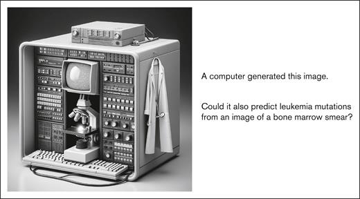
A famous aphorism states that one can choose 2 of these 3—cheaper, faster, or better—but not all at once. AI imaging clearly promises the first 2, but realizing its promise depends on a degree of accuracy still far from reached. Kockwelp et al here demonstrate an impressive ability to predict certain lesions, such as mutations of FLT3 or NPM1, though these are already addressed by rapid PCR assays. To answer whether subtle morphologic cues can be detected by AI to predict other common mutations in myelodysplasia-related genes or RUNX1 or TP53, well annotated training data several orders of magnitude larger will be required. That this feat should be realizable may be at least suggested by intriguing work in other histologies showing that driver mutations, copy number alterations, and bulk gene expression can be predicted from slide sections.8 Such a technology, further refined to predict expression and mutation status at the single-cell level, might truly revolutionize the management of leukemia by allowing therapy to adapt to clonal evolution in real time. Although there remains much work to be done, Kockwelp et al gave us a glimpse into the future of AI-based imaging analytics in AML diagnostics (see figure).
AI-based diagnostics, as “imagined” by AI. Figure generated with DALL-E (OpenAI) and used as per OpenAI terms of service.
AI-based diagnostics, as “imagined” by AI. Figure generated with DALL-E (OpenAI) and used as per OpenAI terms of service.
Conflict-of-interest disclosure: The authors declare no competing financial interests.


