TO THE EDITOR:
Fanconi anemia (FA) is a heritable condition caused by a mutation in 1 of at least 23 genes involved in DNA repair. Progressive loss of hematopoietic stem cells (HSCs) precipitated by environmental and physiologic stressors leads to near-universal bone marrow failure (BMF) by young adulthood.1,2 Allogeneic hematopoietic stem cell transplantation (HSCT) remains the only cure to date. However, patients with FA are predisposed to a range of malignancies, a risk further elevated by most conditioning regimens.3
In utero hematopoietic stem cell transplantation (IUHCT) is a favorable alternative to bone marrow transplantation that takes advantage of the immature fetal immune system and achieves long-term engraftment across human leukocyte antigen (HLA) barriers without myeloablative or immunosuppressive conditioning.4,5 Factors that limit the clinical success of IUHCT include competition of donor HSCs for the fetal hematopoietic niche and the proliferative disadvantage of donor cells compared to endogenous fetal HSCs.6-9 Thus, FA may be particularly favorable for the clinical application of IUHCT due to the presence of prenatal qualitative and quantitative HSC defects along with a previously demonstrated survival advantage of allogeneic or genetically-corrected HSCs over FA-phenotype cells.10-13
Here, we report the competitive engraftment of congenic healthy donor cells and sustained peripheral blood and bone marrow (BM) donor chimerism after IUHCT in a mouse model of FA.
C57BL/6Fancc+/−(H2KbCD45.2+GFP−) mice were provided by A. D’Andrea at the Dana-Farber Cancer Institute. C57BL/6TgN(act-EGFP)OsbY01(CD45.2)(H2KbCD45.2+GFP−,GFP+) mice were provided by M. Okabe at Osaka University. B6.SJL-PtprcaPepcb/BoyJPepBoy(H2KbCD45.1+GFP−, PepBoy) mice were obtained from Jackson Laboratories. Procedures were approved by the Children’s Hospital of Philadelphia Institutional Animal Care and Use Committee.
BM was harvested as described14 with T-cell depletion using CD3ε Microbeads (Miltenyi Biotec Cat#130-094-973) per manufacturer’s instructions. CD3− purity was confirmed by flow (FACS Aria; BD Biosciences). Fetal offspring of heterozygous (Fancc+/− × Fancc+/−) matings were injected with 107 cells on gestational day 14 (E14) via the vitelline vein.14 Donor chimerism (%GFP+/CD45+) in homozygous knockout (Fancc−/−) and wild-type (WT, Fancc+/+) littermates was assessed by FACS at 1, 3, and 6 months of age. Peripheral blood multilineage and BM donor engraftment were analyzed at 6 months.
Secondary transplantation was performed at 6 months, and tertiary transplantation was performed 4 months thereafter. Adult PepBoy recipients were lethally irradiated (10.4Gy in 2 doses) the day before transplantation. A total of 2.5 × 10⁶ whole bone marrow cells, with 0.5 × 10⁶CD45.1 supporting cells, were injected per recipient.
After primary transplantation, some mice received biweekly intraperitoneal injections of 5mg/kg poly(I:C) (Tocris, Bristol, United Kingdom) for 6 months. Mice were subsequently assessed by complete blood count and donor cell chimerism by FACS, with age-matched Fancc+/+ and Fancc−/− controls.
For colony-forming unit (CFU) analysis, 8 × 10⁴ BM cells were plated in triplicate using Methocult M3434 (Stem Cell Technologies, Vancouver, Canada). At 8 days postseeding, CFUs were imaged by EvosFLAuto (Fisher Scientific, Hampton, NH).
For bone histology, femurs were fixed in 10% Formalin (Sigma-Aldrich, St Louis, MO) and stained with green fluorescent protein (GFP)-antibody (Cat# A-11122, Thermofisher, Waltham, MA). Images were captured using the Thunder Imaging System (Leica, Wetzler, Germany).
Data are represented as mean ± standard error of the mean. Differences between groups were determined using unpaired t tests (Welch correction), alpha = 0.05 for statistical significance, using Prism (GraphPad Software Inc, La Jolla, CA). Figures were made using BioRender (Ontario, Canada). Antibodies and gating strategies used for flow cytometry are noted in the supplemental Material.
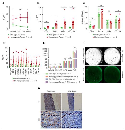
Based on the demonstrated survival advantage of WT cells over Fancc−/− cells in prior studies, we reasoned that IUHCT of WT cells should lead to higher donor chimerism in the Fancc−/− fetus compared with WT littermates.10 We indeed found that Fancc−/− mice injected at E14 had significantly higher donor GFP+ engraftment than WT controls at 1 (6.6% vs 2.9%; P = .01), 3 (7.8% vs 1.7%; P < .001), and 6 months postinjection (7.7% vs 1.3%; P = .007) (Figure 1A). Gains in engraftment were maintained across peripheral blood CD3+ T cells, B220+ B cells, GR1+ granulocytes, and CD11B+ monocytes (Figure 1B). GFP+ cells comprised all lineages in both Fancc−/− and WT mice, excluding donor lineage differentiation bias (Figure 1C). Similar to peripheral blood, BM analysis at 6 months showed high GFP+ engraftment across hematopoietic stem/progenitor subpopulations in the Fancc−/− animals (P = nonsignificant) (Figure 1D). No statistically significant differences in complete blood count metrics were observed among injected and noninjected Fancc−/− and WT animals, as expected (Figure 1E).15
IUHCT results in sustained engraftment in Fancc–/– mice. (A) Flow cytometry of peripheral blood from E14-injected Fancc–/– and WT fetuses. (B) Engraftment observed across cell types. (C) Lineage composition of GFP+ engrafted cells. (D) BM GFP+ cell engraftment. (E) Complete blood counts of injected and noninjected Fancc–/– and WT animals. (F) Representative BM CFU-Cs. (G) Representative femur GFP+ staining from 6 months post-IUHCT. ∗∗∗P < .001; ∗∗P < .002; ∗P < .033; ns, nonsignificant.
IUHCT results in sustained engraftment in Fancc–/– mice. (A) Flow cytometry of peripheral blood from E14-injected Fancc–/– and WT fetuses. (B) Engraftment observed across cell types. (C) Lineage composition of GFP+ engrafted cells. (D) BM GFP+ cell engraftment. (E) Complete blood counts of injected and noninjected Fancc–/– and WT animals. (F) Representative BM CFU-Cs. (G) Representative femur GFP+ staining from 6 months post-IUHCT. ∗∗∗P < .001; ∗∗P < .002; ∗P < .033; ns, nonsignificant.
Assessment of progenitor clonogenicity of GFP+ donor cells via CFU assay revealed higher numbers of GFP+ colonies in the E14-injected Fancc−/− mice compared to WT mice (Figure 1F). Similarly, representative immunohistochemistry staining of GFP showed a larger proportion of GFP+ cells in the BM of E14-injected Fancc−/− mice (Figure 1G). Altogether, these data indicate improved engraftment and early HSCT survival after IUHCT in Fancc−/− embryos.
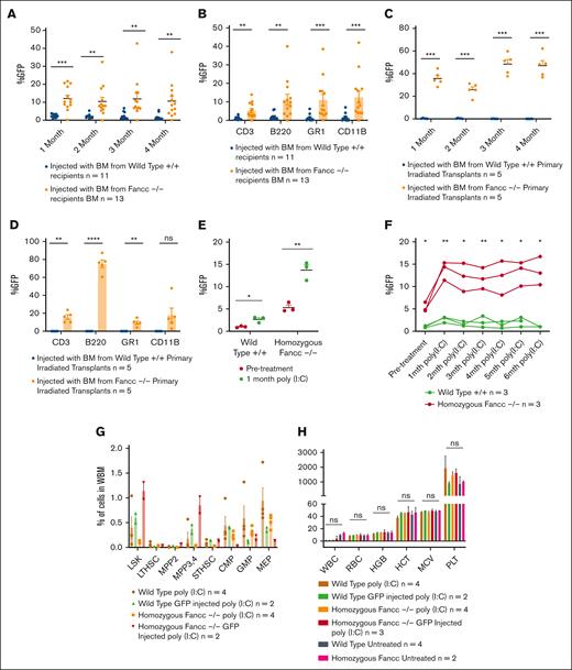
To assess the longevity of engraftment, following primary transplantation, we performed secondary and tertiary transplantation of whole BM from IUHCT-treated mice; as expected, recipient mice transplanted with IUHCT Fancc−/− donor BM had higher peripheral blood GFP+ chimerism than those engrafted with IUHCT WT donor BM at all time points examined (Figure 2A,C). Multilineage analysis further demonstrated that this engraftment advantage of healthy donor-derived GFP+ cells was maintained across cell types (Figure 2B,D). The persistence of GFP+ donor-derived cells through secondary and tertiary transplantation provides evidence for the fetal engraftment of HSCs that contribute to sustained levels of chimerism in the recipients’ BM.
Secondary and tertiary transplantation shows maintenance of engraftment and poly(I:C) treatment leads to selection of GFP+ donor cells. (A) Peripheral blood GFP+ chimerism at indicated time points following secondary transplant with (B) lineage analysis of the peripheral blood postsecondary transplant. (C) Peripheral blood GFP+ chimerism at indicated time points following tertiary transplant with (D) lineage analysis of the peripheral blood posttertiary transplant. (E) Enhanced IUHCT peripheral blood donor chimerism 1 month after poly(I:C) treatment. (F) Peripheral blood donor chimerism following IUHCT at the indicated time points following poly(I:C) treatment. (G) BM analysis of poly(I:C) treated mice at 6 months posttreatment. (H) Complete blood counts 6 months post poly(I:C) treatment. WBM, whole bone marrow.
Secondary and tertiary transplantation shows maintenance of engraftment and poly(I:C) treatment leads to selection of GFP+ donor cells. (A) Peripheral blood GFP+ chimerism at indicated time points following secondary transplant with (B) lineage analysis of the peripheral blood postsecondary transplant. (C) Peripheral blood GFP+ chimerism at indicated time points following tertiary transplant with (D) lineage analysis of the peripheral blood posttertiary transplant. (E) Enhanced IUHCT peripheral blood donor chimerism 1 month after poly(I:C) treatment. (F) Peripheral blood donor chimerism following IUHCT at the indicated time points following poly(I:C) treatment. (G) BM analysis of poly(I:C) treated mice at 6 months posttreatment. (H) Complete blood counts 6 months post poly(I:C) treatment. WBM, whole bone marrow.
Poly(I:C) administration forces the emergence of HSCs from quiescence, which is thought to mimic the physiologic stress that induces BM failure in patients with FA.16 Six months after IUHCT, we treated mice with poly(I:C), which led to a rise in donor chimerism among Fancc−/− recipients from 5.3 ± 0.6% to 13.7 ± 1.2% (P = .04). Chimerism among WT recipients rose from 1.0 ± 0.1% to 2.7 ± 0.4% (P = .008) (Figure 2E) and remained relatively stable thereafter (Figure 2F). Subsequent BM analysis at euthanasia showed progenitor cell depletion in the Fancc−/− mice after poly(I:C) with recovery of some populations in the IUHCT Fancc−/− populations (Figure 2G). Poly(I:C) treatment resulted in no significant differences in hemoglobin at 6 months between the control and IUHCT mice (Figure 2H). Although poly(I:C) is not proposed as a therapy to boost engraftment clinically, the gains in chimerism after poly(I:C) treatment suggests that normal physiologic stressors in patients with FA who undergo IUHCT may be potentially beneficial to increasing engraftment in the long term.
Overall, these data provide formal evidence that healthy donor HSCs engraft and preferentially expand over recipient Fancc−/− HSCs in the fetal liver niche. Our findings suggest that IUHCT may be a therapeutic option in FA siblings, after preimplantation or early gestational FA diagnosis. Notably, the increase in FA pathway proficiency, that is WT chimerism, following poly(I:C) treatment suggests that even low initial levels of donor engraftment may be therapeutic after birth in the setting of physiologic stresses. This is validated by studies showing patients with FA with mosaic phenotypes are protected from BMF and the deleterious aspects of the disease, likely owing to the presence of some WT cells.17 In addition, this conditioning-free IUHCT treatment approach would provide a platform of immunologic tolerance for additional postnatal cell therapies in the form of cellular boosting should engraftment levels fail to meet a therapeutic threshold.18-20
Importantly, a known limitation of this work is the discrepancy in phenotypic manifestation between human patients with FA and most murine models of FA. Unlike human patients, Fancc−/− mice do not show the spontaneous hematopoietic failure seen in FA.15 Nevertheless, the experiments presented here provide strong proof of concept for the efficacy of IUHCT in FA and the selective advantage of WT cells in the fetal environment. Our studies do not address issues of donor-host HLA compatibility applicable to the clinical application of IUHCT.
Acknowledgments: Aaron Weilerstein provided assistance with the management of animals. The University of Pennsylvania Pathology Core provided assistance with bone histology. The Children’s Hospital of Pennsylvania animal facility housed and assisted with the care of animals. Figures were created using GraphPad.
This work was supported by US National Institutes of Health Director's New Innovator award DP2HL152427 (W.H.P.).
Contribution: A.D., S.L., J.S.R., S.R., V.L., C.B., P.M., S.J., and H.L. performed experiments and acquired and analyzed the data; P.K. and W.H.P. provided expert technical guidance and oversaw the project; and P.K., W.H.P., and A.D. wrote the manuscript.
Conflict-of-interest disclosure: The authors declare no competing financial interests.
Correspondence: William H. Peranteau, Perelman School of Medicine, University of Pennsylvania, Children’s Hospital of Philadelphia, 3615 Civic Center Blvd, ARC 1116E, Philadelphia, PA 19104; email: peranteauw@email.chop.edu; and Peter Kurre, Perelman School of Medicine, University of Pennsylvania, Children’s Hospital of Philadelphia, 3615 Civic Center Blvd, ARC 1116E, Philadelphia, PA 19104; email: kurrep@chop.edu.
References
Author notes
Data are available upon reasonable request from the corresponding authors, William H. Peranteau (peranteauw@email.chop.edu) and Peter Kurre (kurrep@chop.edu).
The full-text version of this article contains a data supplement.


