Abstract
CD40 engagement is the major signal that induces B cells to efficiently present antigen to T cells. We previously demonstrated that human peripheral blood–derived CD40-activated B cells (CD40–B cells) function as antigen-presenting cells (APCs). Here, we have established a culture system to generate these APCs under clinically applicable conditions using guanylic acid–grade soluble trimeric CD40 ligand. To monitor APC function and antigen loading for these cells, simple and efficient quality control assays have been developed. Using this approach, we demonstrate that CD40–B cells from healthy donors and cancer patients are fully functional and equally expanded in long-term cultures. These B cells boost robust memory T-cell responses, but more importantly, they also prime naive T-cell responses against neoantigens ex vivo. CD40–B cells overcome current obstacles, such as the difficulty of isolation, generation, and long-term expansion observed with other APCs. Therefore, they are an excellent source of professional APCs for immune assessment, antigen discovery, and antigen-specific immunotherapy.
Introduction
Highly efficient antigen presentation by professional antigen-presenting cells (APCs) is a requisite for the development of T-cell–mediated immunity in vitro and in vivo.1,2 Increasing evidence supports the hypothesis that not all APCs have the same capacity to induce and promote an immune response. For example, only dendritic cells (DCs) have been shown to prime naive T cells and to induce memory T cells to become effectors. For these reasons, many investigators attempting to induce immunity to viral3,4 or tumor antigens5-7 have focused their attention on DCs. In these fields, DCs have been used as cellular adjuvants to present antigen in vivo,8,9 as APCs in vitro to determine immunocompetence, and, finally, in experiments attempting to define immunogenic peptides from viral,10autoimmune,11 and tumor antigens.12-14
Although highly efficient in their capacity to induce T-cell immunity, DCs have several significant drawbacks. First, they are relatively rare in peripheral blood and are therefore usually isolated from apheresis8 or marrow sources.15 Second, DCs are not homogeneous but represent several populations of functionally disparate cell types,2 including tolerogenic DCs.16 Third, it is difficult to expand DCs ex vivo from non–stem cell sources without significant in vivo expansion with cytokines.17 Finally, all of the above approaches are laborious, expensive, and therefore presently restricted in their clinical applicability.
We postulated that it would be advantageous to extend the sources of APCs for antigen discovery, immune assessment, vaccination, and ex vivo T-cell expansion. Ideally, biological characteristics would include T-cell priming and highly efficient presentation of antigen. Generation and expansion should be simple and possible from small amounts of non–stem cell sources without the need to treat donors or patients with biologics or chemotherapeutics prior to cell collection.18,19 The APCs should be easily quality controlled, and their production should be based on the use of recombinant growth factors without the need for xenogeneic cells20 or gene-transfer technology.21 We have recently used CD40-activated B cells (CD40–B cells) as APCs for the ex vivo generation of tyrosinase-specific T cells.22Interactions between CD40 ligand (CD40L) on T cells and CD40 on APCs have been shown to be crucial for the induction of cytotoxic T-lymphocyte (CTL) immunity.23-25 Our earlier experiments using CD40–B cells to stimulate CTLs did not address whether CD40–B cells can prime T-cell responses since one cannot exclude the possibility that a pre-exposed and expanded tyrosinase-specific T-cell pool exists even in healthy individuals. Furthermore, the system was not clinically applicable owing to allogeneic and xenogeneic components, such as pooled human AB serum and human CD40L–transfected murine fibroblasts (tCD40L). Here, we demonstrate that CD40–B cells boost CTL responses to recall antigens, break ignorance to tumor antigens, and prime T-cell responses to viral neoantigens. We also describe the generation and expansion of CD40–B cells from healthy individuals and cancer patients under clinically applicable conditions. Furthermore, we developed protocols to quality-control antigen-loading and APC function that help define rigorous clinical standards for CD40–B cells as APCs.
Materials and methods
Donors and cell lines
All specimens were obtained following approval by our institutional review board (Dana-Farber Cancer Institute, Boston, MA). Informed consent for blood donations was obtained from all volunteers. Peripheral blood mononuclear cells (PBMCs) from healthy donors and cancer patients were obtained by phlebotomy or leukapheresis followed by Ficoll-density centrifugation. Four female and 5 male cancer patients between 29 and 71 years of age were included in this study. The diagnoses included multiple myeloma (n = 3), follicular lymphoma (n = 1), melanoma (n = 1), prostate cancer (n = 2), breast cancer (n = 1), and ovarian cancer (n = 1).
The following tumor cell lines were used for cytotoxicity analysis: U266, HS-Sultan, Malme-3M,26 SK-MEL-2 (ATCC, Manassas, VA), and U2OS.27 COS cells stably expressing A*0201 were transiently transfected with pcDNA1.1amp (Invitrogen, Carlsbad, CA) containing enhanced green fluorescent protein (Clontech, Palo Alto, CA) and a human immunodeficiency virus (HIV) reverse transcriptase (RT)–pol fusion minigene that included the RTpol476 epitope by means of Lipofectamine Plus (Life Technologies, Rockville, MD).
The CD40L system
B cells from whole PBMCs were stimulated via CD40 by means of human good medical practice (GMP)–quality trimeric soluble CD40L (sCD40L) (kindly provided by Immunex, Seattle, WA)28 or NIH3T3 cells transfected with tCD40L.29 CD40L expression on NIH3T3 cells has been stable for more than 5 years with more than 95% of tCD40L cells positive for CD40L. No other human molecules are expressed on tCD40L cells. For B-cell cultures, tCD40L cells were lethally irradiated (96 Gy) and plated on 6-well plates (Costar, Cambridge, MA) at a concentration of 0.4 × 10⁵ cells per well in medium containing 45% Dulbecco modified Eagle medium (Life Technologies), 45% F12 (Life Technologies), 10% fetal calf serum, 2 mM glutamine (Life Technologies), and 15 μg/mL gentamicin (Life Technologies). After 12 to 18 hours of culture, tCD40L cells were adherent and could be used for coculture. Before the addition of PBMCs, tCD40L cells were rinsed with phosphate-buffered saline (PBS). CD40–B cells were generated from PBMCs by coculturing whole PBMCs at 2 × 10⁶ cells per milliliter with tCD40L or 2 μg/mL sCD40L. GMP-grade recombinant human interleukin (IL)–4 (rhIL-4) (2 ng/mL) (R&D Systems, Minneapolis, MN) and clinical-grade cyclosporin A (CsA) (5.5 × 10⁻⁷ M) (Novartis, Basel, Switzerland) were 2 additional, crucial factors necessary to induce B-cell expansion. Iscoves modified Dulbecco medium (IMDM) (Life Technologies) was supplemented with 10% human AB serum or autologous plasma, XXX pM (5 μg/mL) insulin (Sigma Chemical, St Louis, MO), and 15 μg/mL gentamicin. Cultured cells were transferred to new plates and stimulated with either sCD40L (2 μg/mL) or fresh irradiated tCD40L cells in the presence of CsA and IL-4 every 3 to 5 days. Once the cultured PBMCs were greater than 75% CD19+ (Beckman Coulter, Fullerton, CA), they were cultured at concentrations of 0.75 to 1.5 × 10⁶ cells per milliliter. A Coulter Counter Z2 (Beckman Coulter) was used to measure the total number of viable cells, and the number of CD19+ B cells was analyzed by flow cytometry on days 0 and 6 and twice weekly thereafter. By day 14, cultures were negative for CD14 (Dako, Carpinteria, CA), CD11c (Pharmingen, San Diego CA), BDCA-2, BDCA-3, and BDCA-4 (Miltenyi Biotec, Auburn, CA) and consisted of greater than 95% CD19+ B cells that were characterized by small veils.
Dendritic cells
DCs were generated as previously described.22Briefly, monocyte-enriched fractions were obtained from PBMCs by rosetting over sheep red blood cells and subsequently cultured for 6 to 8 days with 50 ng/mL granulocyte-macrophage colony-stimulating factor (GM-CSF) (Genzyme, Cambridge, MA) and 10 ng/mL IL-4 in IMDM supplemented with 2% human AB serum, 2 mM glutamine, and 15 μg/mL gentamicin. GM-CSF and IL-4 were replenished on day 4 of culture. Starting on day 6, DCs were matured for 48 hours with the use of 30 ng/mL tumor necrosis factor (TNF)–α (Genzyme) or 2 μg/mL sCD40L with or without 20 ng/mL interferon [IFN]–γ Genzyme).30 31
Absolute number and density of cell surface molecules
Dual-color fluorescence-activated cell sorter analysis was performed with monoclonal antibodies to CD19 (Beckman Coulter), major histocompatibility complex (MHC) class I and class II, CD58, and CD80 (Pharmingen).22 Quantitative determination of cell surface molecules was performed according to the manufacturer's recommendations by means of DAKO Qifikit (Dako). The antigen quantity is expressed in antigenic binding sites (ABSs). To determine the density of ABSs, we assumed that cells in suspension had a round shape. Using a Coulter Counter Z2, we determined the mean cellular volume of B-cell and DC populations and calculated the cell surface area as A = 4.83 × V2/3, where A stands for area and V for volume. This allows us to calculate the density as DABS = ABS/A, where D stands for density.
Peptides
Peptides were obtained from Sigma Genosys (The Woodlands, Texas). For the MHC class I experiments, an HLA-A*0201–binding peptide (Phe-Leu-Pro-Ser-Asp-Phe-Phe-Pro-Ser-Val) (FLPSDFFPSV) derived from the hepatitis-B core protein was fluorescein isothiocyanate (FITC) conjugated at the internal cysteine (or C) residue after a single amino acid exchange at position 6 (FLPSDC(FITC)FPSV). The following peptides were used in binding studies and for the generation of peptide-specific CTLs: the native hepatitis-B core peptide F18 (FLPSDFFPSV)32; the pan-DR epitope peptide (PADRE) (Ala-Lys-Phe-Val-Ala-Ala-Trp-Thr-Leu-Lys-Ala-Ala-Ala) (AKFVAAWTLKAAA)33; the I540 peptide of human telomerase reverse transcriptase (hTERT) (ILAKFLHWL, in which I is Ile and H is His)27; the RTpol476 peptide of HIV (ILKEPVHGV, in which E is Glu and G is Gly)34; the matrix protein 58 (MP58) peptide of influenza A (GILGFVFTL)35; E26-WT peptide (EAAGIGILTV) and the corresponding heteroclytic peptide E26-HT (ELAGIGILTV) of melanoma antigen recognized by T cells (MART-1).36
Analysis of peptide pulsing
To determine optimal pulsing conditions for exogenous peptide loading, APCs were harvested from culture, washed 3 times, and resuspended in serum-free IMDM at 1.25 × 10⁶ cells per milliliter and seeded into 96-well plates (200 μL per well). Peptide concentration, incubation time and temperature, pH of culture medium, serum concentration, and concentration of β2microglobulin (β2-m) were varied. After incubation with the FITC-conjugated reference peptide under different conditions, cells were harvested, washed, and resuspended in PBS containing 0.1% formaldehyde. Fluorescence analysis was performed immediately on a Coulter EPICS XL flow cytometer. For competition assays, cells were preincubated for 30 minutes at 4°C in a 5% CO2atmosphere with increasing concentrations of the competitor peptide (0.3 to 20 μg/mL) and 127.1/mM (1.5 μg/mL) β2-m. The reference peptide was added at a concentration of 0.2 μg/mL for 150 minutes.
Ex vivoexpansion of CTLs using CD40-activated B cells
Long-term CD40–B-cell cultures (up to 50 days) were used as the source of APCs for repetitive stimulation of autologous T cells beginning at day 14 of B-cell culture. Long term–cultured CD40–B cells maintained their phenotype and efficiently presented antigen to T cells. HLA-A*0201+ CD40–B cells (or CD40L-matured DCs for control experiments) were incubated with peptide (up to 10 μg/mL), irradiated (32 Gy), and added to purified autologous CD8+ T cells (greater than 85%) at a T-cell–to-APC ratio of 4:1 in RPMI containing 10% human AB serum, glutamine, gentamicin, and IL-7 (10 ng/mL) (Endogen, Woburn, MA). On day 7, T-cell cultures were harvested, washed, and restimulated with fresh peptide-pulsed CD40–B cells and IL-7. This was repeated on days 14, 21, and 28. IL-2 (50 IU/mL) (Chiron, Emeryville, CA) was introduced into the cultures at day 8 and every 3 to 4 days thereafter.
Cellular cytotoxicity assay
Expanded autologous CD8+ T-cell lines were analyzed in a standard 4-hour 51Cr-release assay.27Targets were labeled with 51Cr, and 5 × 10³labeled cells per well were plated with various concentrations of effector cells. The percentage of cytotoxicity was calculated as follows: [(cpm{experiment} − spontaneous release)/(maximum cpm − spontaneous release)] × 100%.
Tetramer analysis
Recombinant tetrameric HLA-A2/peptide complexes with MP58, I540, RTpol476, MART-1HT, or human T-cell lymphoma virus (HTLV)–1 Tax 11 were synthesized essentially as described37 and conjugated to streptavidin ALEXA-488 or streptavidin-phycoerythrin (PE) (Molecular Probes, Eugene, OR). For staining, CTL lines were incubated with the tetramer for 15 minutes and with CD8-PE or CD8-PC5 (Coulter) for 20 minutes at room temperature. Dead cells were excluded by costaining with annexin V–FITC (R&D Systems).
Results
CTL responses against viral and tumor antigens with CD40–B cells used as APCs
CD40–B cells from healthy donors and cancer patients were used as the sole APCs to induce CTL responses to HLA-A*0201–restricted epitopes derived from the recall antigen influenza A MP58; the tumor antigens MART-1 (E26) and hTERT (I540); and the neoantigen RTpol (RTpol476) from HIV. By means of peptide-pulsed T2 cells, peptide specificity was demonstrated for all 4 antigens (Figure1A). As expected for memory T-cell responses, CTLs specific for the influenza A–derived peptide MP58 were already highly cytotoxic after one stimulation, and cytolysis was generally higher than for the other CTL. Nevertheless, after 4 stimulations, CTLs specific for the tumor antigens MART-1 or hTERT showed maximum lysis (effector-to-target ratio of 30:1) of peptide-pulsed targets. Lysis of CTLs specific for the neoantigen RTpol was significant although in general lower than lysis of recall antigen-specific T cells. In addition CTLs specific for influenza A, MART-1, hTERT, and RTpol also produced IFN-γ after peptide-specific activation as determined by enzyme-linked immunospot (ELISPOT) analysis (data not shown).
Induction of CTL responses against peptides derived from viral and tumor antigens using CD40–B cells as APCs.
(A) Peptide-specific lysis of T2 cells pulsed with specific peptide (filled circles), irrelevant HLA-A2–binding control peptide (empty circles), or no peptide (empty squares). Influenza A MP58-specific lysis (MP58) is shown for day 18 of T-cell culture; all other experiments for MART-1 (E26), hTERT (I540), and HIV RTpol (RTpol476) were performed on day 25. For each peptide, a representative peptide-specific CTL line is shown here. (B) Tetramer analysis using peptide-specific tetramers was always controlled with the tetramer specific for the immunodominant HTLV-1 tax peptide. The percentage of CD8+ tetramer+ T cells is shown. Analysis was performed after 3 (Influenza A [Inf A]) or 4 weeks of CTL expansion (all other antigens). Shown here is a representative peptide-specific CTL line. (C) Endogenous processing was tested for MART-1 by means of A2+ MART-1+ Malme-3M cells (filled circles) and HLA-A2−SK-MEL-2 cells (empty circles); for hTERT by means of HLA-A2+ hTERT+U266 cells (filled circles) and HLA-A2− HSS cells (empty circles) (second graph), IFN-γ–pretreated (72 hours) HLA-A2+ hTERT-transfected U2OS cells (filled circles), or HLA-A2+ wild-type U2OS cells (empty circles) (third graph); and for RTpol by means of HLA-A2+ RTpol+ COS cells (filled circles) and HLA-A*0201+ RTpol−COS cells (empty circles) as target cells. (D) Generation of peptide-specific CTL lines in healthy donors and cancer patients is expressed as individual T-cell cultures that induced a peptide-specific response out of the total number of T-cell cultures performed. (E) Generation of RTpol476-specific CTL lines using CD40L-matured monocyte-derived DCs. Peptide-specific lysis of T2 cells pulsed with specific peptide (●) and HLA-A2–binding control peptide (○) (upper panel); percentage of CD8+tetramer+ T cells (middle panel); and recognition of endogenously processed peptide with the use of HLA-A2+RTpol+ COS cells (●) and HLA-A*0201+RTpol− COS cells (○) as target cells (lower panel).
MHC/peptide tetramer analysis was performed to assess expansion of peptide-specific CTLs in these cultures (Figure 1B). More than 40% of all CD8+ T cells in the expanded population were MP58-specific (day 18). With the use of autologous CD40L-matured monocyte-derived DCs as APCs,30,31control CTL cultures specific for MP58 (n = 6) did not show significant differences in T-cell expansion and function (data not shown). On day 25 of culture, 21.5% of CD8+ T cells stained with the specific MART-1 tetramer, and 16.8% of the CD8+ T cells stained with the hTERT tetramer. Expansion of RTpol-specific CTLs was significantly less and reached only 0.39% to 3.79% of all CD8+ CTLs after 25 days.
For therapeutic use, it is essential to demonstrate that CTLs can recognize endogenously processed antigen (Figure 1C). CTLs specific for MART-1 lysed the HLA-A2+ MART-1+ melanoma cell line MALME-3M but not the HLA-A2− control cell line SK-MEL-2. Similarly, hTERT-specific CTLs lysed HLA-A2+hTERT+ U266 cells and HLA-A2+ U2OS cells transfected with hTERT, but not HLA-A2− or hTERT− control cells. Cytolysis of surrogate APCs (transiently transfected HLA-A2+ COS cells) endogenously presenting the RTpol476 epitope was demonstrated in 4 of 4 experiments (Figure 1C). The lower cytolysis in this APC screening system depends mostly on the APCs, as control experiments with other antigens have shown (J.L. S, unpublished results, March 2001). As shown in Figure 1D, antigen-specific T-cell responses were induced in 33 of 38 individual cultures, further underlining the robustness of this system to induce T-cell activation and expansion.
Finally, we compared CD40–B cells with CD40-matured DCs as the priming APCs in this culture system (Figure 1A-C, RTpol panel, and Figure 1E). In 4 of 6 experiments comparing autologous CD40L-matured monocyte-derived DCs (6 CTL lines) and autologous CD40–B cells (6 CTL lines) as primary APCs, RTpol-specific CTLs were induced under both conditions. There was no significant difference in peptide-specific lysis (Figure 1A,E, upper panel); number of CTLs staining the RTpol476 MHC/peptide tetramer complex after expansion (Figure 1B,E, middle panel); or lysis of APCs endogenously expressing the RTpol476 epitope (Figure 1C,E, lower panel). Taken together, these data show that CD40–B cells from healthy donors and cancer patients reliably boost viral antigen-specific memory T cells, break T-cell ignorance to tumor antigens, and can also prime neoantigen specific T cells ex vivo.
Clinical applicability of the CD40–B-cell system
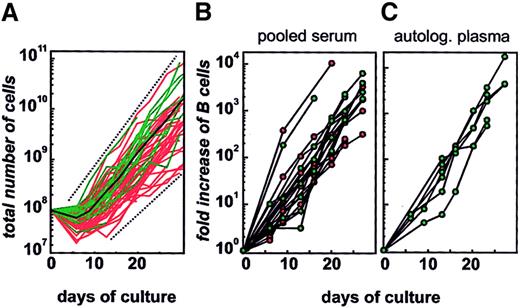
Simple and reliable generation and expansion of APCs from non–stem cell sources determines their clinical applicability. To address this issue, CD40–B cells from PBMCs of 8 cancer patients and 41 healthy individuals were generated by 8 investigators during a period of 18 months. In total, 56 cultures were performed. Three of these 56 cultures were discontinued owing to bacterial contamination. The 90% exact confidence interval for successful expansion was 87% to 99%. Figure 2A shows the total cell count over time in the 53 individual cultures with a culture period of at least 14 days. All cultures showed similar growth kinetics: there was a drop in cell number during the first 5 to 10 days, followed by a significant expansion during the remaining culture period. When the culture started with 108PBMCs, the mean cell count of CD19+ B cells after 27 days of culture was 6.42 × 10⁹ (range, 3.49 × 10⁸ to 5.03 × 10¹⁰). When initial B-cell numbers in the cultures were normalized (11 healthy donors and 8 cancer patients), a continuous expansion of B cells over time was observed, leading to a mean increase of 2032-fold (range, 226- to 6226-fold increase) at day 27 (Figure 2B). There was no difference between cancer patients and healthy donors. CD40–B cells are completely dependent on CD40L and IL-4 since discontinuation of these 2 factors led to cell death by apoptosis in fewer than 4 days (data not shown).
Expansion of CD40–B cells.
(A) Overall expansion of 53 cultures from healthy individuals (red) and cancer patients (green). CD40–B cells were generated by means of CD40L-transfected NIH3T3 cells and rhIL-4. (B) (C) Cultures from 11 healthy individuals (red) and 8 cancer patients (green) were grown in pooled AB serum (panel B) or autologous plasma (panel C), and expansion was normalized for CD19+CD20+cells.
For therapeutic use of CD40–B cells, it is desirable to avoid the use of culture medium supplemented with pooled human AB serum. In 7 cancer patients, we replaced the pooled serum (Figure 2B) with autologous plasma (Figure 2C) and demonstrated no statistically significant difference of B-cell expansion, suggesting that plasma can be used instead of pooled AB serum in this system. Replacing the xenogeneic component (tCD40L) of the CD40–B-cell system with another source of CD40L would significantly improve its clinical applicability. We therefore tested GMP-grade human trimeric soluble CD40L (sCD40L).28 In the presence of rhIL-4 and CsA, sCD40L induced a significant B-cell expansion (3 orders of magnitude), which did not differ from that observed for B cells stimulated with tCD40L (Figure 3). As expected,38rhIL-4 was an important cofactor since B-cell expansion in cultures with tCD40L or sCD40L alone did not expand over the whole culture period and could not be replaced by other cytokines, including IL-6 or TNF-α (data not shown). Further increasing the concentration of IL-4 (more than 2 ng/mL) did not lead to increased B-cell expansion (Figure3).
Replacement of xenogeneic CD40L transfectants with recombinant, trimeric, soluble CD40L.
The generation of CD40–B cells was studied by means of CD40L-transfected NIH3T3 cells (panel A) or GMP-grade soluble trimeric CD40L (panel B) under varying concentrations of clinical-grade rhIL-4 (shown here: 0 [filled circles], 2 [filled squares], and 10 [filled diamonds] ng/mL). A representative experiment of 3 experiments is shown.
Determination of cell surface molecules as surrogates for APC function of CD40–B cells
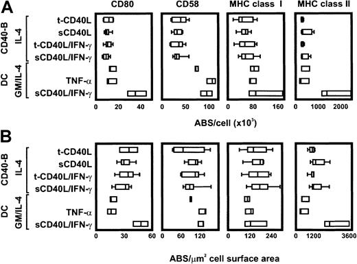
The generation of antigen-specific CTL lines using CD40–B cells cannot be routinely performed as an assay to control for efficient APC function of these cells, particularly in a clinical setting. Since sufficient expression of MHC, adhesion, and costimulatory molecules closely correlates with APC function,39,40 phenotypic analysis of these cell surface molecules was applied as a fast and reliable surrogate readout for APC function of CD40–B cells. For optimal comparison of different culture conditions and for comparison with other cells, the analysis was normalized for number of molecules (ABSs) and cell size. CD40–B cells were generated in the presence of rhIL-4, CsA, tCD40L, or sCD40L, and phenotypic analyses were performed between day 16 and 28. Since IFN-γ is known to increase the expression of MHC molecules,41 the effect of IFN-γ was also measured. Monocyte-derived mature and immature DCs were used as positive controls. As shown in Figure 4A, significant numbers of CD80 (6381 to 15 600 ABSs per cell); CD58 (17 213 to 59 987 ABSs per cell); MHC class I (17 260 to 106 916 ABSs per cell); and MHC class II molecules (294 130 to 1 016 785 ABSs per cell) were expressed on CD40–B cells. No statistically significant difference between sCD40L and tCD40L was observed for these 4 surface molecules, and the addition of IFN-γ did not further increase their expression. The absolute number of CD80, MHC class I, and MHC class II reached that of immature and TNF-α–matured DCs; only CD40L/IFN-γ–matured DCs showed higher levels. Despite significant expression of CD58 on CD40–B cells, DC controls showed a consistently higher absolute expression.
Quantification of cell surface molecules.
CD40–B cells were generated by means of tCD40L or sCD40L in the presence of IL-4 and CsA and were coincubated with or without INF-γ (added for 4 days before analysis). DCs were generated by culture of monocytes with GM-CSF and IL-4 and were matured with TNF-α or CD40L/INF-γ. (A) Surface expression of CD58, CD80, MHC class I, and MHC class II was measured by flow cytometry and normalized by means of the Qifikit to obtain the ABSs per cell. (B) The number of ABSs per cell surface area was calculated to normalize for different sizes, cell types, or culture conditions. Medians (lines), ranges (bars), and 95th percentiles (boxes) of 3 to 9 experiments for each culture condition and cell surface molecule are shown.
After normalization for cell size, the density of CD58 and MHC class I was in the same range for CD40–B cells and all control DCs. Expression of CD80 and MHC class II molecules was denser on CD40–B cells than on immature or TNF-α–matured DCs; however, sCD40L/IFN-γ–matured DCs showed the highest density. Of these cell surface molecules, CD80 showed the lowest density on CD40–B cells (16 to 49 ABSs per square micrometer), whereas the density of MHC class II was the highest (778 to 2359 ABSs per square micrometer; Figure 4B). Taken together, these data suggest that CD40–B cells, generated with either sCD40L or CD40L transfectants, consistently express a high density of cell surface molecules involved in APC function.
Visualization and quantification of antigen delivery to CD40–B cells
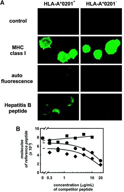
To use APCs therapeutically, it is critical to ensure and monitor efficient antigen delivery. To address this issue for CD40–B cells, we applied direct visualization of peptide loading using a fluorochrome-labeled HLA-A2–binding peptide derived from the hepatitis-B core protein (FITC-conjugated F18).42HLA-A2+, but not HLA-A2−, CD40–B cells showed significant fluorescence after peptide pulsing at concentrations lower than 2 μg/mL (Figure 5A), whereas an HLA-A2–independent increase in fluorescence was observed at concentrations exceeding 2 μg/mL. Peptide-binding analysis using T2 cells revealed no significant difference in HLA-A2 binding between the FITC-conjugated and the native F18 peptide (data not shown). Maximum peptide loading, reaching approximately 10 000 molecules per cell, was achieved at 0.2 μg/mL peptide in 30 minutes at 37°C in serum-free medium without the necessity of β2-m. Control experiments showed comparable peptide loading of DCs (approximately 12 000 molecules per cell for immature DCs, approximately 6000 molecules per cell for mature DCs; data not shown). Optimized peptide-loading also correlated with maximum cytolysis of peptide-loaded CD40–B cells by peptide-specific CTL. A dose-dependent cytolysis of influenza A MP58-pulsed CD40–B cells was determined at peptide concentrations below 2 μg/mL, whereas higher concentrations did not lead to increased cytotoxicity (data not shown).
Antigen loading of CD40–B cells.
(A) Specific binding of hepatitis-B core peptide was demonstrated by means of HLA-A*0201+ (left) and HLA-A*0201−(right) CD40–B cells. As negative control, autofluorescence of CD40–B cells and isotype control are shown. Staining for MHC class I was used as positive control. Identical imaging parameters were used throughout the experiment. (B) Binding affinity of the HLA-A2–binding peptides I540 (filled circles) and F18 (filled diamonds) and the pan-DR–binding peptide PADRE (filled squares) to CD40–B cells was determined by competition with the hepatitis-B core F18-FITC reference peptide. Fluorescence was analyzed by flow cytometry, and molecules of reference peptide were calculated by means of standardization beads (see “Materials and methods”).
For the application of CD40–B cells with other peptides, a competition assay was established with the use of the FITC-conjugated F18 peptide as a reference peptide. Using this assay, we demonstrated binding of 2 peptides with known high affinity for HLA-A2. The native F18 peptide and the I540 peptide from hTERT27 led to significant reduction of fluorescence with the addition of increasing concentrations of peptide (Figure 5B). In contrast, as expected, the HLA-DR–binding peptide PADRE33 did not compete with the FITC-conjugated F18 peptide for HLA-A2 binding. Owing to a significant loss of viability under these experimental conditions, DCs could not be used as controls.
Taken together, CD40–B cells are efficiently loaded with peptide under serum-free conditions without the need of β2-m in a relatively short time at 37°C. Moreover, peptide-loading efficiency of primary APCs for clinical application can be easily monitored by means of FITC-conjugated reference peptides.
Discussion
The CD40–B-cell system is an autologous system for efficiently expanding highly effective APCs without the use of xenogeneic fibroblasts,20 viral components such as Epstein-Barr virus,43 or gene-transfer technology.21 The value of CD40-activated B cells for antigen screening and immune assessment was demonstrated by ex vivo generation of antigen-specific CTLs to recall antigens (eg, influenza A), tumor antigens (eg, hTERT, MART-1), and neoantigens (eg, HIV RTpol in seronegative donors). Applicability of CD40–B cells for clinical use was established by successful replacement of allogeneic, pooled AB serum with autologous plasma and of xenogeneic CD40L transfectants with human GMP-grade soluble trimeric CD40L. The normalized analysis of important cell surface molecules is a simple method to ensure and control the quality of this cellular product as highly efficient APCs. Optimized delivery of peptide antigens, important for clinical applications of any cellular adjuvant, is demonstrated for CD40–B cells by direct visualization of peptide binding in a competition assay using fluorescence-labeled reference peptides. This system is suitable for clinical-grade large-scale expansion of highly efficient APCs in cancer patients.
For an APC to be applicable to antigen discovery and as a cellular adjuvant for vaccination, it is critical to demonstrate that the APC not only boosts T-cell responses to recall antigen but also initiates T-cell responses to neoantigens. DCs are the only APCs that have been reported to prime T cells in vivo1 and in vitro.10,44,45 Priming of human T cells in vitro can be formally addressed only by induction of CTL responses against viral neoantigens in seronegative individuals. Here, we demonstrate that CD40–B cells not only boost T-cell responses to recall antigens such as influenza A but also stimulate T-cell responses to tumor antigens that are ignored (eg, hTERT; R.H.V. and J.L.S., unpublished results, June 2001) and stimulate naive T cells specific for neoantigens, such as RTpol from HIV. On the basis of current knowledge about the T-cell repertoire for HIV10,44 45 in healthy individuals, the successful ex vivo expansion of HIV-specific CTLs in seronegative healthy individuals strongly supports that the CD40–B-cell system can prime naive T cells ex vivo. For further evaluation, the CD40–B-cell system was applied to our tumor antigen discovery program. So far we have tested 14 potential candidate antigens and have been able to identify 23 immunogenic epitopes and 5 heteroclitic peptides using CD40–B cells as APCs (M.S.V.B.-B. and J.L.S., unpublished results, February 2001).
Antigen delivery to APCs is critically important in the development of cellular vaccines, and so far there has been little attention to ensure efficient and reproducible antigen delivery in previous and ongoing trials.46 Fluorescein-labeled peptides have been used mainly to study peptide/MHC interactions on murine cell lines.47-49 We applied this simple technology to visualize peptide binding on primary APCs, such as CD40–B cells and DCs, and to optimize peptide-pulsing conditions. These experiments revealed that the amount of peptide loaded onto CD40–B cells under optimized conditions exceeds the amount of peptide necessary for T-cell activation.50 By using this technique in upcoming vaccine trials, we will be able to determine the impact of insufficient antigen delivery on vaccination efficiency. This technique is not restricted to the most common HLA types. As long as a high affinity–binding peptide is known for the HLA allele of interest, a competition assay using this peptide as a fluorescein-labeled competitor can be established for every patient.
While CD40–B cells generated in coculture with CD40L transfectants are suitable for research purposes and immune assessment, it is preferable to use human GMP-grade soluble trimeric CD40L for therapeutic applications of CD40–B cells to avoid any potential danger from contact with xenogeneic culture components. In fact, cells such as CD40–B cells generated for therapeutic purposes by coculture with xenogeneic-transfected fibroblasts would fall under regulations established for xenotransplantation and gene therapy, thereby significantly limiting clinical applicability.51 This is probably one of the major disadvantages of artificial APCs,20,52-54 which have been suggested as an alternative to human professional APCs. Moreover, unlike DCs and CD40–B cells, artificial APCs lack other human molecules, including cell surface molecules, cytokines, and chemokines, that significantly contribute to the activation and expansion of T cells.55-58
CD40–B cells can be generated in great numbers and might therefore facilitate “dose escalation” of therapeutic vaccination. The beneficial effect of frequent vaccinations with significant numbers of cells in the tumor-bearing host has been clearly demonstrated by Ochsenbein et al.59 In this murine model, tumor regression of well-established tumors was achieved only when antigen-loaded APCs were applied repetitively over a period of 3 weeks. In contrast to DCs, B cells are normally not surveying subcutaneous tissue, and ex vivo–generated CD40–B cells might therefore be incapable of migration into draining lymph nodes. However, in mice, injected B cells are indeed capable of migrating to lymphoid organs even after subcutaneous injection.60 Taken together, CD40–B cells are a powerful source of APCs generated by simple and reliable technology that can be applied to antigen screening, immune assessment, vaccination approaches, and ex vivo T-cell expansion for adoptive therapy.
We thank J. Daley, M. Bedor, K. Hoar, D. Schnipper, and Y. Chen for excellent technical assistance and Dr D. Neuberg for statistical assistance.
Supported by the Deutsche Krebshilfe und Dr Mildred Scheel Stiftung (M.S.v.B.-B.); the Deutsche Forschungsgemeinschaft (B.M.); grant CI2 of the Cancer Research Fund of the Damon Runyon–Walter Winchell Foundation (R.H.V.); the Sankyo Foundation of Life Science (N.H.); National Institutes of Health grants K08-CA-88444-01 (K.S.A.), K08-CA-87720-01 (M.O.B.), P01-CA-66996, and P01-CA-78378 (L.M.N.); a Special Fellowship of the Leukemia and Lymphoma Society (J.L.S.); and a Translational Research Award by the Leukemia and Lymphoma Society (J.L.S.).
R.H.V. and B.M. contributed equally to this study.
The publication costs of this article were defrayed in part by page charge payment. Therefore, and solely to indicate this fact, this article is hereby marked “advertisement” in accordance with 18 U.S.C. section 1734.
References
Author notes
Joachim L. Schultze, Tumor Immunology Laboratory, Dept of Hematology/Oncology, University of Cologne, Joseph-Stelzmann St 9/Haus 16, 50924 Cologne, Germany; e-mail:joachim.schultze@medizin.uni-koelne.de.

![Fig. 1. Induction of CTL responses against peptides derived from viral and tumor antigens using CD40–B cells as APCs. / (A) Peptide-specific lysis of T2 cells pulsed with specific peptide (filled circles), irrelevant HLA-A2–binding control peptide (empty circles), or no peptide (empty squares). Influenza A MP58-specific lysis (MP58) is shown for day 18 of T-cell culture; all other experiments for MART-1 (E26), hTERT (I540), and HIV RTpol (RTpol476) were performed on day 25. For each peptide, a representative peptide-specific CTL line is shown here. (B) Tetramer analysis using peptide-specific tetramers was always controlled with the tetramer specific for the immunodominant HTLV-1 tax peptide. The percentage of CD8+ tetramer+ T cells is shown. Analysis was performed after 3 (Influenza A [Inf A]) or 4 weeks of CTL expansion (all other antigens). Shown here is a representative peptide-specific CTL line. (C) Endogenous processing was tested for MART-1 by means of A2+ MART-1+ Malme-3M cells (filled circles) and HLA-A2−SK-MEL-2 cells (empty circles); for hTERT by means of HLA-A2+ hTERT+U266 cells (filled circles) and HLA-A2− HSS cells (empty circles) (second graph), IFN-γ–pretreated (72 hours) HLA-A2+ hTERT-transfected U2OS cells (filled circles), or HLA-A2+ wild-type U2OS cells (empty circles) (third graph); and for RTpol by means of HLA-A2+ RTpol+ COS cells (filled circles) and HLA-A*0201+ RTpol−COS cells (empty circles) as target cells. (D) Generation of peptide-specific CTL lines in healthy donors and cancer patients is expressed as individual T-cell cultures that induced a peptide-specific response out of the total number of T-cell cultures performed. (E) Generation of RTpol476-specific CTL lines using CD40L-matured monocyte-derived DCs. Peptide-specific lysis of T2 cells pulsed with specific peptide (●) and HLA-A2–binding control peptide (○) (upper panel); percentage of CD8+tetramer+ T cells (middle panel); and recognition of endogenously processed peptide with the use of HLA-A2+RTpol+ COS cells (●) and HLA-A*0201+RTpol− COS cells (○) as target cells (lower panel).](https://ash.silverchair-cdn.com/ash/content_public/journal/blood/99/9/10.1182_blood.v99.9.3319/6/m_h80922505001.jpeg?Expires=1767697553&Signature=onsBVQIq4aZ4nujPRs6EzfZ366lBJTEj0ctjiBLU-dsXOedPAtUWNDWietCtdu0POA3-PjDO~d~OvEysGHQ~~LGMRtq6oG5AiPfqxXJgJpEwIC8A2FcFIOB5m0tZ5anljImbLnqQtL8OHpJErUO7NR0BhNPtkl0kjTSrkvcSLzBhgGUL0B8j3r7bOS4wJW9k6Qg9Ie6NMBGA2gMT0wUlqUSGElrQk7S~rh9JQa392B9wf3bxbOyY9fj9rpA1ZowqtwGfqoDAdzLYDOkiwrAxWzeWBcnB~6-pbH~ZP8fXinplxdPcXO8dXH9-3oUFq24x12bHyayZqxXTulG1VZfAdQ__&Key-Pair-Id=APKAIE5G5CRDK6RD3PGA)

![Fig. 3. Replacement of xenogeneic CD40L transfectants with recombinant, trimeric, soluble CD40L. / The generation of CD40–B cells was studied by means of CD40L-transfected NIH3T3 cells (panel A) or GMP-grade soluble trimeric CD40L (panel B) under varying concentrations of clinical-grade rhIL-4 (shown here: 0 [filled circles], 2 [filled squares], and 10 [filled diamonds] ng/mL). A representative experiment of 3 experiments is shown.](https://ash.silverchair-cdn.com/ash/content_public/journal/blood/99/9/10.1182_blood.v99.9.3319/6/m_h80922505003.jpeg?Expires=1767697553&Signature=xYjcXAv~1wDTOHKTCLnbhPdEu4MSDpniP7LEqR6YDQh3c-i-JRQHzxbHSasFRZ1Kk9NogiIEiqioIp9TMiWl1Q7nqGD4OrjtuT~TEFM47jy~ofKEZVeYndUt4jqBwfMUKBSfArDYLVJ-0~2eoXi9RfbocM~5yWmAVsfQdNxX3STBMWQ8mtTP3y0nUDciR56zqB6CbC0fcKtN8fkpfZQhYDO6uMH6IxqTGrt2~Lh8Lyz8sXRdkxfE1LlvHYxjkTdGuL~u1mDCQIWxVADiCQEMtfZoYV2VmVcnAMtBo6yoqxereWAFNVWongf~KU~BleBqyuG93v6ofFqEOmocHDu9dw__&Key-Pair-Id=APKAIE5G5CRDK6RD3PGA)


This feature is available to Subscribers Only
Sign In or Create an Account Close Modal