This study investigated the efficacy, safety, and clinical benefit of alemtuzumab (Campath-1H) for patients with relapsed or refractory B-cell chronic lymphocytic leukemia exposed to alkylating agents and having failed fludarabine therapy. Ninety-three patients received alemtuzumab in 21 centers worldwide, with the aim to obtain an overall response rate of at least 20%. Dosage was increased gradually (target 30 mg, 3 times weekly, for a maximum of 12 weeks). Infection prophylaxis was mandatory, beginning on day 8, and continuing for a minimum of 2 months after treatment. Responses were assessed at weeks 4, 8, and 12, and patients were followed for 34 months. Overall objective response in the intent-to-treat population (n = 93) was 33% (CR 2%, PR 31%). Median time to response was 1.5 months (range, 0.4-3.7 months). Median time to progression was 4.7 months overall, 9.5 months for responders. At data cut-off, 27 patients (29%) were alive; overall median survival was 16 months (95% CI: 11.8-21.9) and 32 months for responders. Nineteen responders survived more than 21 months. Clinical benefit was observed both in responders and in patients with stable disease. The most common adverse events were related to infusion, generally grade 1 or 2 in severity, occurring mainly in the first week. Grade 3 or 4 infections were reported in 25 patients (26.9%). However, only 3 (9.7%) of 31 patients who responded to alemtuzumab treatment developed grade 3 or 4 infections on the study. Alemtuzumab induced significant responses in these patients with clinical benefit in the majority and with acceptable toxicity in a high-risk group.
Introduction
B-cell chronic lymphocytic leukemia (B-CLL) is the most common adult leukemia occurring in the Western Hemisphere and mainly affects the elderly.1 Although it may have a long clinical course, with survival up to 20 years after diagnosis, once disease progresses, death is almost inevitable. For patients in whom fludarabine treatment has failed, the prognosis is poor, with only approximately 40% surviving beyond 12 months (median survival, 8 months).2
Major response rates in over 20% of patients with refractory B-CLL have been achieved with alkylating agents such as chlorambucil,3 and purine analogs, such as fludarabine4,5 or cladribine.6 Fludarabine is the most effective single agent with response rates ranging between 19% and 71%. It is more successful as front-line therapy,7,8 and as a salvage therapy when the patients have not been heavily pretreated.9-11 However, such responses may be short, and B-CLL often becomes refractory to repeated courses of treatment with the same drug.
Patients who respond to alkylating agents and relapse may fail to respond to fludarabine, and some patients fail to respond to fludarabine even as front-line therapy. Alternative therapy is needed for such patients, preferably using agents with a mode of action that does not overlap with previous chemotherapy. One such alternative is the use of monoclonal antibodies.
Alemtuzumab is a humanized monoclonal antibody directed against CD52, a cell surface protein expressed at high density on most normal and malignant B and T lymphocytes12 but not on hematopoietic stem cells.13 Binding of alemtuzumab to CD52 on target cells may cause cell death by 3 different mechanisms: complement activation,14 antibody-dependent cellular cytotoxicity,15,16 and apoptosis.17
Based on prior reports from small studies demonstrating that alemtuzumab was an effective salvage therapy for patients in whom fludarabine had failed,18 this study was implemented to confirm these encouraging results in a larger cohort of patients with advanced B-CLL. These patients had all been treated previously with alkylating agents and had documented failure to fludarabine therapy.
Patients and methods
A prospective, noncomparative phase II trial was conducted at 21 study centers in the United States and Europe (see for participating investigators and centers). The primary objective of the study was to assess the overall response (OR) rate in patients with B-CLL who had received an alkylating agent and in whom fludarabine treatment had failed, using the 1996 National Cancer Institute Working Group (NCIWG) criteria.19
Secondary objectives were to assess the safety profile of alemtuzumab and to evaluate clinical benefit in this heavily pretreated patient population. The study was conducted in accordance with good clinical practice, and all patients gave informed written consent before commencing treatment.
Patients
Adults with active B-CLL who had received up to 7 previous therapies, including at least one alkylating agent–based regimen, and in whom treatment with fludarabine had failed were eligible for this study. Fludarabine failure was defined as failure to achieve partial response (PR) or complete response (CR) to at least one fludarabine-containing regimen, disease progression while on fludarabine treatment, or disease progression in responders within 6 months of the last dose of fludarabine. Active B-CLL was defined and confirmed according to NCIWG guidelines.19 This study limited recruitment to patients with a performance status (PS) of 2 or less. Patients who had received prior treatment with alemtuzumab, a previous bone marrow transplant, or a prior history of anaphylaxis following exposure to human-rodent (rat or mouse) hybrid monoclonal antibodies were excluded. In addition, patients with an active infection or an active secondary malignancy, with central nervous system involvement, or who were registered as positive for human immunodeficiency virus were also excluded, as were pregnant or lactating women. Any prior therapy had to be completed 3 to 6 weeks before study commencement, depending on hematologic toxicity.
Baseline staging studies were performed within 4 weeks of registration. In addition to physical examination, including assessment of palpable spleen, and a complete blood count, World Health Organization (WHO) performance status, bone marrow aspirate, and trephine biopsies were carried out, as was full laboratory testing. Disease characteristics at enrollment are summarized in Table1. At baseline, 78 patients (84%) had lymphocytosis. Previous therapy for B-CLL is recorded in Table 1. The median number of prior treatments was 3 (range, 2-7). All patients had received prior alkylating agent therapy (median duration of treatment, 11.5 months; range, 0.1-121 months), and all but one had failed fludarabine according to the protocol. Forty-three patients (46%) had received multiple fludarabine treatments; 30 patients (32%) were treated twice, 5 patients (5%) treated 3 times, and 8 patients (9%) treated 4 times.
Disease characteristics at enrollment (n = 93)
| Characteristics . | n (%) . | Overall response (CR + PR, %) . |
|---|---|---|
| Median age (y, range) | 66 (31-86) | 2 + 29 (33) |
| Patients > 70 y | 26 (28) | 1 + 7 (31) |
| Rai stage | ||
| 0 | 1 (1) | 0 |
| I | 5 (5) | 0 + 2 (40) |
| II | 16 (17) | 0 + 7 (44) |
| III | 16 (17) | 2 + 6 (50) |
| IV | 55 (59) | 0 + 14 (25) |
| Lymph node involvement | ||
| No involvement | 22 (23) | 0 + 8 (36) |
| All < 2 cm in diameter | 25 (27) | 1 + 10 (44) |
| At least one node 2-5 cm in diameter | 29 (31) | 1 + 9 (34) |
| At least one node > 5 cm in diameter | 17 (18) | 0 + 2 (12) |
| Hepatomegaly | 34 (37) | 0 + 9 (26) |
| Splenomegaly | 51 (55) | 2 + 14 (31) |
| Splenectomy | 16 (17) | 0 + 5 (31) |
| B-symptoms | 39 (42) | 1 + 8 (23) |
| WHO performance status | ||
| 0 | 24 (26) | 2 + 9 (49) |
| 1 | 50 (54) | 0 + 20 (40) |
| 2 | 19 (20) | 0 |
| Median duration since CLL diagnosis | 6.1 | NA |
| (y, range) | (0.7-37.0) | |
| Hemoglobin ≤ 11.0 g/dL | 57 (61) | 2 + 13 (26) |
| ANC < 1.5 109/L | 24 (26) | 1 + 4 (21) |
| Platelets ≤ 100 × 109/L* | 54 (58) | 0 + 13 (24) |
| Previous therapy | ||
| Never responded to fludarabine | 45 (48) | 1 + 12 (29) |
| Response to fludarabine†,‡ | 48 (52) | 1 + 18 (38) |
| Number of prior therapies1-153 | ||
| 2 | 26 (28) | 1 + 8 (35) |
| 3 | 28 (30) | 1 + 8 (32) |
| 4 | 25 (27) | 0 + 10 (40) |
| > 4 | 14 (15) | 0 + 3 (21) |
All patients included in the study had received alkylating agents and had failed fludarabine.
One patient had a baseline central lab with platelets of 100 × 109/L, but was assessed as Rai IV based on a local lab at baseline of 94 × 109/L and the investigator assessment of Rai IV on inclusion criteria.
Failed fludarabine and/or other purine analog, including cladribine (n = 3) and nelarabine (n = 2) as final treatment.
One patient did not fail by protocol definition, because progression occurred 10 months, not 6 months after therapy.
Each regimen was counted only once, regardless of the number of courses administered or relapse and retreatment.
Treatment and evaluation
Alemtuzumab was diluted into 100 mL 0.9% saline and administered over a 2-hour period through an intravenous infusion line containing a 0.22-μm filter. In the first week the dose was 3 mg, increased to 10 mg, and then to 30 mg as soon as infusion-related reactions were tolerated. If any grade 3 or 4 infusion-related adverse events (AEs) occurred, the lower dose was continued daily until tolerated. Treatment was continued at 30 mg, 3 times a week, up to a maximum of 12 weeks. Premedication with 50 mg diphenhydramine and 650 mg acetaminophen, 30 minutes prior to infusion, was used initially until treatment was well tolerated. If a patient experienced hematologic toxicity or a grade 3 or 4 infection while on treatment, therapy was postponed until the problem was resolved. If postponement lasted longer than a week, treatment was resumed at the lowest dose. Prophylaxis against infection in the form of trimethoprim/sulfamethoxazole (1 tablet twice daily, 3 times a week) and famciclovir (250 mg twice daily), or equivalent, was administered, starting on day 8 of treatment, continuing throughout the course and for a minimum of 2 months after treatment was complete. If venous access was temporarily lost, alemtuzumab was administered subcutaneously in divided doses of no more than 1 mL. Patients were treated for a minimum of 4 weeks provided they did not experience any adverse effects that would preclude continuation in the study. During treatment, weekly procedures included physical examinations, toxicity assessments, and a complete blood count. At weeks 4 and 8, patients were also evaluated for continuation of therapy by additional testing, including WHO performance status, assessment of lymph nodes, liver, and spleen, and full laboratory parameters. At weeks 4 and 8, complete resolution of disease (even if platelet counts and hemoglobin had not completely normalized) or progression of disease indicated treatment should be discontinued, while improvement indicated treatment should be continued. No change in disease status at week 4 indicated treatment should be continued, but if there was still no change at week 8, treatment was discontinued. At the end of treatment, week 12, testing was carried out as for week 4. At the data cut-off point, 93 patients had completed treatment.
Follow-up
After completion of alemtuzumab therapy, patients with CR, PR, or stable disease (SD) were assessed monthly over 6 months for performance status, hematology and all infections, plus documentation of major medical events considered possibly related to alemtuzumab. Disease status evaluation included lymph node measurement, spleen and liver measurement, and flow cytometry in a central laboratory, every 2 months. After 6 months, assessments took place at 3-month intervals until alternative treatment or death. Patients with continuing AEs or abnormal laboratory tests related to alemtuzumab treatment were followed until resolution of these events. Patients developing progressive disease (PD) were followed for survival until alternative treatment or death.
Patients who relapsed during follow-up were not re-entered into the study but were eligible for retreatment under a compassionate-use protocol where considered appropriate.
Statistical analysis
A response rate (RR) of 20% or higher, with a lower limit of the 95% CI of at least 10%, was considered to be meaningful evidence of efficacy in this patient population. Therefore, 75 patients were required to estimate the CI to within 10% of the RR. The intent-to-treat (ITT) population was defined as patients who had received at least one dose of alemtuzumab. The primary end point was the objective response rate (CR + PR) according to 1996 NCIWG criteria.19 According to these criteria, CR is defined as freedom from clinical disease for at least 2 months with a “normal” blood count (hemoglobin > 11g/L, neutrophils > 1.5 × 10⁹/L, lymphocytes < 4 × 10⁹/L, platelets > 100 × 109/L) without transfusion. No constitutional symptoms may be present, with no lymphadenopathy, no hepatosplenomegaly; and less than 30% small lymphocytes in the bone marrow with no nodules. PR is defined as at least a 50% reduction in the number of lymphocytes in the blood and least a 50% reduction in lymphadenopathy or hepatosplenomegaly (or both). At least one of the following should be maintained for at least 2 months: hemoglobin more than 11g/dL or 50% improvement, platelets more than 100 × 109/L, neutrophils greater than 1.5 × 10⁹/L without transfusion. PD is defined as lymphadenopathy, peripheral lymphocyte count, or hepatosplenomegaly increased by 50% or more or histology showing a more aggressive picture. Any response not falling into these categories is defined as SD.
Estimates of duration of response, time to disease progression, and survival were calculated using Kaplan-Meier methodology.20Duration of response was measured from the date the patient first met NCIWG response criteria (confirmation required 2 months later) to the date of progression, alternative therapy, or death. Time to treatment failure was defined as the date from initial administration of alemtuzumab to the first date of objective measurement of disease progression or to alternative therapy or death. Survival was measured from the day of the first dose of alemtuzumab to death; deaths from all causes were included.
An analysis of clinical benefit was also conducted as a secondary end point. Samples were reviewed at a central pathology unit and confirmed by an independent review panel.
Results
Demographics
Between April and October 1998, a total of 93 patients at 21 study sites were registered and treated, of whom 73 were male, 20 female; 86 were white and 7 were black. The median age was 66 years (range, 32-86 years). Seventy-one (76%) of the study participants had advanced disease (Rai stage III/IV).
Median time from preceding therapy in all patients was 4.1 months (range, 0.7-33 months), in responders 2.9 months (range, 0.8-27.1 months), and in patients with CR 2.2 months (range, 2.0-2.4 months). Fludarabine had been the immediate preceding therapy in 57 patients (61%) either as a single agent or in combination with other cytotoxic agents (eg, cyclophosphamide, mitoxantrone, etoposide with or without prednisone). Twenty-nine patients (31%) had received fludarabine or cladribine in combination with other cytotoxic agents, almost always mitoxantrone or cyclophosphamide. Fifteen patients (16%) had been previously treated with the combination of fludarabine or cladribine and cyclophosphamide, regimens that are used for salvage of patients in whom fludarabine alone has failed. All 15 of these patients had failed the combination therapy by the protocol definition. Almost half the patients had never responded to any nucleoside analog-based regimen (45 of 93, 48%).
Sixty-five patients (70%) completed treatment according to the protocol, including some patients who discontinued because of PD. Treatment took up to 16 weeks to complete in 2 patients (2%) who received subcutaneous alemtuzumab injections due to loss of venous access, and in a further 12 patients (13%) due to dose interruptions. In 4 cases (4%), dose interruption prolonged treatment to between 17 and 28 weeks. Treatment was prematurely discontinued in 28 patients, 3 (3%) of whom were nonevaluable due to termination in the first week of therapy, and were therefore considered failures.
Response
Thirty-one (33%) of the 93 patients responded to alemtuzumab therapy (Table 2), significantly exceeding the target of 20% (95% CI: 10%-30%). Although only 2 patients (2%) achieved a CR using NCIWG criteria, 6 additional patients (7%) had clearing of B-CLL from all sites, but had persistent anemia and/or thrombocytopenia. At the last on-treatment assessment of these 6 patients, anemia was grade 1 in 2 patients and grade 2 in 2 patients, and thrombocytopenia was grade 2 in 4 patients, grade 3 in 1 patient, and grade 4 in 1 patient. Grades 3 and 4 thrombocytopenia resolved completely during long-term follow-up.
Summary of disease response (n = 93)
| Response . | NCIWG assessment n (%) . |
|---|---|
| Overall response | 31 (33) |
| Complete response | 2 (2) |
| Partial response | 29 (31) |
| CR except for cytopenia | 6 (7) |
| Nodular PR | 5 (5) |
| Stable disease | 50 (54) |
| Response . | NCIWG assessment n (%) . |
|---|---|
| Overall response | 31 (33) |
| Complete response | 2 (2) |
| Partial response | 29 (31) |
| CR except for cytopenia | 6 (7) |
| Nodular PR | 5 (5) |
| Stable disease | 50 (54) |
Responses by disease status are shown in Table 1. Responses to alemtuzumab were seen in all prognostic subsets and were similar in patients who had failed treatment or had previously had a short response to fludarabine. Patients were less likely to respond if they had Rai stage IV disease or at least one lymph node more than 5 cm in diameter, or WHO performance status of 2. Seven patients (7%) had previously been treated with the anti-CD20 monoclonal antibody, rituximab, which resulted in 1 PR, 2 SD, and 4 PD; alemtuzumab treatment of this group produced 1 PR, 4 SD, and 2 PD.
Advanced age did not preclude response; the patients who achieved CR were 68 and 71 years of age, and the median age of responding patients was 66 (range, 46-78 years).
Overall median time to response was 1.5 months (range, 0.4-3.7 months); 1.8 months in the case of the 2 complete responders and 1.2 months in the case of the 29 partial responders. Median duration of response was 8.7 months (range, 2.5-22.6+ months). Median time to progression in the ITT population was 4.7 months (range, 0.2-23.6+ months, including patients who discontinued early without formal assessment of progression) and 9.5 months (range, 3-23.6+ months) for the responding subgroup, with 3 patients (3%) still in remission (response durations of 17+ to 22.6+ months) at last follow-up.
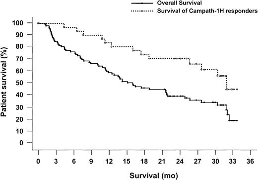
The Kaplan-Meier plot of overall survival is shown in Figure1. Twenty-seven (35%) of the 93 patients were alive with a median follow-up of 29 months, 3 (3%) had been lost to follow-up, and 63 (68%) had died. In the majority of cases (n = 37), cause of death was a consequence of malignancy, with an additional 2 deaths due to autoimmune hemolytic anemia or idiopathic thrombocytopenia purpura. Death was due to, or complicated by, infection in 17 cases. The remaining deaths were due to inanition, cerebrovascular accident, respiratory distress, and pulmonary embolism, with 3 patients lost to follow-up. Death occurred during the study or within 30 days after treatment in 9 patients, between days 30 and 180 after treatment in 19 patients, and more than 180 days after treatment in 35 patients. Median survival was 16 months (95% CI: 11.8-21.9). Nineteen (61%) of 31 responders were still alive 21 to 30 months after alemtuzumab therapy, with median survival of 32 months.
Kaplan-Meier survival curve.
Twenty-seven (29%) of 93 patients were alive at 29 months of follow up. Median survival was 16 months (95% CI: 11.8-21.9 months). Nineteen (61%) of 31 responders were still alive 21 to 34 months after alemtuzumab therapy, with median survival of 32 months. In total, 21 (68%) of 31 responders were still alive at the end of the study, and 42 (45%) of 93 patients survived more than 18 months.
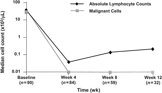
Response in blood and bone marrow is shown in Table3, demonstrating substantial disease reduction at these sites. Peripheral blood data were supplemented by flow cytometry in 90 patients at baseline showing that clonal CD19+ lymphocytosis in 89 patients had rapidly reduced with alemtuzumab therapy (Figure 2). Median number of malignant lymphocytes in the peripheral blood fell from 33.6 × 10³/μL at baseline to 0.003 × 10³/μL at week 4, and 0.001 × 10³/μL at weeks 8 and 12 (Figure 3).
Summary of response in peripheral blood and bone marrow (NCIWG criteria) (n = 93)
| . | Lymphocytosis (n, %) . | Bone marrow (n, %) . | ||||
|---|---|---|---|---|---|---|
| All patients . | Responders . | Stable disease . | All patients . | Responders . | Stable disease . | |
| Patients with involvement at baseline | 783-150 | 28 | 40 | 853-151 | 31 | 44 |
| Patients evaluable at last assessment | 67 | 26 | 35 | 62 | 31 | 27 |
| Response | ||||||
| Resolved3-152 | 65 (83%) | 26 (93%) | 34 (85%) | 22 (26%) | 15 (48%) | 7 (16%) |
| ≥ 50% improved3-153 | 2 (2.6%) | 0 | 1 (2.5%) | 16 (19%) | 7 (23%) | 7 (16%) |
| Stable3-155 | 0 | 0 | 0 | 23 (27%) | 9 (29%) | 12 (27%) |
| > 50% increased3-154 | 0 | 0 | 0 | 1 (1.2%) | 0 | 1 (2.3%) |
Fifteen patients did not have lymphocytosis at baseline but had CD19+ clonal expansion in peripheral blood, were Rai stage III/IV, or both.
Determined by biopsy. Six of 7 patients with inadequate biopsies at baseline showed a predominance of malignant lymphocytes by aspirate.
Blood response defined as less than 30% lymphocytosis, bone marrow defined as normal biopsy.
Defined as 50% or greater decrease in malignant cells in blood/biopsy.
Defined as no change in malignant cells in blood/biopsy.
Defined as greater than 50% increase in malignant cells in blood/biopsy.
CD19+/CD5+ cells in peripheral blood assessed by flow cytometry.
The ITT population included 93 patients. Clonal CD19+lymphocytosis in 89 patients was rapidly reduced with alemtuzumab therapy. Median number of malignant lymphocytes in the peripheral blood fell from 33.6 × 10³/μL at baseline to 0.003 × 10³/μL at week 4, and 0.001 × 10³/μL at weeks 8 and 12.
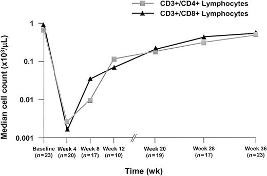
T-lymphocyte subset counts in peripheral blood as a percentage of baseline levels assessed by flow cytometry.
The ITT population included 93 patients.
T-lymphocyte subset counts in peripheral blood as a percentage of baseline levels assessed by flow cytometry.
The ITT population included 93 patients.
Alemtuzumab also induced responses at other major disease sites. Of 66 patients with lymphadenopathy at enrollment 49 (74%) responded; 18 patients (27%) achieved CR and 31 patients (47%) achieved at least a 50% reduction in enlarged nodes. Responses by size of lymph node at enrollment are shown in Table 4 and demonstrate a lower response rate in the largest lymph nodes. Of 31 patients with hepatomegaly at enrollment, at the last treatment of alemtuzumab, hepatomegaly was completely resolved in 16 patients (52%) and 7 patients (23%) had at least a 50% reduction. Of 46 patients with splenomegaly, 54% had complete resolution, and an additional 28% showed at least a 50% reduction in size of the palpable spleen.
Responses in lymph nodes
| Baseline maximum node diameter . | n4-150 . | Resolved . | 50% improved . |
|---|---|---|---|
| > 0-< 2 cm | 22 | 14 (63.6%) | 6 (27.3%) |
| > 2-< 5 cm | 27 | 4 (14.8%) | 15 (55.6%) |
| > 5 cm | 17 | 0 | 10 (58.8%) |
| Baseline maximum node diameter . | n4-150 . | Resolved . | 50% improved . |
|---|---|---|---|
| > 0-< 2 cm | 22 | 14 (63.6%) | 6 (27.3%) |
| > 2-< 5 cm | 27 | 4 (14.8%) | 15 (55.6%) |
| > 5 cm | 17 | 0 | 10 (58.8%) |
Patients with data available.
At enrollment, 76 patients (82%) had various cytopenias (hemoglobin ≤ 11g/dL, absolute neutrophil count (ANC) < 1500, platelet count ≤ 100 000). This subgroup was analyzed for changes in hemoglobin, ANC, and platelet count at the end of treatment and at the 2-month follow-up. Improvement was defined as an increase over baseline values of 50% or more, or achievement of normal hematologic parameters. Fifty-seven patients (61%) had anemia at baseline. At the 2-month follow-up, 25 (44%) of 57 patients with anemia at baseline had improved, with or without blood transfusion or erythropoietin, and of 8 patients who did not receive a blood transfusion or erythropoietin through 70 days after the study, 63% had improved (including 4 of 4 responders [100%], and 1 of 3 patients [33%] with SD). Fifteen patients (16%) received erythropoietin during (n = 8) and/or after the study (n = 12). Seven patients entered the study while receiving erythropoietin, and 4 lost this requirement during the study. Similarly, of 24 patients with neutropenia at baseline, 50% improved, and of 54 patients with thrombocytopenia, 39% improved. Thirty-three patients (35%) received granulocyte colony-stimulating factor (G-CSF) or granulocyte-macrophage colony-stimulating factor (GM-CSF) or both during or after the study, of whom 17 (18%) started growth factor treatment during the study and 16 (17%) started after the study. Of the 17 patients, 12 received on-study CSFs for longer than 7 days, including 3 patients with PR and 2 with SD. In total, 42 (55%) of 76 patients with hematologic abnormalities at baseline had improvements in at least one parameter including 17 (74%) of 23 responders and 19 (44%) of 43 patients with SD.
Evidence of clinical benefit
At the end of treatment, clinical benefit was also assessed (Table5). Of the 31 responders, 17 had B-symptoms on entry, such as fever, night sweats, or weight loss, which resolved in all 17 cases. In total, 59 patients had B-symptoms or fatigue that resolved entirely in 31 patients (52.5%), including those with SD. In a selected group of patients with SD who were assessed for 6 categories of clinical benefit, the most common improvement was an increase in chemotherapy-free time; before alemtuzumab, the median was 3.8 months, and after alemtuzumab treatment, 7.6 months. The median chemotherapy-free periods prior to and following alemtuzumab treatment in the group of responders (n = 31) were 4.0 months and 12.4 months, respectively. Seventeen of the responders had chemotherapy-free periods of more than 1 year following alemtuzumab treatment. Other disease-related symptoms also resolved. In total, 42 (45%) of 93 patients survived more than 18 months, and 21 (68%) of 31 responders were still alive at the end of the study (Figure 1). Massive splenomegaly (> 6 cm) resolved in 90% of responders and 25% overall. In patients with anemia (NCIWG hemoglobin grades 1-4) at baseline, 73% of responders and 49% of patients overall experienced improvement in hemoglobin by at least one grade by follow-up.
Assessments of clinical benefit
| Clinical benefit . | Responders n/N (%) . | Nonresponders n/N (%) . | All patients n/N (%) . |
|---|---|---|---|
| Resolution of B-CLL symptoms or fatigue | 17/17 (100) | 28/42 (67) | 45/59 (76) |
| Resolution of massive splenomegaly | 9/10 (90) | 7/19 (37) | 16/29 (55) |
| Improvement in WHO PS | 8/20 (40) | 9/49 (18) | 17/69 (25) |
| Improvement in anemia | 11/15 (73) | 14/36 (39) | 25/51 (49) |
Infusion-related toxicity
In 75 (81%) of 93 patients, the complete duration of alemtuzumab administration, including dose delays and interruptions, occurred within 12 calendar weeks. In 14 patients (15%), between 13 and 16 calendar weeks were required to complete therapy, and in only 4 patients (4%) did this period exceed 16 weeks. Two patients had treatment postponed for approximately 3.5 months by the investigator. A median daily dose of 30 mg was achieved by 87 patients (93%), and the median cumulative dose (MCD) was 673 mg (range, 200-1106 mg), with 2 patients reaching an MCD of less than 200 mg, 18 reaching 200 to 399 mg, 16 reaching 400 to 599 mg, 24 reaching 800 to 999 mg, and 19 reaching 1000 to 1106 mg.
The most commonly reported AEs were infusion related and were generally of grade 1 or 2 in severity. They included rigors (90% in total, 14% grade 3), fever (85% in total, 17% grade 3, 3% grade 4), nausea (53% in total, all grade 1 or 2), vomiting (38% in total, 1% grade 3) and rash (33% in total, all grade 1 or 2). Infusion-related toxicity decreased with time, and, except for rashes, there was a substantial decrease in incidence from week 1 to weeks 2 to 4, with a further decrease reported for treatment beyond week 4. Fewer than 0.5 events, per patient at risk (receiving infusions), were reported from week 2 until the end of the study. This decrease in the number of events over time on treatment was also apparent when only grade 3 or 4 acute infusion-related events were evaluated.
During the study, 26 patients (28%) had dyspnea (11 patients, grade 3 or 4), 16 patients (17%) had hypotension (2 patients, grade 3 or 4) and 3 patients (3%) had hypoxia (2 patients, grade 3 or 4).
Overall, 46 patients (49%) reported 65 episodes of serious AEs during the study, and in 49 of these reports, the primary event was considered to be related to alemtuzumab. Twenty-two patients (24%) discontinued because of AEs judged likely to be related to the treatment, the most common being infection (n = 8). The majority of these 22 patients had not responded to alemtuzumab.
During long-term follow-up (median, 28 months), an additional 49 patients (53%), the majority of whom were nonresponders, died of complications attributable to disease, including disease progression (n = 26) and infection (n = 13). Of these 49 deaths, 30 occurred more than 6 months after treatment, and 4 were considered likely to be related to alemtuzumab treatment.
Infectious complications
A prior history of infection was reported in 49 (53%) of 93 patients, and 31 (33%) of 93 patients had an infection reported in the month prior to alemtuzumab administration, all mild except for 2 grade 3 infections. Overall, 51 patients (55%) experienced at least one infection during the study (Table 6). Infections were mild or moderate in 26 patients, and more severe (grade 3 or 4) in 25 patients. Septicemia occurred in 14 patients (15%). In 10 patients (10%), sepsis was grade 3 or 4, caused by gram-positive bacteria in 4 cases, Escherichia coli in 1 case, andPseudomonas in 3 cases, with no causative organism identified in the other 2 cases. Two cases of sepsis led to death, on or within 30 days of study end. Superficial moniliasis occurred in 9 patients; and viral reactivation such as cytomegalovirus (CMV; n = 7) or Herpes simplex (n = 6) occurred in 13 patients. Significantly fewer patients who responded to alemtuzumab developed grade 3 or 4 infections during the study, 3 (10%) of 31 patients, compared with 22 of 62 nonresponding patients (36%,P < .01).
Patients reported with infections (all grades) by month
| Month . | Before treatment . | On treatment . | After treatment6-150 . | |||||||
|---|---|---|---|---|---|---|---|---|---|---|
| −1 . | 1 . | 2 . | 3 . | 1 . | 2 . | 3 . | 4 . | 5 . | 6 . | |
| N . | 93 . | 93 . | 73 . | 43 . | 92 . | 88 . | 71 . | 60 . | 45 . | 34 . |
| Infection | ||||||||||
| Bacterial | 3 | 5 | 5 | 1 | 8 | 2 | 3 | 2 | 1 | 1 |
| (3%) | (5%) | (7%) | (2%) | (9%) | (2%) | (4%) | (3%) | (2%) | (3%) | |
| Fungal | 2 | 4 | 6 | 1 | 6 | 1 | 0 | 0 | 1 | 1 |
| (2%) | (4%) | (7%) | (2%) | (7%) | (1%) | — | — | (2%) | (3%) | |
| Protozoan | 1 | 0 | 0 | 0 | 1 | 0 | 0 | 0 | 0 | 0 |
| (1%) | — | — | — | (1%) | — | — | — | — | — | |
| Viral | 7 | 8 | 0 | 1 | 5 | 0 | 3 | 0 | 2 | 0 |
| (8%) | (9%) | — | (2%) | (5%) | — | (4%) | — | (4%) | — | |
| Unspecified | 22 | 8 | 9 | 0 | 14 | 8 | 10 | 3 | 6 | 6 |
| (24%) | (9%) | (12%) | — | (15%) | (9%) | (14%) | (5%) | (13%) | (18%) | |
| Any infection(s)6-151 | 31 | 24 | 15 | 2 | 27 | 11 | 14 | 4 | 10 | 8 |
| (33%) | (26%) | (21%) | (5%) | (29%) | (13%) | (20%) | (7%) | (22%) | (24%) | |
Data cut-off at start of next treatment.
Patients may have multiple infections.
A total of 11 patients, all of whom had advanced disease, developed opportunistic infections (OIs) during treatment, some patients experiencing multiple OIs. A further 7 OIs occurred in the follow-up period. OIs included Pneumocystis carinii pneumonia (n = 1), no prophylaxis administered as per protocol;Aspergillus pneumonia (n = 1); rhinocerebral mucormycosis, a technically considered “on-study” fatal infection (n = 1); systemic candidiasis (n = 1); cryptococcal pneumonia (n = 1, fatal); Herpes zoster (n = 4 after study); pulmonary aspergillosis (n = 1 after study, fatal); invasive aspergillosis; andListeria meningitis (n = 1, after study). The most commonly reported OI was CMV reactivation (n = 7), with 3 cases of grade 2 and 4 cases of grade 3 infection reported on study. There were no cases of CMV reported in the poststudy period. Five cases resolved and in 2 cases, CMV infection was the reason for treatment discontinuation. OIs were infrequently associated with concomitant use of systemic corticosteroids; of the cases above, 7 OIs occurred in patients who were treated with steroids for longer than 5 days. Patients who developed OIs were also, in general, heavily pretreated; 2 prior chemotherapy regimens had been used in only one case, while 3 regimens were previously used to treat 5 patients, and 12 patients had received between 4 and 7 prior regimens.
Nine deaths occurred during treatment or within 30 days of the last administration of alemtuzumab. Three deaths were due to PD, 3 were due to pneumonia, and pulmonary embolism, sepsis, and rhinocerebral mucormycosis caused the remaining 3 deaths. Seven of these patients had advanced disease (Rai stage III or IV) at study entry, and all 9 had failed to respond to alemtuzumab. Five of the on-study deaths were caused by infection judged likely to be related to treatment. A further 19 deaths occurred between days 30 and 180, due to PD (n = 11), including one case of hemolytic anemia and one of idiopathic thrombocytopenia purpura), infection (n = 6), respiratory distress (n = 1), and inanition (n = 1).
Cytopenias and immunosuppression
Most patients experienced transient cytopenias during treatment. Neutropenia was most common during weeks 5 and 6 (30% patients) and resolved in the majority of patients by 2-month follow-up. Thrombocytopenia was most common during the first 2 weeks of therapy and by 2-month follow-up, recovery to or above baseline grade, was seen in 89% of patients' platelet counts and 85% of hemoglobin counts.
Immunoglobulin A, G, and M shifts from baseline are shown in Table7. No significant change was noted at any level.
Immunoglobulin data: shift from baseline to worst grade on study
| Nephelometry . | Baseline . | Worst grade on study . | ||
|---|---|---|---|---|
| n (%) . | Normal . | High . | Low . | |
| Serum IgA | ||||
| Normal | 27 (31.8) | 20 | 1 | 5 |
| High | 1 (1.2) | 0 | 1 | 0 |
| Low | 55 (64.7) | 2 | 0 | 49 |
| Serum IgG | ||||
| Normal | 21 (22.6) | 7 | 4 | 9 |
| High | 1 (1.1) | 0 | 1 | 0 |
| Low | 69 (74.2) | 0 | 1 | 64 |
| Serum IgM | ||||
| Normal | 15 (16.1) | 7 | 0 | 7 |
| High | 6 (6.5) | 2 | 4 | 0 |
| Low | 72 (77.4) | 0 | 0 | 68 |
The median absolute CD3+/CD4+ T-cell count was lowest for the first assessment at week 4 (0.002 × 10³/μL) but significantly increased (0.084 × 10³/μL) by the assessment at week 12. Median CD4+ T-cell counts declined through week 4 and then began to rise (Figure 3). Similar results were observed for the CD3+/CD8+ T-cell count.
Discussion
As treatment for B-CLL has evolved, patients treated with fludarabine have become more common, as has the number of patients having a relapse (progression within 6 months) or refractory to fludarabine treatment (fludarabine failed). This status is associated with a poor prognosis.2 21 In treating these patients with escalating doses of alemtuzumab, we observed a rapid OR rate of 33%, exceeding the expectations of 20% OR set at the start of the study, and also improved disease-related symptoms in patients with SD.
A high proportion of patients achieved responses in the blood (83%) and bone marrow (26%). Responses were less common in Rai stage IV patients with low platelet counts, or patients of an advanced age. Although some patients with massive lymphadenopathy saw dramatic improvements in disease bulk, responses in bulky lymph nodes overall were less marked as has been previously observed with alemtuzumab therapy.22
Median time to progression for responders was 9.5 months, with 3 patients continuing in remission as of February 28, 2001. This study demonstrated a longer survival for all patients (median, 16 months) than other historical controls.2 21
Infusion-related toxicity such as rigors and fever became less common with time and remained below 0.5 events per patient at risk from week 2 until the end of the study. Some delays in treatment were necessary due to cytopenias and infections. Infections are already common in these patients, and it was anticipated that there would be a relatively high infection rate during treatment in this severely myelosuppressed patient population. The spectrum of infections was primarily bacterial, despite the T-cell depletion, which is a consequence of treatment. The incidence of OIs was lower than has been observed in other studies without trimethoprim/sulfamethoxazole and famciclovir prophylaxis. Famciclovir does not prevent CMV reactivation, which was the most common OI observed. No cases of grade 4 CMV infection were reported, and most lower-grade CMV infections responded rapidly to ganciclovir.
Although, as expected, most of the patients had low baseline values for one or more of the immunoglobulin groups, shifts from baseline for IgG showed no significant changes.
A total of 9 deaths occurred during treatment or within 30 days of last dose of alemtuzumab, of which 5, all associated with infections, were considered likely to be related to treatment. Early death rates and infectious death rates compare favorably to fludarabine pivotal studies, which reported an on-study death rate of 22% with half of these associated with infection.23
This study investigating patients with a poor prognosis indicates that there are some patients, namely those with bulky nodes or PS of 2, who are less likely to achieve a response following alemtuzumab therapy. Other salvage therapies that have been recently tested in refractory B-CLL include the combination of fludarabine and cyclophosphamide, which produced 38% response rates (3% CR) in patients refractory to fludarabine as a single agent,24and rituximab, which achieved a 40% response rate (0% CR), in less heavily treated patients than reported in this study.25 26This alemtuzumab study shows that patients who had failed treatment with rituximab were still able to respond to alemtuzumab.
Future studies include the investigation of alemtuzumab in combination with other agents. Preliminary results indicate that the combination of alemtuzumab with fludarabine is effective in patients who are refractory to each agent alone, with 5 of 6 patients (83%) responding (4 PR, 1 CR).27 All patients had been heavily pretreated and in the 5 responders, bone marrow was cleared of morphologic evidence of B-CLL, including 3 cases that were negative by flow cytometry. These results should be followed up by larger scale studies. Current trials are also investigating the use of alemtuzumab in untreated patients using subcutaneous injection protocols and preliminary results in 23 evaluable patients show a high response rate (16 PR, 4 CR) with a good toxicity profile.28 More extensive studies may establish a role for alemtuzumab, alone or in combination, in previously untreated patients.
In conclusion, alemtuzumab was effective and acceptably safe in a population of 93 high-risk patients with advanced B-CLL. Alemtuzumab was equally effective in patients who had failed purine analog therapy as in those who had never responded, and increased the median survival of these patients compared with historical controls. In addition, clinical benefit was shown in patients with SD as well as in those who responded, with improvements in hematologic parameters, reduction in bone marrow infiltration, and improvement in, or complete resolution of, disease-related symptoms.
Principal investigators and participating centers were J-L. Binet, Hôpital Pitié Salpetrière, Paris, France; R. B. Bolin, Bay Area Hospital, Coos Bay, OR; J. C. Byrd, Walter Reed Army Medical Center, Washington, DC; B. Cazin, Hôpital Claude Huriez, Lille, France; V. Diehl, Klink I für Innere Med Universistat, Koln, Germany; M. Divine, Hôpital Henri Monder, Créteil, France; M. J. S. Dyer, The Royal Marsden Hospital, Surrey, United Kingdom; C. Emmanouilides, UCLA, Los Angeles (one patient was enrolled but not treated at this site); I. W. Flinn, Johns Hopkins Institute, Baltimore, MD; P. Hillmen, Leeds General Infirmary, Leeds, United Kingdom; R. M. Hutchinson, Leicester Royal Infirmary, Leicester, United Kingdom; V. Jain, PRN, Dallas, TX; S. Johnson, Taunton & Somerset Hospital, Musgrove Park, Taunton, United Kingdom; M. J. Keating, M. D. Anderson Cancer Center, Houston, TX; M. Leporrier, Centre Hospitalier Universitaire, Caen, France; M. Milder, Swedish Medical Center, Seattle, WA; K. R. Rai, Long Island Jewish Medical Center, New Hyde Park, NY; A. Saven, Scripps Clinic, La Jolla, CA; B. Silver, University Hospitals of Cleveland, Cleveland, OH; P. Travade, Hôpital de l'Hotel Dieu, Clermont-Ferrand, France; D. Winfield, Royal Hallamshire Hospital, Sheffield, United Kingdom; L. Zehngebot, Walt Disney Cancer Center, Orlando, FL.
An independent international panel of experts who had not participated in the study reviewed the key eligibility criteria and data related to disease response for all patients entered in this study. The following members of the panel are gratefully acknowledged: John M. Bennett, University of Rochester, Rochester, NY; Martin S. Tallman, Northwestern University, Chicago, IL; and Federico Caligaris-Cappio, University of Torino, Torino, Italy.
Supported in part by research funding from Millennium Pharmaceuticals and ILEX Oncology. K.R.R. gratefully acknowledges a research grant from the Chemotherapy Foundation.
The publication costs of this article were defrayed in part by page charge payment. Therefore, and solely to indicate this fact, this article is hereby marked “advertisement” in accordance with 18 U.S.C. section 1734.
References
Author notes
Michael J. Keating, M. D. Anderson Cancer Center, 1515 Holcombe Blvd, Houston, TX 77030; e-mail:mkeating@mdanderson.org.


This feature is available to Subscribers Only
Sign In or Create an Account Close Modal