Abstract
Monocytes/macrophages play a predominant role in the immunologic network by secreting and reacting to a wide range of soluble factors. Human immunodeficiency virus (HIV) infection leads to deep immunologic dysfunctions, also as a consequence of alterations in the pattern of cytokine release. Recent studies on in vivo models demonstrated that the expression of HIV Nef alone mimics many pathogenetic effects of HIV infection. In particular, Nef expression in monocytes/macrophages has been correlated with remarkable modifications in the pattern of secreted soluble factors, suggesting that the interaction of Nef with monocytes/macrophages plays a role in the pathogenesis of acquired immunodeficiency syndrome (AIDS). This study sought to define possible alterations in intracellular signaling induced by Nef in monocytes/macrophages. Results demonstrate that HIV-1 Nef specifically activates both α and β isoforms of the signal transducer and activator of transcription 1 (STAT1). This was observed both by infecting human monocyte-derived macrophages (MDMs) with HIV-1 deletion mutants, and by exploiting the ability of MDMs to internalize soluble, recombinant Nef protein (rNef). STAT1-α activation occurs on phosphorylation of both C-terminal Tyr701 and Ser727 and leads to a strong binding activity. Nef-dependent STAT1 activation is followed by increased expression of both STAT1 and interferon regulatory factor-1, a transcription factor transcriptionally regulated by STAT1 activation. It was also established that Nef-induced STAT1- α/β activation occurs through the secretion of soluble factors. Taken together, the results indicate that HIV-1 Nef could interfere with STAT1-governed intracellular signaling in human monocytes/macrophages.
Introduction
The pathology of acquired immunodeficiency syndrome (AIDS) should be considered as the sum of effects generated by human immunodeficiency virus (HIV) replication, with direct T-cell destruction, and by a deep alteration in the pattern of soluble factors. The latter largely depends on the extent of viral replication, because it is hindered by effective antiretroviral therapy. HIV/simian immunodeficiency virus (SIV) Nef, a multifunctional 27- to 34-kd protein expressed early in viral replication, was demonstrated to be a strong candidate for many of the pathogenic effects of HIV/SIV. This was first highlighted by the evidence that monkeys failed to develop the disease on infection with nef-deleted SIV,1and later confirmed by the observation that nef transgenic mice developed a syndrome strictly related to AIDS.2Furthermore, Nef perturbs the pattern of secreted factors in different cell types. As an example, the engagement of Nef with the ζ chain of T-cell receptor leads to release of Fas-ligand from T cells in an antigen-independent manner.3 In addition, the expression of Nef in monocytes/macrophages leads to a massive release of macrophage inflammatory protein 1α/β (MIP-1α/β) chemokines and of still unknown factor-activating lymphocytes.4 These soluble factors possibly promote recruitment and activation of T lymphocytes, which become susceptible to HIV replication. Nef is also able to alter some cellular functions, that is, CD4 internalization/recycling,5-7 major histocompatibility complex class I surface expression,8 and srctyrosine kinases signaling (reviewed in Herna Remkema9) through intracellular mechanisms.
We were interested in investigating whether the expression of Nef influences the activation of signal tranducers and activators of transcription (STAT) molecules (reviewed in Schindler and Darnell,10 Leaman et al,11Darnell,12 Stark et al,13 and Bromberg and Darnell14). STAT-governed pathways were first described by Darnell and colleagues by studying the interferon (IFN)–induced intracellular signal transduction.15 Seven different STATs have been characterized so far. Activation of STATs is involved in the response of a wide number of cytokines, growth factors, and hormones. Typically, binding of cytokines with specific receptors lacking intrinsic kinase activity in its cytoplasmic tail induces receptor aggregation and recruitment of members of Janus kinases. These become activated by phosphorylating themselves and tyrosine residues of the receptor cytoplasmic tails. The receptor phosphotyrosines serve as docking sites for the binding of inactive STAT through the Src-homology 2 (SH2) domains. STAT monomers become phosphorylated at a constant tyrosine residue and dimerize. The activated dimers translocate to the nucleus thereby binding to specific DNA response elements, ultimately influencing gene expression programs. Hence, alterations in the STAT pathways could substantially influence cellular homeostasis. Few reports regarding the effects of HIV-1 infection on STAT activation/induction have been published. Pericle and coworkers reported diminished levels of STAT-1α, STAT-5A, and STAT-5B in T lymphocytes from 5 patients with HIV, whereas a selective reduction of STAT-5B only was observed in lymphocytes on in vitro infection with some, but not all, HIV-1 isolates.16 More recently, constitutive activation of both STAT1-α and a C-terminal truncated STAT5 (ΔSTAT5) has been described in the peripheral blood mononuclear cells (PBMCs) of the majority of individuals with progressive HIV disease, being ΔSTAT5 activation restricted to CD4+lymphocyte cells.17 Such activation was observed also on in vitro infection of PBMCs.17
We were interested in establishing the contribution of monocytes/macrophages in STAT activation induced by HIV-1. Monocytes/macrophages play a crucial role in the defense against microbial agents, and their altered behavior is considered a hallmark of AIDS.18,19 Monocytes/macrophages replicate preferentially HIV strains (defined as M-tropic) more represented in the early phases of disease20,21 but are resistant to the cytocidal effect of HIV, thus representing a reservoir for HIV infection.22 Long-lasting detrimental effects induced by released HIV and by an altered pattern of secreted soluble factors assign to infected monocytes/macrophages a central role in the development of AIDS pathogenesis.
Here, we demonstrate that the expression of either the whole viral genome or the Nef protein alone is able to activate both STAT1-α and -β isoforms in monocyte-derived macrophages (MDMs) through the action of released soluble factors. This indicates that monocytes/macrophages respond to Nef expression by modifying the pattern of released soluble factors, possibly leading to pathologic effects both in monocytes/macrophages and in all cells functionally correlated.
Materials and methods
Cell cultures
The PBMCs were isolated from the buffy coat obtained from healthy male donors, aged 20 to 40 years. Monocytes were isolated by 1-hour adherence of PBMCs followed by immunodepletion performed by using anti-CD2, anti-CD3, and anti-CD19 monoclonal antibodies (mAbs) (DAKO, Glostrup, Denmark) coupled, in a second incubation step, with anti–mouse IgG Dynabeads (Dynal, Oslo, Norway). The purity of recovered cell populations was assayed by fluorescence-activated cell sorter (FACS) analysis by means of phycoerythrin (PE)–conjugated anti-CD14 mAbs (Becton Dickinson, Mountain View, CA) labeling. Cell preparations staining below 95% positive for CD14 (a cell surface marker specific for monocyte/macrophage cell populations) were discarded. MDMs were obtained by culturing monocytes in 48-well plates for 7 days in RPMI 1640 (Life Technologies, Milan, Italy) supplemented with 20% heat-inactivated fetal calf serum (FCS). Peripheral blood lymphocytes (PBLs) were obtained as described,23 activated with 0.5 μg/mL phytohemagglutinin (PHA), and treated with 100 U/mL interleukin-2 (IL-2) (Roche, Nutley, NJ). Both HeLa and 293 cells were grown in Dulbecco modified minimum essential medium supplemented with 10% heat-inactivated FCS. For the IFN treatments of cell culture controls, human recombinant (r) IFN-α2b (Intron-A, 2 × 10⁸ IU/mg protein; Schering-Plough, Kenilworth, NJ), human rIFN-β (Rebif, 3 × 10⁸ IU/mg protein; Ares-Serono, Geneva, Switzerland), or human rIFN-γ (EC LTD, 3 × 10⁷ IU/mg protein; Peprotech, London, United Kingdom) were used. To inhibit active protein synthesis, cells were treated with 5 μg/mL cycloheximide (Sigma-Aldrich, Milan, Italy).
Plasmids, virus preparations, infections, and detection
Full-length pNL4-324 and its derivative defective for nef expression (Δnef)25have been described. The Δenv HIV-1 construct was obtained by inserting in SalI and BamHI sites of the pNL4-3 plasmid the SalI/BamHI fragment from the Δenv HXB2 HIV-1 molecular clone.26 This fragment was also inserted in the SalI and BamHI sites of the Δnef pNL4-3 molecular clone to obtain the Δenv/nef pNL4-3 double mutant. The envelope glycoprotein from the vesicular stomatitis virus (VSV-G) was expressed in a pcDNA3 vector (Invitrogen, Groningen, The Netherlands) under the control of the immediate early cytomegalovirus promoter.
Pseudotyped HIV-1 virus preparations were obtained from supernatants of 293 cells 48 hours after cotransfection with different NL4-3 derivative and VSV-G–expressing plasmids (molar ratio 5:1) performed by the calcium phosphate method.27 Supernatants were clarified and concentrated by ultracentrifugation as described.28Virus preparations were titrated by measuring HIV-1 p24 contents by quantitative enzyme-linked immunosorbent assay (ELISA; Abbott, Abbott Park, IL). Ten nanograms per 106 cells of pseudotyped HIV-1 were used to infect 7-day-old MDMs. Percentages of cells expressing intracytoplasmic HIV-1 Gag-related products were evaluated by FACS analyses after treatment with Permeafix (Ortho Diagnostic, Raritan, NJ) for 30 minutes at room temperature and labeling for 1 hour at room temperature with a l:50 dilution of KC57-RD1 PE-conjugated anti–HIV-1 Gag mAb (Coulter, Hialeah, FL). Reverse transcriptase assay was performed as described.29
Recombinant protein preparation and immunodepletions
The Nef and Uvp-1 recombinant (r) proteins were obtained as 6 × His-tagged fusion proteins as previously described.23,30 As for rNef, the gene from the NL4-3 HIV-1 strain24 was amplified by polymerase chain reaction (PCR) and cloned in-frame with the 6 × His tag into the 5′BamHI/3′ SalI sites of pQE 30 vector (Qiagen, Chatsworth, CA). rNef was purified by lysing bacteria in an 8-M urea buffer, and using Ni-NTA resin (Qiagen) following the manufacturer's recommendations. rNef was eluted stepwise with 100, 250, and 500 mM imidazole, and the fractions were collected and analyzed by 12% sodium dodecyl sulfate–polyacrylamide gel electrophoresis (SDS-PAGE). The rNef-containing fractions were pooled and dialyzed stepwise against gradually decreasing concentrations of urea (ie, 3-fold dilutions, 24 hours' dialysis each) in 1 × phosphate-buffered saline (PBS) to both completely remove the denaturing agent and allow a correct refolding of the protein. Finally, 10-μg aliquots of rNef were analyzed by SDS-PAGE and found devoid of nonspecific bands through Coomassie brilliant blue staining. Recombinant protein preparations were scored as negative for the presence of bacterial endotoxins by using the Limulus amebocyte lysate assay (Biowhittaker, Walkersville, MD). To ensure a total and specific depletion of rNef, complete medium supplemented with 100 ng/mL rNef was incubated for 8 hours at 4°C with a 1:50 dilution of a cocktail containing 6 different monoclonal and polyclonal anti-Nef antibodies (all obtained from the National Institutes of Health AIDS Research and Reference Program). As a control, rNef-complemented medium was incubated with equal amounts of irrelevant isotype- and species-matched antibodies. Then, immunocomplexes were reacted with detergent-free protein A-G agarose beads (Pierce, Rockford, IL) overnight at 4°C. Afterward, immunocomplexes bound to protein A-G agarose were discarded through centrifugation; supernatants were filtered (0.22-μm pore diameter) and added to MDM cultures. MIP-1α and IL-10 amounts in supernatants of rNef-treated MDMs were measured by ELISA (R & D Systems, Minneapolis, MN). MIP-1α and IL-10 immunodepletions were performed as for rNef by using specific neutralizing mAbs (R & D Systems). Checks for the complete clearing of soluble factors were carried out by ELISA.
Western blot assay
The MDMs were washed twice with PBS pH 7.4 and lysed in 20 mM HEPES pH 7.9, 50 mM NaCl, 10 mM EDTA, 2 mM EGTA, 0.5% nonionic detergent IGEPAL CA-630 (Sigma), 0.5 mM dithiothreitol (DTT), 20 mM sodium molibdate, 10 mM sodium orthovanadate, 100 mM sodium fluoride, 10 μg/mL leupeptin, 0.5 mM phenylmethylsulfonyl fluoride (PMSF) for 20 minutes in ice. Whole-cell lysates were centrifuged at 6000g for 10 minutes at 4°C and the supernatants frozen at −80°C. The protein concentration of cell extracts was determined by the Bio-Rad (Hercules, CA) protein assay. Aliquots of 30 μg cell extracts were resolved on 7% to 10% SDS-PAGE and transferred by electroblotting on polyvinylidene difluoride (PVDF; Immobilon-P, Millipore, Bedford, MA) membranes for 60 minutes at 100 V with a Bio-Rad transblot. For the immunoassay, PVDF membranes were blocked in 3% bovine serum albumin (BSA) fraction V (Sigma) in TTBS/EDTA (10 mM Tris pH 7.4, 100 mM NaCl, 1 mM EDTA, 0.1% Tween-20) for 30 minutes at room temperature, then incubated for 1 hour at room temperature with specific antibodies diluted in 1% BSA/TTBS-EDTA. Antibodies used in the different immunoblotting were the following: polyclonal anti–phosphotyrosine STAT1, polyclonal anti–human phosphoserine STAT1, and monoclonal anti–phosphotyrosine STAT5 A-B from UBI (Lake Placid, NY); monoclonal anti-STAT1 and monoclonal anti-STAT5 from Transduction Laboratories (San Diego, CA); polyclonal anti–interferon regulatory factor 1 (IRF-1) from Santa Cruz Biotechnology (Santa Cruz, CA); and monoclonal anti–β-tubulin from ICN Biomedicals (Costa Mesa, CA). Immune complexes were detected with horseradish peroxidase–conjugated goat anti–rabbit (Calbiochem, Darmstadt, Germany) or goat anti–mouse (NEN, Boston, MA) antiserum followed by enhanced chemoluminescence reaction (ECL; Amersham Pharmacia Biotech, Milan, Italy). The efficiency of rNef immunodepletions was checked on the protein A-G agarose fraction by an anti-Nef quantitative Western blot analysis performed by running in parallel decreasing amounts of rNef. This assay allowed us to detect less than 100 pg recombinant protein (not shown).
DNA electrophoretic mobility shift assay
Whole-cell extracts as prepared for Western blot assays were used. To measure the association between DNA binding proteins and different DNA sequences, the double-stranded oligonucleotides (12 pmol) described below were end-labeled with γ[32P]ATP (1.11 MBq, 222 TBq, NEN) by T4 polynucleotide kinase (Biolabs, Beverly, MA). The labeled oligonucleotide probes (74-130 mBq) were incubated for 1 hour at 4°C and 20 minutes at room temperature in a final volume of 20 μL containing 20 μg cell extract proteins prepared as above in a binding buffer containing 20 mM Tris (pH 7.5), 75 mM KCl, 1 mM DTT, 6 μg/mL BSA, 2 μg/mL poly(dI)-poly(dC) (Sigma), and 13% glycerol. To control the specificity of the DNA-protein binding, cold competitors were added in 200-fold molar excess of the radiolabeled probe. Antibody treatment for supershifts was performed by adding 1 μg specific antibodies to 20 μg cell extract proteins. In this regard, anti-STAT1 mAbs from Transduction Laboratories, and anti-STAT2, -STAT3, –IRF-1, and -p48 polyclonal antibodies from Santa Cruz Biotechnology were used. Cell extract proteins (10 μg) from HeLa cells treated with 100 IU/mL human rIFN-β for 30 minutes were used as positive controls for the detection of STAT1- and STAT3-containing complexes. Cell extract proteins (20 μg) from HeLa cells treated with 500 IU/mL human rIFN-α for 2 hours were used as controls for detecting complexes containing IFN-α–stimulated gene factor 3 (ISGF-3) and IRF-1. The analysis of the DNA-protein complex was carried out on 5% nondenaturating gel (acrylamide/bisacrylamide 29:1) in 1 × TBE buffer pH 8.3 (100 mM Tris, 97 mM boric acid, 2.5 mM EDTA).
The following double-stranded oligonucleotides were used: γ-activated sequence (GAS) of IRF-1 promoter 5′GCCTGATTTCCCCGAAATGATGAGG 3′),31c-sis-inducible element (SIE) sequence ofc-fos promoter 5′GTCGACATTTCCCGTAAATCGTCGA3′),32 and interferon α stimulated response element (ISRE) sequence of 2′-5′Oligo(A) Synthetase promoter 5′CTCCTCCCTTCTGAGGAAACGAAACCAACAGCAGTCCAAG 3′).33
Results
HIV-1 infection induces STAT1 activation in human MDMs: role ofenv and nef genes
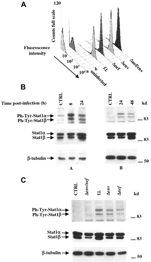
Activation of STATs in PBMCs infected either in vivo or in vitro by HIV-1 has been recently described.17 We attempted to define the contribution of monocytes/macrophages in such an activation as well as the HIV-1 proteins involved. Usually, specific STAT activation occurs in a very short time (ie, 15-30 minutes) after cell treatment with the stimulating factors. Afterward, overcoming of specific inhibitors gradually switches off STAT activation.34 Thus, to appropriately monitor and characterize possible effects on STAT activation of HIV-1 infection, a protocol of highly efficient, single-cycle HIV infection was required. Because in vitro infection of MDMs with even high multiplicity of infection (MOI) of wild-type HIV-1 leads to the virus expression in a minority of challenged cells,35 36 we decided to infect MDMs with HIV-1 preparations pseudotyped with the membrane glycoprotein from VSV (VSV-G). In our hands, a viral inoculum containing 10 ng HIV-1 p24 was sufficient to infect 106cells with an efficiency of more than 95%. This was demonstrated through the intracellular FACS analysis for the expression of HIV-1 Gag-related products performed 24 hours after the infection (Figure1A).
MDMs infected by HIV activate STAT1 by means of either
nef or env expression. (A) FACS analysis for the expression of HIV-1 in MDMs 24 hours after challenge. Peripheral blood human MDMs from healthy donors were infected with 10 ng p24 Gag/106 cells of different VSV-G pseudotyped HIV-1 strains, and the percentages of cells expressing HIV-1 were scored 24 hours afterward through intracytoplasmic FACS analysis performed by using a PE-conjugated anti-Gag HIV-1 mAb on cells infected with full-length (fl) HIV-1, or with env,nef, or env/nef deletion mutants. As controls, uninfected cells were labeled with either isotype-matched, unspecific PE-conjugated immunoglobulins (lane a) or with anti-Gag HIV-1 mAbs (lane b). (B) Western blot analysis of phosphotyrosine-STAT1, STAT1, and β-tubulin levels performed on total cell extracts assayed at different times after infection with VSV-G pseudotyped full-length HIV-1 strain. Cell extracts analyzed (A,B) were obtained by pooling simultaneous cell cultures from 4 healthy donors each.(C) Phosphotyrosine-STAT1, STAT1, and β-tubulin levels assayed through Western blots performed on total extracts of cells harvested 8 hours after infection with indicated VSV-G pseudotyped HIV-1 and indicated mutants thereof. Analyses were performed on cell extracts obtained by pooling simultaneous cell cultures from 4 healthy donors. In both panels B and C, specific signals are marked on the left side; kilodaltons of molecular size markers are reported on the right.
In such experimental conditions, the infection of MDMs with full-length HIV-1 induces activation of STAT1. STAT1 exists as 2 protein products: the 91-kd (α) and the 84-kd (β) isoforms. The latter originates from an alternative messenger RNA (mRNA) splicing, leaving out 38 amino acids from the COOH terminal of the α isoform. Both proteins contain the 701Tyr residue, whose phosphorylation is required for activation, dimerization, and nuclear translocation (reviewed in Ramana et al37). Figure 1B shows that HIV-1 infection reproducibly leads to a strong STAT1-α/β activation as early as 8 hours after challenge, which apparently decreased over the time, being still detectable 48 hours after infection. Of note, HIV-1 challenge did not apparently influence cell viability up to the last time point assayed, as proved by the cytofluorometric analysis of infected cell cultures after propidium iodide labeling (not shown). By challenging MDMs with different deletion mutants of VSV-G pseudotyped HIV-1, most interesting results were obtained with env- and nef-deleted HIV-1 strains (Figure 1C). In particular, whereas STAT1 activation detected 8 hours after the infection with either Δnef or Δenv HIV-1 was comparable to that observed on the infection with the full-length HIV-1, the infection with theenv/nef double-defective HIV-1 was ineffective, also at the later time points tested (ie, 24 and 48 hours after challenge, not shown). Of note, we reproducibly detected a slight constitutive STAT1 activation that should correlate with the monocyte-to-macrophage maturation of the cell cultures, as previously described.38 For this reason, analysis on uninfected control cell cultures was carried out on cells harvested concurrently with the later time points considered for challenged cells.
We conclude that HIV-1 infection is indeed able to activate STAT1 in MDMs and that this activation correlates with the expression ofnef and env genes.
Soluble recombinant Nef mimics the effects of the viral-encoded protein
The Nef-dependent STAT1 activation appeared new and of potential great interest, whereas STAT1 activation as a consequence of the action of HIV-1 Env gp120 has been described in cells strictly related to monocytes/macrophages.39 It is conceivable that an alteration of the STAT pathway could be part of the contribution of Nef in the development of AIDS pathogenesis.
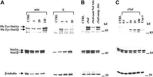
We were able to reproduce STAT1 activation by adding recombinant soluble Nef (rNef) to MDM cultures. We recently demonstrated that rNef is able to enter primary human MDMs, thereby inducing effects superimposable to those described for ectopically expressed Nef.23 We treated 7-day MDMs from different healthy donors with 100 ng/mL rNef and analyzed STAT1 activation through Western blot at different times (Figure 2A). We observed STAT1-α/β activation starting at 2 hours after the addition of rNef. This was also the time of maximal activation that, however, was clearly detectable until 20 hours after treatment (Figure2A). Such a clear-cut activation signal was observed in MDM cultures obtained from 29 of 31 healthy donors tested. Of note, the rNef treatment induced also a reproducible increase in the levels of STAT1-α/β proteins, which became clearly detectable 5 hours after the treatment (Figure 2A).
Human MDMs specifically activate STAT1 on treatment with rNef.
(A) Western blot analysis of phosphotyrosine-STAT1, STAT1, and β-tubulin levels detected on total cell extracts assayed at different times after addition of 100 ng/mL rNef. (B) Phosphotyrosine-STAT1, STAT1, and β-tubulin amounts detected through Western blots performed on total cell extracts of MDMs treated for 2 hours with medium complemented with 100 ng/mL rNef or with the same medium after treatment with anti-Nef–specific antibodies or unspecific, species-matched antibodies, and clearing with protein A-G agarose. (C) Western blot analysis of phosphotyrosine-STAT1, STAT1, and β-tubulin levels detected on total cell extracts of human MDMs after treatment for 2 hours with different doses (ie, 0.1-100 ng/mL) of rNef or with 100 ng/mL rUvp-1. In all panels, analysis on control cell cultures was carried out on cells harvested concurrently with the later time points considered for treated cells. Specific signals are indicated on the left side; molecular size markers (in kilodaltons) are reported on the right.
The specificity of the rNef effect was first demonstrated by treating MDMs with the rNef-complemented medium after Nef immunodepletion (Figure 2B). The complete rNef removal was checked by Western blot (not shown). Also, we did not detect STAT1 activation in MDMs treated with 100 ng/mL recombinant Uvp-130 (Figure 2C), a plasmid DNA invertase-resolvase protein recovered through an experimental procedure identical to that used for rNef production. Hence, we could formally exclude that STAT1 activation was a consequence of the presence of even undetectable amounts of products contaminating rNef preparations.
Extracellular Nef has been detected in the majority of AIDS patients' sera analyzed at a concentration of 5 to 10 ng/mL.40 To define whether rNef-induced STAT1 activation observed in MDMs could have an in vivo relevance, we analyzed STAT1 activation in MDMs treated with rNef doses from 0.1 to 100 ng/mL. As shown in Figure 2C, STAT1 was strongly activated starting at a concentration of 10 ng/mL, pointing out the possibility that our observations reflect phenomena actually occurring in vivo.
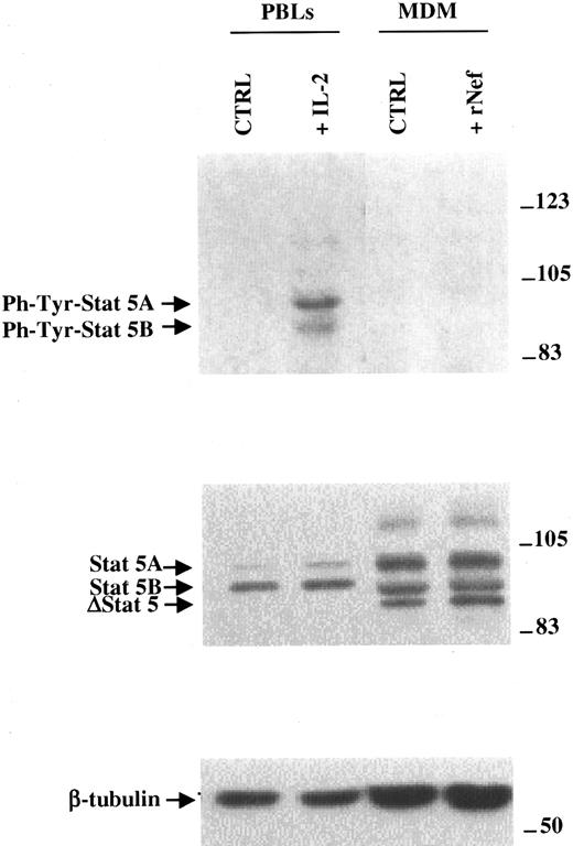
It has been reported that HIV-1 infection of PBMCs leads to activation of a truncated form of STAT5 (ΔSTAT5).17 To determine whether the interaction of Nef with monocytes/macrophages contributes to PBMC STAT5 activation, we performed Western blot analysis on purified MDMs treated with 100 ng/mL rNef. No signals were reproducibly detectable by using antibodies anti–phosphotyrosine STAT5A and STAT5B (Figure 3). This suggests that the STAT5 activation previously described in HIV-1–infected human PBMCs referred chiefly to lymphocytes or other viral products.
STAT5 is not activated on rNef treatment of human MDMs.
Phosphotyrosine-STAT5, STAT5, and β-tubulin amounts detected through Western blots performed on total cell extracts of MDMs treated for 2 hours with medium complemented with 100 ng/mL rNef. As controls, analyses of total cell extracts from PBLs nonstimulated or PHA activated and treated for 30 minutes with IL-2 are included. Specific signals are indicated on the left side; molecular size markers (in kilodaltons) are reported on the right.
Characterization of rNef-induced STAT1 activation in MDMs
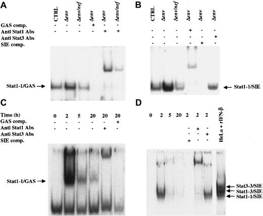
We characterized the rNef-induced STAT1 activation in terms of the ability to bind specific target sequences. In particular, we tested by electrophoretic mobility shift assay (EMSA) the binding with the GAS element from IRF-1 promoter and the SIE of c-fos promoter. It has been described that GAS element binds STAT1 homodimers,10-15 and that SIE, a GAS-like element,41 binds either STAT1-1 homodimers, STAT3 (ie, a member of the STAT family having target sequences similar to those recognized by STAT1) homodimers, and STAT1-3 heterodimers.42
Figure 4 shows EMSA carried out on total cell extracts of MDMs. We detected the formation of a single complex with both sequences tested. Cells infected with Δenv or Δenv/nef (VSV-G) HIV-1 pseudotypes were tested for STAT1 binding activity 8 hours after infection. As shown in Figure 4, panels A and B, infected MDMs showed a significant increase in STAT1 binding activity in the presence of Nef expression. Anti-STAT1 antibodies were able to shift the signals obtained with either probe, thus confirming the presence of STAT1 in the detected DNA/protein complexes. Conversely, anti-STAT3 antibodies did not alter the signal from SIE-bound complexes (Figure 4B). This might be due to either the lack of STAT3 activation or to an excess of STAT1 homodimers in SIE-bound complexes that masks possible STAT3 binding. Similar results were obtained when the assay was reproduced in rNef-treated MDMs (Figure4C,D). The peak of this binding activity (ie, 2 hours after treatment) nicely correlated with the time of maximal STAT1 activation as detected by Western blot (Figure 2A).
Nef-dependent activated STAT1 forms homodimers and binds specific DNA-responsive elements.
(A) EMSA performed by using total cell extracts from MDMs 8 hours after the infection with either Δenv or Δenv/nef(VSV-G) pseudotyped HIV-1 (A,B) or after treatment with 100 ng/mL rNef for the indicated times (C,D), and the 32P-labeled GAS element of the human IRF-1 promoter (A-C), or the 32P- labeled SIE element of the human c-fos promoter (B-D) as a probe. As controls, GAS (A-C) or SIE competitors (B-D) were used. Supershifts were performed with anti-STAT1 antibodies, and also with anti-STAT3 antibodies (B-D). As an additional control, SIE-specific EMSA was also performed on total extracts of HeLa cells treated with 100 IU/mL IFN-β (D). Electrophoretic mobility is indicated on the left side for STAT1-1/GAS complexes and on the right side for STAT1-1, 1-3, and 3-3/SIE complexes.
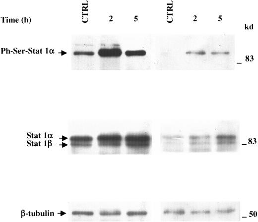
Besides the phosphorylation at the Tyr701 residue, phosphorylation at the Ser727 of activated STAT1-α has been reported.43,44Optimal transactivation driven by STAT1 homodimer depends on Ser727 phosphorylation.45 As shown in Figure5, rNef treatment of MDMs was able to induce STAT1 phosphorylation in Ser also, with a kinetic overlapping that was already observed for the Tyr701 phosphorylation. This strongly suggests that Nef-induced STAT1 activation leads to a strong transcriptional activation of target gene promoters. The slight constitutive STAT1 activation, as detectable in the assays for phosphotyrosine also (Figure 2A), should correlate with the maturation of cell cultures.38
rNef treatment of MDMs induces Ser727 phosphorylation of STAT1.
Western blot analysis of phosphoserine-STAT1, STAT1, and β-tubulin levels performed on total MDM extracts from 2 healthy donors after treatment for 2 or 5 hours with 100 ng/mL rNef. Analysis on control cell cultures was performed on cells collected concurrently with the later time point considered for treated cells. Specific signals are indicated on the left side; molecular size markers (in kilodaltons) are reported on the right. Analyses on MDMs from 2 different donors are reported.
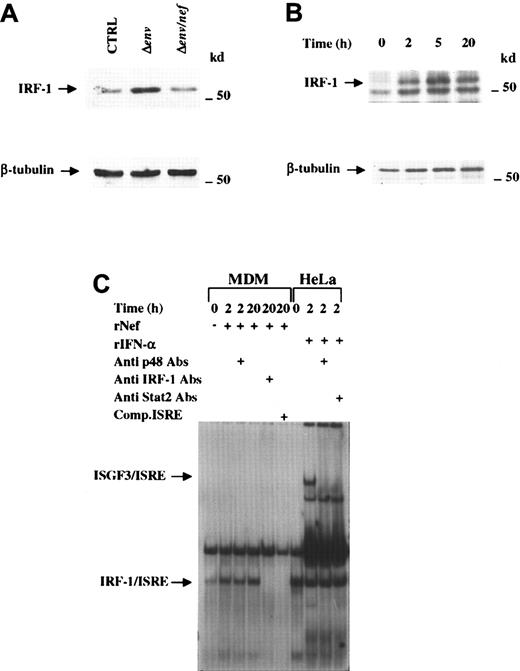
To assess whether the rNef-induced STAT1 activation leads effectively to gene activation, we tested the expression of IRF-1 protein, a transcription factor member of the IRF family of transcriptional regulators (reviewed by Mamane et al46) encoded by a gene transcriptionally regulated by STAT1 activation.47 MDMs were infected with Δenv or Δenv/nef (VSV-G) HIV-1 pseudotypes, and 8 hours after infection cell extracts were assayed for the induction of IRF-1. Figure6A shows that IRF-1 amounts increased in cells infected with a Nef expressing HIV-1. Similarly, IRF-1 was induced in rNef-treated MDMs (Figure 6B). The amounts of IRF-1 increased with the times of rNef treatment, with maximal expression at 5 hours.
IRF-1 is induced by Nef.
(A) Western blot analysis of IRF-1 and β- tubulin levels on total cell extracts of MDMs 8 hours after the infection with either Δenv or Δenv/nef (VSV-G) pseudotyped HIV-1. (B) Western blot analysis of IRF-1 and β-tubulin levels on total MDM extracts after treatment with 100 ng/mL rNef for the indicated time intervals. Specific signals are indicated on the left side; molecular size markers (in kilodaltons) are reported on the right. (C) EMSA performed by using total cell extracts from either MDMs treated with 100 ng/mL rNef or HeLa cells treated with 500 IU/mL rIFN-α2b and the32P-labeled ISRE element of the human 2′-5′A synthetase promoter as a probe. Supershifts were performed with anti-STAT2, –IRF-1 or -p48 antibodies. Electrophoretic mobility of both ISGF-3/ISRE and IRF-1/ISRE complexes are indicated on the left side.
IRF-1 binds specific elements (ie, ISRE and positive regulatory domain 1) present in the promoter of, respectively, IFN-regulated genes and IFN-β gene. EMSA performed by using the ISRE element of the 2′-5′A synthetase gene promoter showed that rNef-induced IRF-1 is indeed able to bind target sequences (Figure 6C). Through the same analysis, we noticed the lack of ISGF-3/ISRE complex formation in cell extracts from rNef-treated MDMs. ISGF-3 is a protein complex composed by the oligomerization of activated STAT1, activated STAT2 (another member of the STAT family specifically activated by type I IFN), and p48.48 This strongly suggests that rNef treatment did not induce activation of STAT2.
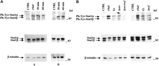
STAT1 is activated through the release of soluble factors
STATs are typically activated as a consequence of the JAK phosphorylation that follows the engagement of cytokines or growth factors to the specific receptors. The evidence that a regulatory viral protein apparently not recognizing a secretory pathway is able to activate the STAT1 signaling appeared at first glance intriguing. In fact, we already reported that rNef apparently does not recognize specific receptors on the membrane of human monocytes/macrophages.23 To establish whether Nef acts through an intracellular pathway or, differently, the Nef-induced STAT1 activation depended on the release of soluble factors, supernatants from MDMs treated for 2 hours with rNef were added to fresh MDMs from the same donors and STAT1 activation assayed shortly thereafter (ie, 30 and 60 minutes). Importantly, supernatants were tested only after immunodepletions that efficiently cleared the rNef originally added. Figure 7A shows that MDMs treated with Nef-immunodepleted medium conditioned by rNef-treated MDMs (ΔrNef CM) efficiently activated STAT1. This indicates that soluble factors induced by Nef are responsible for the STAT1 activation. The fact that the activation occurs more rapidly (ie, 30 minutes) than following rNef treatment (2 hours) further excludes that it depended on the presence of residual rNef after immunodepletion. Of note, STAT1 activation was observed also by treating for 1 hour fresh MDMs with supernatants collected 8 hours after MDM infections with wild-type,nef-, or env-deleted pseudotyped HIV-1, but not with the env/nef double-defective HIV-1 (Figure 7B). The STAT1 activation observed in MDMs treated with supernatants from Δnef HIV-1–infected MDMs likely originated at least in part from HIV-1 Env gp120 shed from infected cultures.39In these experiments, virus particles residual from challenging inoculum were cleared from supernatants of infected MDMs by ultracentrifugation. The effective removal of virus particles was checked by reverse transcriptase assay (not shown).
Nef-induced STAT1 activation in MDMs is mediated by soluble factor release.
(A) Western blot analysis of phosphotyrosine-STAT1, STAT1, or β-tubulin levels performed on total cell extracts of MDMs treated for 30 and 60 minutes with supernatants collected from MDMs of the same donors treated with 100 ng/mL Nef for 2 hours after immunodepletion for the presence of residual rNef. Cell extracts from MDMs treated with supernatants from untreated MDMs (Ctrl) or directly treated for 2 hours with 100 ng/mL rNef served as controls. Cell extracts analyzed (A,B) were obtained by pooling simultaneous cell cultures from 4 healthy donors each. (B) Western blot analysis of phosphotyrosine-STAT1, STAT1, or β-tubulin in MDMs treated with supernatants collected from MDMs infected with VSV-G pseudotyped HIV-1 expressing the full length (fl) or defective HIV-1 genomes. Eight hours after infection, supernatants were clarified from residual viral particles through ultracentrifugation and added to fresh MDMs of the same donors. After an incubation period of 60 minutes, cells were collected and total cell extracts assayed. Cell extracts from MDMs treated with supernatants from uninfected cells (Ctrl) or incubated for 2 hours with 100 ng/mL rNef were used as controls. Analyzed cell extracts were obtained by pooling simultaneous cell cultures from 3 healthy donors. In both panels, specific signals are indicated on the left side; molecular size markers (in kilodaltons) are reported on the right.
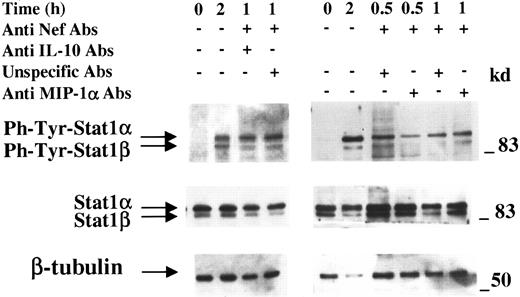
We also attempted to identify the Nef-induced soluble factor(s) responsible for the STAT1 activation. It was reported that either the ectopic expression of Nef or the rNef treatment of human MDMs induces the release of cytokines49,50 and chemokines.4,23 Among such soluble factors, those known to activate STAT1 are MIP-1α51 and IL-10.52 53Thus, we tested ΔrNef CM after additional immunodepletion of IL-10 or MIP-1α. Effective and complete removals of each factor were checked by ELISA (not shown). Figure 8 shows that neither IL-10 nor MIP-1α appeared involved in the Nef-induced STAT1 activation. Hence, an analytical molecular approach appears necessary to individuate the soluble factor(s) involved in the Nef-dependent STAT1 activation.
IL-10 and MIP-1α are not involved in the rNef-dependent STAT1 activation in MDMs.
Western blot analysis of phosphotyrosine-STAT1, STAT1, or β-tubulin carried out on total cell extracts from MDMs incubated for indicated times with supernatants from MDMs treated for 2 hours with 100 ng/mL rNef after rNef and IL-10 or MIP-1α double immunodepletions. After rNef treatment, supernatants of MDMs were collected, clarified, and immunodepleted of residual rNef and of IL-10 or MIP-1α. As control, unspecific, species-matched antibodies were added in part of the supernatants. Immunodepleted supernatants were added to fresh MDMs from the same donors and, after incubations of 30 (for MIP-1α only) and 60 minutes, cells were harvested and total cell extracts assayed. Cell extracts from MDMs treated for 2 hours with supernatants from untreated MDMs (zero time) or with 100 ng/mL rNef served as controls. Analyses were performed on cell extracts obtained by pooling simultaneous cell cultures from 4 healthy donors. Specific signals are indicated on the left side; molecular size markers (in kilodaltons) are reported on the right.
Active protein synthesis is required for the rNef-induced STAT1 activation
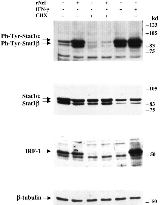
Here, we show that MDMs respond to the presence of Nef by releasing soluble factors activating STAT1. We investigated the mechanism through which Nef induces the release of the STAT1-activating factor. It is conceivable that Nef induces a proteolytic maturation of a presynthesized factor, as occurs in IL-1 secretion. Alternatively, Nef could induce a de novo synthesis of the STAT1-activating factor. In this respect, we analyzed the rNef-dependent STAT1 activation in the presence of an inhibitor of the active protein synthesis. We pretreated MDMs with 5 μg/mL cycloheximide, and, after 2 hours, rNef was added for 2 additional hours. As control, cells were also treated with 100 U/mL human IFN-γ, inducing a cycloheximide-independent STAT1 activation. We analyzed by Western blot the STAT1 Tyr701 phosphorylation, as well as the levels of STAT1, IRF-1, and β-tubulin proteins. As shown in Figure 9, cycloheximide treatment abolished the ability of rNef, but not of IFN-γ, to activate STAT1. The total expression of STAT1 was slightly affected by cycloheximide treatment and tubulin levels were not influenced, whereas IRF-1 expression dropped dramatically, as expected in view of its short half-life (about 30 minutes).54
Cycloheximide pretreatment blocks rNef-induced STAT1 activation.
Western blot analysis of phosphotyrosine-STAT1, STAT1, IRF-1, or β-tubulin on total cell extracts of MDMs pretreated or not with 5 μg/mL cycloheximide and incubated for 2 hours with either 100 ng/mL rNef or 100 IU/mL human IFN-γ for 2 hours. As controls, the analyses were carried out on cell extracts of MDMs treated either for 2 hours with 100 ng/mL rNef or with cycloheximide only. Cell extracts were obtained by pooling simultaneous cell cultures from 4 healthy donors. Specific signals are indicated on the left side; molecular size markers (in kilodaltons) are reported on the right.
This result demonstrates that active protein synthesis is required for the autocrine-regulated, rNef-dependent STAT1 activation.
Discussion
The use of a highly efficient, single-cycle infection protocol allowed us to demonstrate that HIV-1 infection of MDMs leads to the activation of both STAT1 isoforms. By using HIV-1 deletion mutants, the independent involvement of both HIV-1 nef and envgene expression in STAT1 activation has been established. Essentially overlapping results were obtained either by infecting MDMs with Nef expressing HIV-1 or by exploiting the ability of monocytes/macrophages to internalize rNef.23 This latter represents a useful tool for a more detailed characterization of the Nef-dependent activation of STATs and of its mechanism. In addition, this model seems to be relevant in that we demonstrated STAT1 activation by treating MDMs with rNef concentrations close to those detected in the serum of the majority of patients with AIDS (ie, 5-10 ng/mL).40
We observed that the extent of STAT1 activation diminished over the time in both rNef-treated and HIV-1–infected cells; the intracellular Nef levels in the latter case remained unaltered at all times considered (not shown). This is likely the consequence of either the raising of STAT-specific inhibitors,14,34 37 or of a block in the induction of the still unidentified Nef-dependent STAT1-inducing soluble factor.
Nef induced STAT1-α phosphorylation at both Tyr701 and Ser727 residues. The Ser phosphorylation is suggestive of a strong STAT1 transcriptional activity,43 44 enforcing the idea that Nef substantially influences the gene expression program in monocytes/macrophages via STAT1 activation. This is also indicated by the observed induction of both IRF-1 and STAT1.
Nef-dependent STAT1 activation has been observed by adding supernatants from rNef-treated MDMs to fresh MDMs and overlapping results were obtained by using supernatants from MDMs infected by Nef-expressing HIV-1 strains. These data are consistent with the hypothesis that STAT1 activation largely depends on the effects of soluble factors released on Nef stimulation. However, we cannot formally exclude that at least part of STAT1 activation could be mediated by an endogenous mechanism. The fact that Nef-dependent STAT1 activation appears to be a cycloheximide-sensitive event implies that the presence of Nef induces an ex novo synthesis of the factors directly or indirectly activating STAT1.
We excluded that release of MIP-1α or IL-10 could participate in the Nef-induced STAT1 activation in MDMs. These are soluble factors released on Nef stimulus4,49 known as STAT1 inducers.51-53 In the same manner, the absence of antiviral activity in the supernatants from rNef-treated MDMs (not shown) strongly suggests that rNef-dependent STAT1 activation was independent from type I (as also indicated by the lack of ISGF-3/ISRE complex formation) or type II IFN action. Identification of the soluble factors involved in the STAT1-α/β activation could lead to the discovery of factors possibly participating in the evolution of AIDS.
It is well established that Nef can interact through its SH3 homology domain with cellular Src kinases, such as Lck (in T cells)55 and Hck (in monocytes/macrophages).56,57 Also, the interaction of Nef with Vav, leading to the activation of JNK/SAPK pathway, has been recently described.58 It should be of interest to establish whether some of the already described interactions are involved in the reactions leading to the synthesis and release of STAT1-activating soluble factors.
As for the significance of the Nef-induced STAT activation in terms of AIDS pathogenesis, it has been described that infection by different viruses or even the expression of single viral proteins could generate an altered pattern of STAT activation. In particular, it has been reported that Epstein-Barr virus,59 Friend spleen focus-forming virus,60 or human hepatitis B virus X protein61 activates cell signaling cascades involving JAK/STAT pathways. Also for HIV-1, previous reports demonstrated an altered pattern of STAT1 and STAT5 response as a consequence of the viral infection.16,17,39 Even if it is known that STAT1 could be activated by several cytokines, experiments with knock-out mice revealed that STAT1 activation seems mainly involved in the response to type I and II IFNs.62,63 Thus, alterations in STAT1 biochemical pathways induced by Nef in monocytes/macrophages may lead to both a hyporesponsiveness to a possible anti-HIV effect of type I IFN and a dysregulation in the response to type II IFN, with an overall decay in the immune reactivity toward the infection. Results reported here suggest that the effects of HIV-1 infection on STAT5 activation, formerly observed in PBMCs,17 seem to involve mainly lymphocytes or viral products other than Nef. As indicated by studies performed with constitutively activated mutants, STAT5 promotes cell cycle progression and survival64 and contributes to factor-independent cell growth.65 Clearly, inappropriately prolonged cell survival of HIV-infected lymphocytes could favor HIV spread and diffusion, thus strongly influencing the progression of AIDS pathogenesis.
An unresolved debate has developed around the role that Nef plays in the development of AIDS in humans. In fact, although the use of both SIV-infected Macaca and transgenic mouse systems generated consistent conclusions on the critical role of Nef in AIDS pathogenesis,1,2 much more controversial data regarding the role that HIV-1 Nef expression plays in humans have been published.66-70 We have already described how the internalization of extracellular Nef in MDMs could induce a preferential replication of T tropic, which is the HIV quasi species more represented in the later stages of the disease,22compared to M-tropic HIV variants.23 Data presented here describe an additional consequence of Nef expression, that is, the alteration of the pattern of soluble factors released by monocytes/macrophages, which, in turn, could lead to modifications in the cross-talk between monocytes/macrophages and cells of the immune system.
Our results indicate that Nef induces similar effects in MDMs when expressed endogenously or on extracellular protein internalization. This supports the hypothesis that in vivo Nef acts similarly either in cells infected by Nef-expressing HIV strains, or in monocytes/macrophages picking up extracellular Nef possibly released by apoptotic, HIV-infected lymphocytes in those tissue districts (ie, lymph nodes) where high levels of HIV replication couple with tight interactions between monocytes/macrophages and lymphocytes. The effects of Nef expression deserve great interest considering that monocytes/macrophages resist the cytopathic effect of HIV infection and, hence, an altered soluble factor pattern may influence the immune system as long as infected monocytes/macrophages survive HIV infection.
Taken together, our data support the concept that Nef expression might induce detrimental effects in both infected and uninfected, neighboring cells. In addition, our model seems useful for the individuation of soluble factors involved in AIDS pathogenesis, as well as for new intracellular partners or functions of Nef in monocytes/macrophages.
This paper is dedicated to the memory of the late Professor Giovanni Battista Rossi and of the late Professor Franco Tatò. Monoclonal and polyclonal anti-Nef were obtained from the AIDS Research and Reference Program, Division of AIDS, National Institute of Allergy and Infectious Diseases, National Institutes of Health, Bethesda, MD. We thank Drs M. Gabbianelli and E. Pelosi, Istituto Superiore di Sanità, Rome, for kindly providing peripheral blood mononuclear cell preparations. We are indebted to A. Lippa and F. M. Regini for the excellent editorial assistance.
Supported by grants from the AIDS project of the Ministry of Health, Rome, Italy.
The publication costs of this article were defrayed in part by page charge payment. Therefore, and solely to indicate this fact, this article is hereby marked “advertisement” in accordance with 18 U.S.C. section 1734.
References
Author notes
Maurizio Federico, Laboratory of Virology, Istituto Superiore di Sanità, Viale Regina Elena, 299, 00161-Rome, Italy; e-mail: federico@iss.it.

This feature is available to Subscribers Only
Sign In or Create an Account Close Modal