Continuous hematopoietic cell lines have become important research tools, contributing significantly to a better understanding of the pathophysiology of hematopoietic tumors.1,2 A relatively large number of cell lines have also been derived from patients with multiple myeloma and related diseases such as plasma cell leukemia, plasmacytoma, etc.3,4 Unfortunately, a high percentage of hematopoietic cell lines seem to have been cross-contaminated with cells from other cell lines, which, as they have a growth advantage, overgrew completely the original culture.5
Cell line Jijoye was established from the tumor tissue of a 7-year-old boy with Burkitt lymphoma in Africa in 1963.6,7 The cell line HS-Sultan was allegedly established from the plasmacytoma of a 56-year-old Caucasian man who had IgG multiple myeloma.8
In a recent study by Davies et al (and also in a large number of previous studies by this group), the continuous cell line HS-Sultan was used as a “multiple myeloma” cell line.9 However, it has been clearly established that this cell line is in reality a subclone of the Burkitt lymphoma–derived cell line Jijoye and not a myeloma cell line.3,8,10 11 The evidence is 5-fold: most importantly DNA fingerprinting, but also viral, cytogenetic, immunologic, and morphologic findings. Due to insufficient documentation of the allegedly “new” cell line and hence caused by the lack of a proper karyotype of the original HS-Sultan cells, the circumstances of the cross-contamination can no longer be traced back. Most likely, during the attempts to establish a cell line, the cell culture was cross-contaminated with Jijoye cells, so there probably never was an “individual” HS-Sultan.
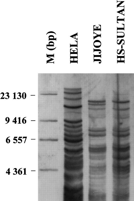
DNA fingerprinting using different methodologic approaches at the cell line banks American Type Culture Collection (ATCC) and the German Collection of Microorganisms and Cell Cultures (German abbreviation DSMZ) document independently and unequivocally that cell lines HS-Sultan and Jijoye carry identical DNA fingerprints (Figure1).
High-resolution DNA fingerprint analysis of cell lines Jijoye and HS-Sultan.
A multiple-locus DNA profile of the indicated cell lines was generated using 10 μg of DNA digested to completion (HinfI), size-separated on agarose gels and blotted on positively-charged nylon membranes. After hybridization with a digoxigenin-labelled (GTG)5 oligonucleotide as probe, a chemoluminescent detection of (GTG)5 was carried out and the blot exposed for 15 minutes to X-ray films. Molecular weight markers were used for size determination. Cervix carcinoma cell line HELA was used as positive control. The methods used have been described in details elsewhere.11 13
Both Jijoye and HS-Sultan are Epstein-Barr virus–positive (EBV+).8 While all African-type Burkitt lymphomas are EBV+ (including the cell lines derived from these tumors), myeloma is clearly not an EBV-associated disease; neither EBV DNA nor EBV nuclear antigen (EBNA) have been detected in myeloma cells in the tumor. Myeloma cells are usually refractory to EBV immortalization. There is no bona fide EBV+ myeloma cell line.4
Our cytogenetic analysis showed that both Jijoye and HS-Sultan have a similar though not identical hyperdiploid karyotype including t(8;14)(q24;q32), occurring in 75%-85% of all Burkitt lymphoma cases,12 together with partial or complete trisomy of chromosome 7. Their karyotypes are as follows: Jijoye, 46/47〈2n〉X/XY,dup(7)(q35q36),t(8;14)(q24;q32),der(14)t(9;14)(q11;q32), −18,+2mar; and HS-Sultan, 48〈2n〉XY,+7,t(8;14)(q24;q32), +mar. EBV is, of course, a polyclonal activator and any karyotypic differences may be either in vitro artefacts or attributable to the known propensity of Jijoye to give rise to phenotypically distinct subclones or a combination of both. It should be mentioned that the t(8;14)(q24;q32) is not specific for Burkitt lymphoma and was also found in some 6% of the bona fide myeloma cell lines.4
A comparative immunophenotyping analysis of myeloma and nonmyeloma cell lines showed that HS-Sultan does not express a immunomarker profile compatible with that of canonical myeloma cell lines.4 10
Finally, on May-Grünwald-Giemsa–stained cytospin slides, HS-Sultan cells do not display the morphologic features that are typical of myeloma cell lines (deep basophilic cytoplasm, excentrically located nucleus, perinuclear clear hof, dense or clumped chromatin). On the contrary, HS-Sultan cells show an abundance of vacuolization (typical for B-acute lymphoblastic leukemia and Burkitt lymphoma, also described as FAB L3), large nuclei and prominent nucleoli.
Cross-contamination of cell lines is a wide-spread problem and invalidates many research results.13 DNA fingerprinting, which is the method of choice, has documented that about 15%-20% of the currently “circulating” cell lines are cross-contaminated and overgrown by other cells.5,11 Unfortunately, cell line HS-Sultan had been used indiscriminately in a large number of studies (even after the true nature of this cell line had become known), rendering some of the conclusions irrelevant to the biology of multiple myeloma.10 Unless Anderson et al and other investigators wish to assign Burkitt lymphoma to the entity of multiple myeloma and related diseases, cell line HS-Sultan should not be employed any longer as a model system for multiple myeloma.


This feature is available to Subscribers Only
Sign In or Create an Account Close Modal