Aguayo and colleagues recently reported elevated levels of vascular endothelial growth factor (VEGF) in the cells of B-cell chronic lymphocytic leukemia (CLL).1 When discussing the possible significance of the elevated VEGF levels in CLL, the authors cite their prior abstract in which they report no significant increase in vascularity in bone marrow biopsies from patients with CLL.2
The relationship of VEGF and other growth factors to bone marrow angiogenesis is important in determining their possible role in the pathogenesis of CLL. Therefore, we wish to make the readers aware of our study that reports significantly increased angiogenesis in bone marrow biopsies from patients with CLL.3
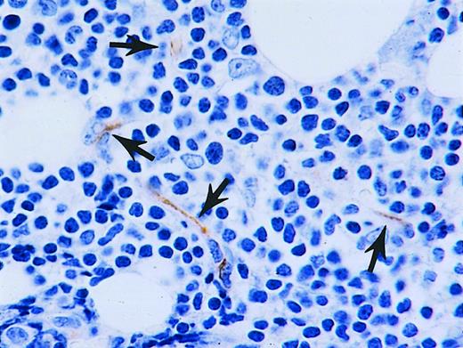
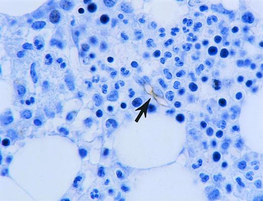
In our study, we quantified the degree of angiogenesis in CLL by measuring microvessel density in bone marrow biopsy trephine sections from controls and CLL patients using a method previously described.4 The control marrows were of comparable age and were negative for any infiltrating lesions. The microvessels in the bone marrow sections were delineated by immunohistochemistry using antibodies to CD34. The bone marrow sections from the CLL patients (Figure 1) had a mean microvessel density of 7.64 per high power field (hpf), in contrast to the control samples (Figure 2), which had a mean microvessel density of 2.11/hpf (P = .0001). The mean hot-spot microvessel density, the area with the highest microvessel density, was also significantly higher in CLL than in the control biopsy sections (P = .0008). Importantly, we also noted significant elevations of another vascular growth factor, bFGF, in the urine of CLL patients compared to controls. Thus based on the recent paper by Aguayo and our own work, there is evidence for increased or abnormal levels of 2 vascular growth factors in CLL.
Increased vascularity in CLL bone marrow.
Bone marrow trephine biopsy from CLL patient with several microvessels (arrows) highlighted by immunohistochemistry using antibodies to CD34.
Increased vascularity in CLL bone marrow.
Bone marrow trephine biopsy from CLL patient with several microvessels (arrows) highlighted by immunohistochemistry using antibodies to CD34.
Vascularity in control bone marrow.
Bone marrow biopsy from control with one microvessel (arrow) highlighted by immunohistochemistry using antibodies to CD34.
Vascularity in control bone marrow.
Bone marrow biopsy from control with one microvessel (arrow) highlighted by immunohistochemistry using antibodies to CD34.
The paper by Aguayo is important because it contributes to our knowledge about the possible factors that might be important in the growth and proliferation of the B-cells in CLL. Correlation of angiogenesis in bone marrow trephine biopsies and other tissues typically involved in CLL,5 such as lymph nodes with the levels and functions of other growth factors and cytokines, is critical in further defining the nature of these relationships. We believe that our findings add to the evidence that angiogenic factors are dysregulated in CLL and that one endpoint of this may be the abnormal angiogenesis found in CLL marrows.


This feature is available to Subscribers Only
Sign In or Create an Account Close Modal