Abstract
Most insights into the molecular mechanisms underlying transformation by the p210BCR/ABL oncoprotein are derived from studies in which BCR/ABL cDNA was introduced into hematopoietic or fibroblast cell lines. However, such cell line models may not represent all the features of chronic myelogenous leukemia (CML) caused by additional genetic abnormalities and differences in the biology of cell lines compared with primary hematopoietic progenitor and stem cells. A primary human hematopoietic progenitor cell model for CML was developed by the transduction of b3a2 BCR/ABL cDNA in normal CD34+cells. Adhesion of BCR/ABL-transduced CD34+ cells to fibronectin was decreased, but migration over fibronectin was enhanced compared with that of mock-transduced CD34+ cells. Adhesion to fibronectin did not decrease the proliferation of BCR/ABL-transduced CD34+ cells but decreased the proliferation of mock-transduced CD34+ cells. This was associated with elevated levels of p27Kip in p210BCR/ABL-expressing CD34+ cells. In addition, the presence of p210BCR/ABLdelayed apoptosis after the withdrawal of cytokines and serum. Finally, significantly more and larger myeloid colony-forming units grew from BCR/ABL than from mock-transduced CD34+ cells. Thus, the transduction of CD34+ cells with the b3a2-BCR/ABL cDNA recreates most, if not all, phenotypic abnormalities seen in primary CML CD34+ cells. This model should prove useful for the study of molecular mechanisms associated with the presence of p210BCR/ABL in CML.
Introduction
Chronic myelogenous leukemia (CML) is a malignant disease of the human hematopoietic stem cell, characterized by the Philadelphia chromosome (Ph) and a rearrangement between the breakpoint-cluster region (BCR) gene on chromosome 22 and the Abelson leukemia (ABL) gene on chromosome 9.1 The presence of p210BCR/ABL tyrosine kinase is essential and sufficient for the malignant transformation of hematopoietic cells.2-7 Studies have shown p210BCR/ABL binding or activation of molecules for a number of intracellular signaling pathways, including cytoskeletal proteins,8,9 RAS,10,11 phosphatidylinositol 3′-kinase,12 and Janus kinase–signal transducer and activator of transcription (Jak/Stat).13 However, how p210BCR/ABL causes the clinical CML syndrome is still unclear.
CML is characterized by the abnormal accumulation of immature myeloid progenitors and precursors in blood and marrow.14 At the cellular level, CML progenitors show predominant myeloid differentiation, decreased adhesion, enhanced migration, uncontrolled proliferation, and delayed apoptosis,11,15-21 all of which contribute to the clinical phenotype.14 Cell lines derived from patients with CML or generated by the transfection of the BCR/ABL cDNA in existing cell lines have been used extensively to correlate molecular effects of BCR/ABL with the CML phenotype.3,22 23 Even though hematopoietic cell lines have been used to evaluate the effect of p210BCR/ABL on cell function, these cells do not represent normal hematopoietic stem and progenitor cells, where clinical CML originates. Additional genetic abnormalities may occur in the cell lines, either as a result of the underlying transformed cell type or of the long-term presence of p210BCR/ABL, and may interfere with the interpretation of changes thought to be the result of p210BCR/ABL. Finally, the use of primary hemopoietic cells from patients with chronic-phase CML, though informative, may be complicated by the fact that additional genetic changes may already have occurred that influence the interpretation of p210BCR/ABL- mediated effects. To avoid such complicating factors, we here developed a primary CD34+ CML model by transducing normal CD34+cells with b3a2 BCR/ABL cDNA, and we demonstrate that this recreates the phenotype of CML.
Materials and methods
Preparation of primary progenitors
Heparinized cord blood was obtained from 20 full-term pregnancies. Alternatively, we used cells from normal marrow (n = 6) or marrow from patients with BCR/ABL+ chronic phase CML (n = 4). Cells were obtained after informed consent was obtained, in accordance with guidelines from the Human Subjects Committee at the University of Minnesota. Mononuclear cells were separated by Ficoll-Hypaque centrifugation (specific gravity, 1077) (Sigma Chemical, St Louis, MO). CD34+ cells were selected by 2 passages over the MACS CD34 Isolation Kit (Miltenyi Biotec, Sunnyvale, CA). CD34+-enriched populations were usually greater than 95% pure.
p210-eGFP retroviral vector
Full-length b3a2 BCR/ABL cDNA (a kind gift from Dr J. Y. Wang, University of California at San Diego, California)23was cloned upstream from the internal ribosomal entry site (IRES) of the murine stem cell virus (MSCV)–IRES–enhanced green fluorescent protein (eGFP) vector (a kind gift from Dr W. Pear, University of Pennsylvania, Philadelphia).24 The BCR/ABL-containing vector was termed p210-eGFP. The correct sequence of the p210-eGFP construct was determined by DNA sequence mapping. eGFP and p210-eGFP plasmids were cotransfected with the pCL-Ampho packaging plasmid into 293kj cells (a kind gift from Dr Haas, University of California at San Diego)25 using a modification of the HEPES-buffered saline calcium-phosphate method. Then 293kj cells were cocultured with DNA containing the precipitate for 6 hours and were shocked by the addition of 15% glycerol solution. Cells were cultured in Dulbecco minimum essential medium with high-glucose (Gibco/BRL, Gaithersburg, MD), virus-containing supernatant harvested on days 1 and 2. Titer of the eGFP vector was 5 × 10⁵ plaque-forming units (PFU)/mL and titer of the p210-eGFP vector was 5 × 10⁵PFU/mL, as determined by infecting 3T3 cells in the presence of 8 μg polybrene.
Transduction of primary CD34+ cells
CD34+ cells obtained from umbilical cord blood or bone marrow (n = 2) were cultured in serum-free medium (Stem Cell Technologies, Vancouver, Canada) with 50 ng/mL stem cell factor (SCF; Amgen, Thousand Oaks, CA), 50 ng/mL fetal liver–tyrosine kinase-3 ligand (Flt3-L; Immunex, Seattle, WA), and 50 ng/mL thrombopoietin (TPO; Amgen) for 2 days. 105 CD34+ cells were then resuspended in 200μL retroviral supernatant supplemented with SCF, Flt3-L, and TPO and were placed in collagen-coated transwells (Transwell-COL; Corning Costar, Cambridge, MA) of 24-well plates that had previously been incubated with 50μg/mL recombinant CH296 fibronectin (Takara Shuzo, Otsu, Japan). Once the viral medium was drained through the transwell, the filtrate was removed from the bottom compartment, and 200 μL fresh viral supernatant with cytokines was added to the transwell until 1 mL p210-eGFP or eGFP containing retroviral supernatant was filtered.26 When the last 200 μL viral supernatant was added, serum-free medium with cytokines was added to the bottom compartment, and cells were cultured with virus for 8 hours at 32°C in 5% CO2.21 Afterward, all viral supernatant was drained, and fresh serum-free medium with cytokines was added to the transwell and bottom compartment for 16 hours. This was repeated once or twice. Twenty-four hours after the last transduction, cells were labeled with anti-CD34–phycoerythrin (Becton Dickinson, Palo Alto, CA), and eGFP+CD34+ cells were selected by FACS (FACStar Plus with Consort 32 computer) (Becton Dickinson) using isotype control-labeled cells.
Adhesion assays
For adhesion assays, 104 p210-eGFP– or eGFP-transduced GFP+ CD34+ cells suspended in 150 μL serum-free medium were plated in fibronectin-coated (50 μg/mL) or bovine serum albumin (BSA)-coated (5 mg/mL) (both from Sigma) wells of 48-well plates for 2 hours. Nonadherent and adherent cells were recovered and plated in short-term methylcellulose assay as described.18 The percentage of adherent colony-forming cells (CFCs) was calculated as: [no. adherent CFCs/(no. adherent CFCs + no. nonadherent CFCs)] × 100.
Migration assay
For migration assays, 104 p210-eGFP– or GFP-transduced eGFP+ CD34+ cells, or normal or CML marrow, were plated in fibronectin-coated (50 μg/mL) or BSA-coated (5 mg/mL) dishes and tilted to 80° for 1 hour, which allowed cells to accumulate in the lower rim. Dishes were lowered to 20° for 12 hours, and migration was assessed after enumerating the number of cells that had migrated beyond the middle line. In some experiments a blocking anti–β1-integrin antibody (P4C10) or a control IgG was added (both from Gibco-BRL).
Proliferation inhibition assay
For proliferation inhibition assays, eGFP- or p210-eGFP–transduced GFP+ CD34+ cells were treated with 5 μg/mL β1–integrin-activating antibody 8A2 (a kind gift from Dr N. Kovach, University of Washington, Seattle), as previously described.27 Then 104CD34+ cells were plated in fibronectin-coated (50 μg/mL) or poly-L–lysine-coated (Sigma) (5 mg/mL) dishes for 12 hours. Nonadherent and adherent cells were collected as described. Cell cycle status was assessed using propidium iodide as described.28
Determination of p27Kip levels in transduced CD34+ cells
The levels of p27Kip in eGFP- and p210-eGFP–transduced CD34+ cells were determined by FACS 24 hours after transduction.28 Transduced cells were fixed and permeabilized with 1% paraformaldehyde, 80 μg/mL lysolecithin (all from Sigma) in phosphate-buffered saline (PBS; Gibco) for 5 minutes at 4°C. Cells were labeled with anti-CD34–antigen-presenting cells (Becton Dickinson) and then incubated with anti-p27Kip antibodies (Pharmingen), washed, and incubated with phycoerythrin-conjugated goat antimouse immunoglobulin (Pharmingen) for 30 minutes. CD34+ eGFP+ cells and CD34+ eGFP− cells were gated, and p27Kip levels were compared.
Progenitor survival assay
CD34+ cells were plated in serum-free medium with or without 500 pg/mL recombinant human interleukin-3 (rhIL-3) for 2 days. The absolute number of viable CFCs was determined by plating culture progeny in methylcellulose assay. Survival of CFCs was determined by comparing the number of CFCs in culture progeny with those in fresh, uncultured cells.
Colony-forming assay
CD34+ cells were plated in methylcellulose containing Iscoves modified Dulbecco medium supplemented with 30% fetal calf serum, 3 IU erythropoietin (Amgen), 10 ng/mL each rhIL-3 (R&D Systems, Minneapolis, MN), SCF, granulocyte-macrophage–colony-stimulating factor (GM-CSF; Immunex), and 3 IU erythropoietin (Amgen), as previously described.18
Western blot analysis for p210BCR/ABL
Western blot analysis was performed as previously described.29 Briefly, 5 × 10⁵ p210-eGFP– or eGFP-transduced GFP+ CD34+ cells were lysed directly in SDS sample buffer (50 mM/L Tris-HCl, pH 6.8, 2% SDS, 10% glycerol, and 5% β-mercaptoethanol; all from Sigma). After boiling, the lysates were separated by SDS-PAGE and transferred onto polyvinylidene difluoride. Blots were incubated with Blocker Blotto in TBS (Pierce, Rockford, IL) and 0.1 μg/mL anti-ABL–mouse IgG1 antibody (clone 24-11; Santa Cruz Biotechnology, Santa Cruz, CA), washed, and incubated for 1 hour with a goat antimouse horseradish peroxidase–conjugated antibody (1:10 000 dilution; Jackson ImmunoResearch Laboratories, West Grove, PA). Protein bands were visualized using an ECL detection system (du Pont de Nemours, Boston, MA).
Immunohistochemistry
For immunochemistry eGFP- or p210-eGFP–transduced GFP+ CD34+ cells were cytospun on glass slides and incubated in 100% ethanol (Sigma) for 20 minutes. Endogenous peroxidase was blocked with 0.3% H2O2 in 50% methanol for 5 minutes (Sigma). Slides were stained using an established avidin–biotin complex (ABC) protocol (Vectastain Elite Kit; Vector Laboratories, Burlingame, CA) according to the manufacturer's recommendation. Slides were incubated with 20% horse serum (Stem Cell Technologies) for 60 minutes, then incubated with 4 μg/mL anti-BCR mouse IgG1 (clone 7C6; Santa Cruz Biotechnology) at 4°C overnight. Slides were rinsed in PBS, and goat antimouse secondary antibody (Vector Laboratories) was added at room temperature for 30 minutes. After another PBS rinse, slides were incubated in freshly prepared ABC solution, rinsed again, and developed with diaminobenzidine. All slides were lightly counterstained with hematoxylin (Sigma).
Statistical analysis
Results of experimental points obtained from multiple experiments were reported as mean ± SEM. Significance levels for differences between different samples were determined using the 2-tailed Student t test.
Results
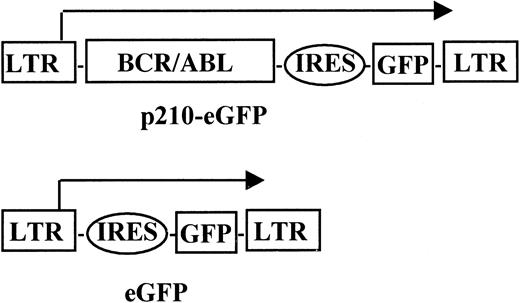
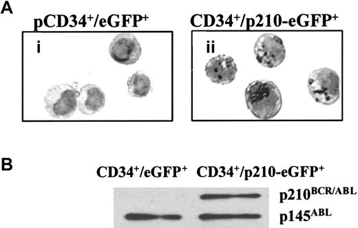
We constructed the p210-eGFP retroviral vector by cloning the b3a2 BCR/ABL cDNA in the multicloning site upstream from the IRES sequence of the MSCV–IRES–eGFP (eGFP) vector. Because p210BCR/ABLis toxic to fibroblast,23 we performed transient cotransfection of the p210-eGFP or eGFP plasmid with the pCL plasmid in 293kj cells to generate retroviral vector–containing supernatants.25 Umbilical cord blood CD34+cells were transduced with p210-eGFP or eGFP supernatant on 2 or 3 consecutive days using a transwell transduction procedure as described previously.26 Twenty-four hours after the last transduction, eGFP+ CD34+ cells were selected by FACS, and adhesion, migration, proliferation, and apoptosis were evaluated (Figure 1). Between 40% and 65% of all cells recovered from transduction cultures were CD34+. Thirty percent to 40% of CD34+ cells transduced with the eGFP vector expressed eGFP, whereas 5% to 10% of CD34+ cells transduced with the p210-eGFP vector were eGFP+. We assessed the expression of p210-eGFP transduced CD34+ cells by immunohistochemistry and Western blot analysis. As shown in Figure 2A, the cytoplasm of p210-eGFP–transduced CD34+ cells stained considerably stronger with an anti-BCR antibody than did control eGFP-transduced CD34+ cells, indicating the expression of p210BCR/ABL in the cytoplasm of transduced cells. Similarly, Western blot analysis showed the presence of p210BCR/ABL at levels similar to those for endogenous p145ABL in p210-eGFP–transduced, but not in eGFP-transduced, CD34+ cells (Figure 2B). We then examined whether the transduction of normal CD34+ cells with BCR/ABL would recreate the cellular phenotype of CML. Specifically, we examined the effect on cell adhesion, cell migration, cell proliferation and differentiation, and cell survival.
p210-eGFP vector.
Full-length b3a2 BCR/ABL cDNA was cloned upstream from the IRES of the MSCV–IRES–eGFP vector. The correct sequence of the p210-eGFP construct was determined by DNA sequence mapping.
p210-eGFP vector.
Full-length b3a2 BCR/ABL cDNA was cloned upstream from the IRES of the MSCV–IRES–eGFP vector. The correct sequence of the p210-eGFP construct was determined by DNA sequence mapping.
p210BCR/ABL is expressed in p210-eGFP–transduced GFP+ CD34+ cells.
(A) p210-eGFP– or eGFP-transduced GFP+ CD34+cells were cytospun onto glass slides and fixed in 100% ethanol for 20 minutes. Endogenous peroxidase was blocked with 0.3% H2O2 in 50% methanol for 5 minutes. Immunohistochemistry was performed using an established avidin-biotin complex with specific primary anti-BCR antibody. Staining is significantly greater in the cytoplasm of p210-eGFP–transduced eGFP+ CD34+ cells (ii) than in eGFP-transduced eGFP+ CD34+ cells (i). Representative example of 2 individual experiments is shown. (B) Proteins were extracted from 5 × 10⁵ p210-eGFP– or eGFP-transduced GFP+CD34+ cells. Proteins were resolved by electrophoresis on 8% polyacrylamide gels and transferred to polyvinylidene difluoride. Blots were incubated with anti-ABL antibody and a goat antimouse horseradish peroxidase–conjugated antibody, as described in “Materials and methods.” Protein bands were visualized using an ECL detection system. p210BCR/ABL was present in p210-eGFP– but not in eGFP-transduced eGFP+ CD34+ cells at levels similar to those of endogenous p145ABL. Representative example of 2 individual experiments is shown.
We performed adhesion assays of p210-eGFP– or eGFP-transduced GFP+ CD34+ cells to fibronectin or BSA.18 We found that 24.4% ± 4% of eGFP-transduced CFCs adhered to fibronectin, whereas only 3.4% ± 1% of p210-eGFP–transduced CFCs adhered to fibronectin (n = 4) (Figure3). Thus, the presence of p210BCR/ABL in normal CD34+ cells caused decreased adhesion. This is similar to what we have previously described for primary, chronic-phase CML (10% ± 4% adhesion) CD34+ cells versus normal bone marrow (22% ± 5% adhesion) CD34+ cells.30
Presence of p210BCR/ABL in normal CD34+ cells inhibits adhesion of CFCs to fibronectin.
p210-eGFP– (▪) or eGFP-transduced (■) GFP+CD34+ cells (104) from 4 umbilical cord blood samples were plated in 24-well plates coated with 100 μg/mL fibronectin (FN, left) or 5% BSA (right) for 2 hours. Nonadherent cells and adherent cells were collected separately and plated in methylcellulose progenitor assay. The percentage of adherent CFCs was calculated as [no. adherent CFCs/(no. adherent CFCs + no. nonadherent CFCs)] × 100. The number of CFCs in the nonadherent + adherent population was 66 ± 6/1000 cells for eGFP-transduced GFP+ CD34+ cells and 268 ± 25/1000 cells for p210-eGFP–transduced GFP+CD34+ cells. Comparison between p210-eGFP– and eGFP-transduced GFP+ CD34+ cells (P = .001).
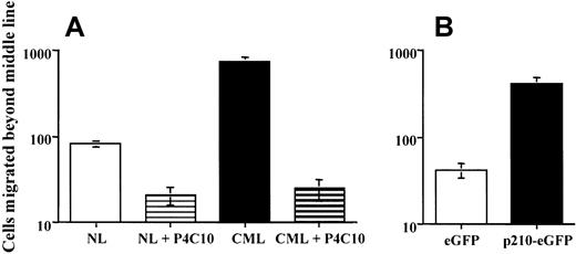
We next tested the effect of p210BCR/ABL on CD34+ cell migration. We plated normal bone marrow CD34+ cells (n = 4), CML bone marrow CD34+cells (n = 4), p210-eGFP– or eGFP-transduced GFP+CD34+ cells in fibronectin- or BSA-coated wells. Wells were then tilted to 80° for 1 hour and lowered to 20°. After 12 hours, we enumerated the fraction of cells that had migrated at least halfway across the well. When 104 primary CML CD34+cells were allowed to migrate on fibronectin-coated tilted dishes, 758 ± 79 cells migrated beyond the middle line, and this could be inhibited by pretreating the cells with a blocking anti–β1-integrin antibody (25 ± 7 cells) (Figure4A). In contrast, when 104CD34+ cells from normal marrow were allowed to migrate, only 83 ± 7 cells migrated beyond the middle line. Of eGFP+ CD34+ cells transduced with eGFP, 42 ± 8 cells crossed the middle line, whereas 418 ± 66 eGFP+ CD34+ cells transduced with p210-eGFP (n = 4) (Figure 4B) migrated beyond the middle line. Thus, the introduction of BCR/ABL in normal CD34+ cells recreates the abnormal migration characteristics of primary CML CD34+cells.
Presence of p210BCR/ABL in normal CD34+ cells enhances migration.
eGFP+ CD34+ cells were plated in fibronectin- or BSA-coated dishes and tilted to 80° for 1 hour: 104normal bone marrow (n = 4) or CML bone marrow (n = 4) (A) or p210-eGFP–transduced (n = 4) or eGFP-transduced (n = 4) (B). For normal and CML bone marrow, studies were performed in the presence or absence of the blocking anti–β1-integrin antibody, P4C10. Dishes were then lowered to 20° for 12 hours, and cells that had migrated past the middle line were enumerated under the microscope. (A) Comparison between NL and CML CD34+ cells (P = .001). Comparison between NL CD34+ cells with and without P4C10 (P < .01). Comparison between CML CD34+ cells with and without P4C10 (P = .001). (B) Comparison between p210-eGFP– and eGFP-transduced GFP+CD34+ cells (P = .001).
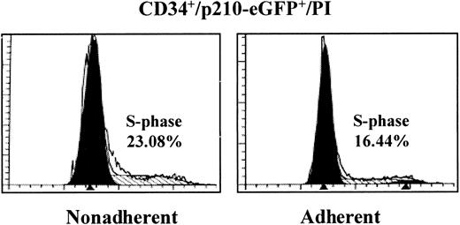
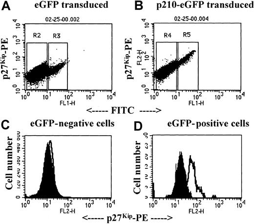
Engagement of β1 integrins inhibits the proliferation of normal but not chronic-phase CML CD34+ cells.28,31 To examine whether CD34+ cells transduced with BCR/ABL cDNA have similar characteristics, we cultured p210-eGFP– or eGFP-transduced GFP+ CD34+ cells for 12 hours with fibronectin or the nonspecific adhesive substrate poly-L-lysine and assessed the cell cycle status of adherent and nonadherent cells using propidium iodide. To enhance the adhesion of p210BCR/ABL+ cells, p210-eGFP– or eGFP-transduced cells were treated with the β1-integrin–activating antibody, 8A2. We have previously shown that this increases cell adhesion without affecting integrin-mediated proliferation inhibition.27 As shown in Figure 5, 23.7% ± 1.7% fibronectin-nonadherent and 17.4% ± 2.3% fibronectin-adherent p210-eGFP–transduced eGFP+ cells were in S-phase (n = 3; difference not significant). In contrast, 17% ± 1% fibronectin-nonadherent cells but only 9% ± 1% fibronectin-adherent eGFP-transduced GFP+fibronectin-adherent cells were in S-phase (P = .001). These results are consistent with previous studies from our group, demonstrating 50% inhibition of proliferation of normal CD34+ cells after adhesion to fibronectin and less than 25% inhibition in CML CD34+ cells.28 We have also shown that the lack of adhesion-mediated growth inhibition after the engagement of β1 integrins is, at least in part, due to the lack of p27Kip-mediated inhibition of Cdk2 activity.28 Furthermore, we showed that the expression of p27Kip is abnormally elevated but mislocated in the cell cytoplasm. To determine whether p210BCR/ABL causes elevated levels of p27Kip in CML, we analyzed p27Kiplevels by FACS 24 hours after the last transduction with the eGFP or the p210-eGFP vector. As we saw for primary CML cells, p27Kip levels were significantly higher in eGFP+ CD34+ that had been transduced with p210-eGFP (Figure 6) (n = 3) than eGFP+ CD34+ cells that had been transduced with eGFP.
Presence of p210BCR/ABL in normal CD34+ cells prevents adhesion-mediated proliferation inhibition.
Cord blood CD34+ cells (5 × 10⁶) were transduced on 3 consecutive days with the control vector, eGFP, or with p210-eGFP and were plated in fibronectin or poly-L-lysine–coated dishes for 12 hours in the presence of the β1-integrin activating antibody, 8A2. Nonadherent and adherent cells were collected separately, cells were fixed and incubated with propidium iodide, and the cell cycle status of CD34+/eGFP+ cells was analyzed by FACS using Modfit-LT software. A representative example of fibronectin-nonadherent and fibronectin-adherent p210-eGFP–transduced cells is shown.
Transduction of p210BCR/ABL in NL CD34+ cells causes elevated levels of p27Kip.
Cord blood CD34+ cells (5 × 10⁶) were transduced on 3 consecutive days with the control vector, eGFP (A: R2, eGFP−; R3, eGFP+) or with p210-eGFP (B: R3, eGFP−; R4, eGFP+). Twenty-four hours after the last transduction, cells were labeled with anti-CD34 APC, fixed, permeabilized, and incubated with antibodies against p27Kip, and washed and incubated with phycoerythrin-conjugated goat antimouse immunoglobulin. CD34+ eGFP− cells (C: R2, eGFP transduced; R4, p210-eGFP transduced) and CD34+ eGFP+ cells (D: R3, eGFP transduced; R5, p210-eGFP transduced) were gated, and p27Kip levels were compared. A representative example of 3 separate experiments is shown.
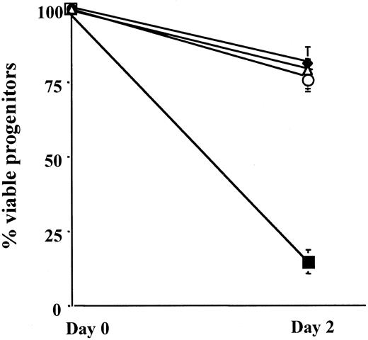
Considerable evidence shows that p210BCR/ABL has antiapoptotic effects.11,21 32-34 Therefore, we assessed whether the transduction of CD34+ cells with p210BCR/ABL would render cells resistant to apoptosis induced by serum and cytokine withdrawal. p210-eGFP– or eGFP-transduced GFP+ CD34+ cells were cultured in serum-free medium with or without 500 pg/mL IL-3 for 2 days, and CFCs were enumerated in fresh and cultured cells. We found that 78% ± 5% of eGFP-transduced CFCs remained viable after 48 hours in culture with IL-3, whereas only 15% ± 2% of eGFP-transduced CFCs were recovered after culture for 48 hours without IL-3 (Figure7) (n = 3). In contrast, 81% ± 6% of p210-eGFP–transduced CFCs were maintained after 2 days of culture without IL-3, and 77% ± 5% were maintained when cultured with IL-3.
Presence of p210BCR/ABL in normal CD34+ cells delays apoptosis.
p210-eGFP– or eGFP-transduced GFP+ CD34+ cells (104) from 4 cord blood samples were plated in serum-free medium with or without 500 pg/mL rhIL-3 for 2 days. The number of CFCs was determined in fresh and 2-day culture cells by plating 104 cells or their culture progeny in methylcellulose assay. Percentage CFCs that survived the 2-day culture was calculated as (no. CFCs in 2-day progeny/no. CFCs in fresh cells) × 100. Comparison between p210-eGFP– and eGFP-transduced GFP+CD34+ cells (P = .001). ▪, eGFP/no IL-3; ▵, eGFP/+IL-3; ♦, p210-eGFP/no IL-3; ○, p210-eGFP/+IL-3.
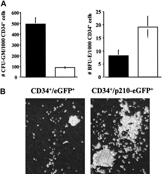
Finally, CML is characterized by extensive expansion of the myeloid cell pool and significantly smaller expansion of the erythroid cell pool.15,16 35 To determine whether transduction with BCR/ABL would recreate this last characteristic, we assessed the number of granulocyte-macrophage colony-forming units (GM-CFU) and burst-forming units-erythroid (BFU-E) generated per 1000 eGFP- or p210-eGFP–transduced GFP+ CD34+ cells cultured in methylcellulose progenitor assay supplemented with SCF, GM-CSF, and IL-3, each at a final concentration of 5 ng/mL and erythropoietin 3 IU/mL. In cultures initiated with 1000 p210-eGFP–transduced CD34+ cells, we detected 493 ± 58 GM-CFU and 8 ± 2 BFU-E (n = 4) (Figure8A,B). In contrast, in cultures initiated with 1000 eGFP-transduced CD34+ cells, we detected 89 ± 10 GM-CFU and 19 ± 4 BFU-E (n = 4).
Presence of p210BCR/ABL in normal CD34+ cells increases number of GM-CFU.
p210-eGFP– or eGFP-transduced GFP+ CD34+ cells (103) from 4 umbilical cord blood samples were plated in methylcellulose assay with SCF, GM-CSF, and IL-3 each at a final concentration of 5 ng/mL and erythropoietin at 3 IU/mL. After 14 days, GM-CFU and BFU-E were enumerated separately, and the size of colonies was assessed visually. (A) Significantly greater numbers of GM-CFU in 103 p210-eGFP– (▪) than eGFP-transduced (■) GFP+ CD34+ cells and similar numbers of BFU-E in either population. Comparison between GM-CFU in 103p210-eGFP– and eGFP-transduced GFP+ CD34+cells (P = .001). (B) In addition, the size of GM-CFU was significantly greater in p210-eGFP– than in eGFP-transduced GFP+ CD34+ cells.
Discussion
We have developed a human model of p210BCR/ABL+ CML by transducing normal, human umbilical cord blood CD34+ cells with a retroviral vector containing the b3a2 BCR/ABL cDNA. The model recreates most, if not all, features of CML. Like chronic-phase CML CD34+ cells, BCR/ABL-transduced CD34+ cells adhere less well to,18,30 but migrate better over, fibronectin than normal or mock-transduced CD34+cells.8,36 BCR/ABL-transduced CD34+ cells, but not untransduced or mock-transduced CD34+ cells, continue to proliferate after the engagement of β1 integrins.19,31 Consistent with our recent study of primary CML CD34+ cells, we found that the transfer of BCR/ABL in CD34+ cells is associated with elevated levels of the cell cycle inhibitor p27Kip.28 We also show that BCR/ABL-transduced CD34+ cells are less sensitive to apoptotic cell death after the withdrawal of cytokines and serum.11,21,32,33 Finally, as is seen in primary CML,15,16 35 there is excess production of myeloid progenitors and precursors.
We show that BCR/ABL transduced CD34+ cells differentiate chiefly to the myeloid lineage, which is consistent with findings demonstrated by Clarkson et al16 and Marley et al.15,35 In addition, the size of GM-CFU in cultures with BCR/ABL-transduced CD34+ cells was greater than that of mock-transduced GM-CFU. The increase in the number of GM-CFU generated may be due to the increased sensitivity of BCR/ABL-containing progenitors to cytokines, resulting in the recruitment of otherwise quiescent progenitors in cycle.33 In addition, we found that p210-eGFP–transduced CD34+ cell survival under cytokine-depleted conditions is significantly better than that of mock-transduced cells, consistent with a series of studies indicating that p210BCR/ABL has antiapoptotic activity.11,21,32 33 However, survival and number of GM-CFU was not different when mock- or p210BCR/ABL-transduced cells were cultured in the presence of cytokines for 2 days. Nevertheless, the increased number and size of GM-CFU in cultures of BCR/ABL-transduced CD34+ cells may also be due to increased proliferation or decreased apoptosis of progenitors, which becomes obvious only after a longer period in culture under cytokine-replete conditions. Single-cell assays will be needed to discriminate between these possibilities.
Compared with what we have observed for primary CML CD34+cells,27,30,37,38 the excess GM-CFU growth is more pronounced in the model described here. Similarly, the increased size of GM-CFU in cultures of BCR/ABL-transduced CD34+ cells is more pronounced than what we have commonly seen in cultures of primary CML CD34+HLA-DR+ cells. Finally, the degree of resistance against apoptosis, when cultured in serum-free and cytokine-free cultures, was greater in the model system than in primary CML. It is possible that differences seen between our CML model and primary chronic-phase CML may be related to the level of p210BCR/ABL in transduced cord blood cells. In the p210-eGFP model, p210BCR/ABL expression is regulated from the MSCV-LTR promoter and not the endogenous BCR promoter, as in chronic-phase CML. Levels of p210BCR/ABL in the p210-eGFP model are similar to those of endogenous p145ABL, which is 3- to 5-fold higher than p210BCR/ABL levels in primary CML CD34+ cells. Our finding that the effects of BCR/ABL are exaggerated is similar to what has been reported by a number of investigators who generated a murine model of CML-like disease by transducing mouse stem cells with a BCR/ABL-containing retroviral vector.2,6,7 They, too, speculated that this was the result of high levels of p210BCR/ABL oncoprotein in the hematopoietic stem and progenitor cells. This was elegantly illustrated by Cambier et al,33 who found that effects on cell survival and in vivo tumorigenicity of cell lines varies significantly with different levels of p210BCR/ABL expression. Use of an inducible vector will be needed to determine whether the higher expression of p210BCR/ABL in the model system is responsible for these differences.
An alternative explanation may be that we introduced the BCR/ABL cDNA in cord blood CD34+ cells and not in adult marrow cells. We chose umbilical cord blood rather than marrow because the transduction of primary normal marrow-derived CD34+ cells with the p210-eGFP construct was usually less than 1%, which precludes the studies described here. We have previously shown that cord blood CD34+ cells adhere to fibronectin, migrate over fibronectin, and are regulated by adhesion to fibronectin in a manner similar to that of marrow-derived CD34+cells.39 Whether differences in proliferative potential between cord blood and adult bone marrow CD34+cells40 41 contribute to the “exaggerated” proliferation and resistance to apoptosis seen between p210-eGFP–transduced cord blood CD34+ cells and primary CML CD34+ cells is unknown.
Despite these differences, we believe that the model in which normal CD34+ cells are transduced with p210-eGFP recreates the cardinal clinical features of CML and should be invaluable in studies aimed at further characterizing the molecular mechanisms through which BCR/ABL causes functional defects in CML CD34+ cells. Several groups have shown that the transplantation of murine progenitors transduced with a BCR/ABL cDNA-containing retroviral vector creates a myeloproliferative disease akin to chronic-phase CML in 100% of recipient mice, with a latency of 4 to 6 weeks.2,6 7Whether functional abnormalities resulting from the transduction of human progenitors with p210-eGFP at the cellular level identified in the human model described in this article are also seen in the murine models is yet to be evaluated.
Transplantation of human chronic-phase CML marrow or blood in immunodeficient mice leads to the development of a CML-like disease in the marrow and spleen of engrafted mice.42-44 However, the frequency and level of engraftment are variable when different patient samples are examined, somewhat compromising the usefulness of the model. This variability may be due to the fact that some, but not all, primary chronic-phase CML samples have already acquired additional genetic abnormalities that contribute to the final phenotype seen for each patient.45 This would be circumvented in the model developed here.
Finally, our study confirms that the p210BCR/ABLoncoprotein is necessary and sufficient for the abnormal functional characteristics seen in CML. To prove that functional abnormalities in CML are caused by the p210BCR/ABL oncoprotein, several studies used antisense29,46,47 or ribozyme48strategies to suppress p210BCR/ABL expression, or they used tyrosine kinase inhibitors such as STI57149,50 or AG95751 to inhibit the p210BCR/ABL kinase activity. The fact that we can recreate all cellular functional defects ascribed to CML by transferring the BCR/ABL gene in normal primary hematopoietic cells further confirms the causal relation between p210BCR/ABL and the CML phenotype.
In conclusion, this model recreates most characteristics of chronic-phase CML in vitro. It should, therefore, prove a useful tool for investigators interested in determining the molecular mechanisms underlying p210BCR/ABL-mediated abnormalities in progenitor function.
We thank Valerie McCullar, Erjin Fan, and the members of the Stem Cell Laboratory for their excellent technical assistance.
Supported by National Institutes of Health grants RO1-HL-49930, RO1-DK-53673, and RO1-CA 74887; the Leukemia and Lymphoma Society of America; University of Minnesota Bone Marrow Transplant Research Fund; Minnesota Medical Foundation; University of Minnesota Graduate School; and the Institute of Hematology at the Chinese Academy of Medical Science.
The publication costs of this article were defrayed in part by page charge payment. Therefore, and solely to indicate this fact, this article is hereby marked “advertisement” in accordance with 18 U.S.C. section 1734.
References
Author notes
Catherine M. Verfaillie, Stem Cell Institute, University of Minnesota, Mayo Mail Code 806, 420 Delaware St SE, Minneapolis, MN 55455; e-mail: verfa001@tc.umn.edu.

![Fig. 3. Presence of p210BCR/ABL in normal CD34+ cells inhibits adhesion of CFCs to fibronectin. / p210-eGFP– (▪) or eGFP-transduced (■) GFP+CD34+ cells (104) from 4 umbilical cord blood samples were plated in 24-well plates coated with 100 μg/mL fibronectin (FN, left) or 5% BSA (right) for 2 hours. Nonadherent cells and adherent cells were collected separately and plated in methylcellulose progenitor assay. The percentage of adherent CFCs was calculated as [no. adherent CFCs/(no. adherent CFCs + no. nonadherent CFCs)] × 100. The number of CFCs in the nonadherent + adherent population was 66 ± 6/1000 cells for eGFP-transduced GFP+ CD34+ cells and 268 ± 25/1000 cells for p210-eGFP–transduced GFP+CD34+ cells. Comparison between p210-eGFP– and eGFP-transduced GFP+ CD34+ cells (P = .001).](https://ash.silverchair-cdn.com/ash/content_public/journal/blood/97/8/10.1182_blood.v97.8.2406/5/m_h80810938003.jpeg?Expires=1770836707&Signature=jl6eoTNV7j8UMS1CoGQDrYl6g3znqvTcodtzWL8ueNi9XMi5HVnDkYZHzjVoPMnmFJ4n6f5mbXVuYNNUHVS8LrWFP7ZlbAfspVLTCWKHeaCWEW3auxOlul3PG~qbyecH7f47YNa0Dt-SUnQhJ2w~aECzUU9AOCOVoAOInwpLJ~glaR6UNzqBSxrZdFR45DPshKa1g13myjcvnJmoWRkNn~4Yi5iOoRnJ5cW3Sfb8QOU~EXkrZVR9~7LK-lFMwW2wM8rhq7y8a8ZG1ZdvR1rhTgyrhVKqD5~soElaSq9HMXJIoKfQU2Nc-F99oMBGJgpX1qzlYk3S2o4BFaZENfSUYQ__&Key-Pair-Id=APKAIE5G5CRDK6RD3PGA)
This feature is available to Subscribers Only
Sign In or Create an Account Close Modal