Abstract
AML1 is a transcription factor that is essential for normal hematopoietic development. It is the most frequent target for translocations in acute leukemia. Recently, fluorescence in situ hybridization was used to identify a novel syndrome of radiation-associated secondary acute myelogenous leukemia that had AML1 translocations. Using polymerase chain reaction, the AML1 fusion transcript was isolated from the patient who had a t(19;21) radiation-associated leukemia. The AML1gene is fused out of frame to chromosome 19 sequences, resulting in a truncated AML protein bearing the DNA binding domain but not the transcriptional activation domain. This fusion AML1 protein functions as an inhibitor of the normal AML1 protein.
Introduction
The AML1 gene (also known asRUNX1 and CBFΑ2) codes for a protein (CBFα2) that complexes with CBFβ to form the core binding factor (CBF) complex. Disruption of CBF is the most common alteration in acute leukemia.1,2 The AML1 protein is essential for the normal induction of myeloid cell differentiation.2 AllAML1 translocations for which the partner gene has been identified result in in-frame fusion genes, except for the t(3;21), AML1/EAP.3-7
We recently described data on 3 patients with secondary acute myelogenous leukemia (AML) after close exposure to nuclear explosions that had translocations in the AML1 gene by fluorescence in situ hybridization (FISH).8 One of the patients had a t(19;21)(q13;q22) clonal cytogenetic abnormality involvingAML1. Using rapid amplification of complementary DNA (cDNA) ends (RACE) polymerase chain reaction (PCR) the AML1 fusion transcript from this translocation was isolated.
Study design
RACE PCR
Total RNA was isolated using Trizol (Life Technologies, Bethesda, MD). After DNase treatment, AML1 sequences were amplified using a modification of a 3′ RACE kit (Life Technologies).
The AML1 specific primer no. 1 was 5′-ctggccgaccacccgggcgagctggtgcgc, and AML1 specific primer no. 2 was 5′-caacaagaccctgcccatcgctttcaaggt. Both nested amplifications took place at 95°C for 2 minutes for 1 cycle, followed by 95°C for 30 seconds and 68°C for 3 minutes for 35 cycles using Advantage 2 Taq polymerase (Clontech, Palo Alto, CA). The AUAP 3′ tail primer was added after the 10th cycle at 95°C. PCR product subclones were screened by Southern analysis using radiolabeled AML1 specific primer no. 3: 5′-aggtttgtcggtcgaagtggaagagggaaa. Once the fusion transcript was identified, the entire coding sequence was amplified using the following primers, 5′-ttgttgtgatgcgtatccccgtagatgcca and 5′-gtataaattgggactttatttattttc.
Cotransfection assays
The AML1-AMP19 fusion cDNA was subcloned into the cytomegalovirus expression vector CB6.9 One million 293 cells were transfected using calcium phosphate with CB6-AML1/AMP19, CBFβ, AML1B, and the macrophage colony-stimulating factor (M-CSF) receptor promoter-luciferase reporter construct as we previously described.10
Immunohistology
Cellular localization of the fusion AML1 protein was performed using AML1 polyclonal antisera (Calbiochem) and immunohistology as we described11 on transiently transfected 293 cells.
Results and discussion
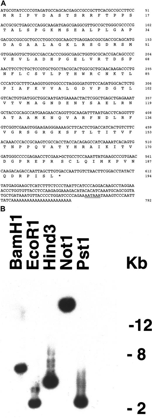
Using RACE PCR fusion transcripts from a t(19;21) leukemia that had a translocated AML1 were isolated and characterized (Figure 1A). In the most common transcript, the 5′ end of the AML1 gene, after exon 5, containing the DNA binding domain, was fused to a 236-nt sequence on chromosome 19 DNA termed AMP-19 (Figure 1B). However, rare splice variants had breakpoint junctions after exon 6 ofAML1, indicating that the translocation occurred in intron 6.
Nucleotide and amino acid sequences.
(A) Nucleotide and amino acid sequences of the most commonAML1-AMP19 transcript. The polyadenylation signal is underlined. The AML1-AMP19 nucleotide fusion junction is indicated by a bold arrow. (GenBank no. AY004251, rare splice variants are no. AF312386 and no. AF312 387). (B) Southern analysis of the restriction map of AMP19 on chromosome 19q13.3 BAC CTC-518O7 (GenBank no. AC012310).
Although the two chromosome 19 nucleotide segments (4423 nt apart) that make up AMP19 have classic exon-intron sequences at their boundaries, a BLAST search of the EST database, Northern analysis, hybridization screening of 4 cDNA libraries, and PCR screening of 18 cDNA libraries did not reveal an expressed gene. There is an EST (AI204456) on chromosome 19 within 51 nt of theAMP19 sequence. However, isolating and sequencing the 3.2-kB gene this EST represents revealed that AMP19 is located in opposite orientation within an intron of this EST, and the EST exons around AMP19 are 3′ untranslated sequences, making it unlikely that this EST is part of AMP19.
Multiple 5′ RACE analyses and screening a PCR constructed marrow cDNA library did not reveal a reciprocal der21 messenger RNA (mRNA) product. The untranslocated AML1 transcript was amplified using reverse transcription-PCR,12 subcloned, and sequenced. There were no changes in the amino acid sequence.
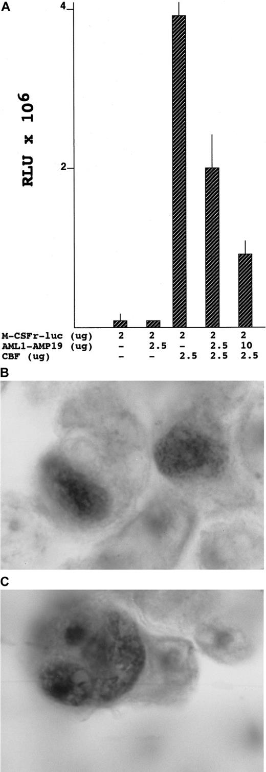
AML1 is fused out of frame with the AMP19sequence, which would produce a truncated CBFα2 protein containing only the DNA binding domain. Because this protein could compete with normal CBFα2, it is possible that AML1-AMP19 could function as a dominant negative inhibitor of promoters that CBF activates. This hypothesis was tested in a cotransfection assay using the M-CSF receptor promoter as a reporter. Figure2A shows that CBF activation of the M-CSF receptor promoter was inhibited an average of 3.1-fold by AML1-AMP19. Because AML1-AMP19 inhibits normal AML1 protein function, it is possible that it functioned in this patient's secondary leukemia to block differentiation.
Results of CBF cotransfection assays.
(A) CBF cotransfection assays with or without expression of AML1-AMP19. RLU-106 β-galactosidase normalized relative luciferase units. (B) Peroxidase immunohistology of the cellular location of normal control AML1B transiently transfected into 293 cells. (C) Peroxidase immunohistology of the cellular localization of AML1-AMP19 transiently transfected into 293 cells.
We analyzed the cellular location of AML1-AMP19 using immunohistology.11 12 We found that AML1-AMP19 was located both in the cytoplasm and in the nucleus in discrete bundles (Figure 2B,C).
The AML1-AMP19 fusion transcript is reminiscent of theAML1-EAP fusion.4 Both are fused out of frame, resulting in truncated AML1 species that function as inhibitory transcription factors.
The publication costs of this article were defrayed in part by page charge payment. Therefore, and solely to indicate this fact, this article is hereby marked “advertisement” in accordance with 18 U.S.C. section 1734.
References
Author notes
Robert Hromas, Indiana University Cancer Center, R4-202, 1044 W Walnut St, Indianapolis, IN 46202.


This feature is available to Subscribers Only
Sign In or Create an Account Close Modal