Endocytosis and recycling of coagulation factor VIIa (VIIa) bound to tissue factor (TF) was investigated in baby hamster kidney (BHK) cells stably transfected with TF or TF derivatives. Cell surface expression of TF on BHK cells was required for VIIa internalization and degradation. Approximately 50% of cell surface–bound VIIa was internalized in one hour, and a majority of the internalized VIIa was degraded soon thereafter. Similar rates of VIIa internalization and degradation were obtained with BHK cells transfected with a cytoplasmic domain-deleted TF variant or with a substitution of serine for cysteine at amino acid residue 245 (C245S). Endocytosis of VIIa bound to TF was an active process. Acidification of the cytosol, known to inhibit the internalization via clathrin-coated pits, did not affect the internalization of VIIa. Furthermore, receptor-associated protein, known to block binding of all established ligands to members of the low-density lipoprotein receptor family, was without an effect on the internalization of VIIa. Addition of tissue factor pathway inhibitor/factor Xa complex did not affect the internalization rate significantly. A substantial portion (20% to 25%) of internalized VIIa was recycled back to the cell surface as an intact and functional protein. Although the recycled VIIa constitutes to only approximately 10% of available cell surface TF/VIIa sites, it accounts for 65% of the maximal activation of factor X by the cell surface TF/VIIa. In summary, the present data provide evidence that TF-dependent internalization of VIIa in kidney cells occurs through a clathrin-independent mechanism and does not require the cytoplasmic domain of TF.
Introduction
Tissue factor (TF) is the cellular transmembrane receptor for factor VIIa (VIIa). Fibroblasts, pericytes, smooth muscle cells, and epithelial cells constitutively express TF, whereas cells in direct contact with the blood, such as endothelial cells and monocytes, express TF only when activated by specific pathophysiological stimuli.1 Injury to the vessel wall or pertubation of endothelium or monocytes, which could occur in various diseased states, results in exposure of blood to TF, thereby leading to the formation of TF/VIIa complexes that trigger the coagulation cascade.2In addition to its established role as an initiator of the coagulation process, TF was recently shown to function as a mediator of intracellular activities either by interactions of the TF cytoplasmic domain with the cytoskeleton or by supporting the VIIa-protease–dependent signaling.3,4 Such activities may be responsible, at least in part, for the implicated role of TF in tumor development,5-7 metastasis,8-11 and angiogenesis.12-14
Cellular exposure of TF/VIIa activity is advantageous in a crisis of vascular damage, but it may be fatal when exposure is sustained as it is in various diseased states. Thus, regulation of TF/VIIa activity plays a key role in vascular health conditions. Tissue factor pathway inhibitor (TFPI) is pivotal in regulating TF/VIIa function by inhibiting its enzymatic activity.15,16 TFPI was also shown to down-regulate TF/VIIa activity on monocytes17 and fibroblasts18 by a mechanism whereby it induces the internalization and degradation of the complex upon binding to TF/VIIa. Recent studies with fibroblasts18 provide evidence for the existence of an additional, TFPI-independent internalization of TF/VIIa. At present, it is unknown whether TF cytoplasmic domain is required for TF-mediated VIIa endocytosis. Although none of the known sorting sequences for association to clathrin-coated vesicles19 are present in the cytoplasmic tail of TF, however, it contains a cysteine (C)245 available for palmitoylation and 3 serine residues susceptible to phosphorylation.1 Both of these posttranslational modifications of TF may result in an altered affinity for membranes and proteins and thereby affect its subcellular location and the endocytosis pattern.
In the present investigation we examined the role of TF cytoplasmic domain and C245 in TF-mediated endocytosis of VIIa by using baby hamster kidney (BHK) cells transfected with wild-type TF and TF variants. We also evaluated whether the observed VIIa internalization results from normal membrane turnover or involves an active internalization process. Our results show that the TF-dependent internalization of VIIa is an active process that occurs through a clathrin-independent mechanism and does not require the cytoplasmic domain of TF. A substantial portion of the internalized VIIa returns to the cell surface, and this recycled VIIa is functionally fully active.
Materials and methods
Reagents
We used the following materials: Dulbecco modified Eagle medium (DMEM), fetal bovine serum (FBS), trypsin-EDTA (ethylenediamine tetraacetic acid), and penicillin-streptomycin (all from Gibco-BRL Life Technologies, Paisley, Scotland); tissue culture flasks and plates (Nunc A/S, Roskilde, Denmark); other chemicals, reagent grade or better, (Sigma Chemical, St Louis, MO); monospecific polyclonal sheep antihuman VII antibodies (Affinity Biologicals, Hamilton, ON, Canada); human factor X (X) and Xa (Enzyme Research Laboratory, South Bend, IN); and receptor-associated protein (RAP) (gift from Frederik Vilhardt, University of Copenhagen, Denmark). Recombinant human VIIa,20 VIIa blocked in the active site with phenylalanyl-phenylalanyl-arginyl chloromethyl ketone (FFR-VIIa),21 recombinant sTF,22 monospecific polyclonal rabbit antihuman TF immunoglobulin G (IgG),23and recombinant full-length TFPI24 were prepared as previously described.
Cell culture
The BHK cell line BHK-21 tk-ts13 (CRL 1632; American Type Culture Collection, Bethesda, MD) was maintained in DMEM (4.5 g/L glucose) with GlutaMAX 1 supplemented with 10% FCS, 10 IU/mL penicillin, and 100 μg/mL streptomycin. Generation of BHK cells that were stably transfected with full-length TF (BHK(TF)), a cytoplasmic domain–deleted construct of TF (BHK(TFΔcyto)), or a substitution of serine for cysteine at amino acid residue 245 (C245S) construct of TF (BHK(TFC245S)) was previously described.25
Radiolabeling of proteins
VIIa, FFR-VIIa, and ricin were labeled with iodine-125 (125I) using IODO-GEN–coated (Pierce, Rockford, IL) tubes and sodium (Na)125I according to the manufacturer's technical bulletin and as described previously.26 Briefly, the labeling reaction was performed in tubes coated with 10 μg IODO-GEN for 4 minutes on ice. The reaction was quenched by the addition of 1% KI, and free iodine was removed by extensive dialysis against 10 mM HEPES (4-(2-Hydroxyethyl)-1-piperazineethanesulfonic acid) (pH 7.5) and 150 mM sodium chloride (NaCl). The concentration of the labeled proteins was determined by measurements of the absorbance at 280 nm.125I-VIIa retained about 90% to 100% of the functional activity of unlabeled VIIa.
Internalization of 125I-VIIa
Confluent monolayers of parental BHK cells, BHK cells transfected with wild-type TF and TF variants, BHK(TF), BHK(TFΔcyto), and BHK(TFC245S) were incubated with 10 nM 125I-VIIa for varying times (5 minutes to 4 hours) at 37°C in buffer B, which comprised 10 mM HEPES, 150 mM NaCl, 4 mM potassium chloride (KCl), and 11 mM glucose (pH 7.5) buffer containing 5 mM CaCl2 and 1 mg/mL bovine serum albumin. In some experiments 100 nM RAP, 1 U/mL heparin, or 10 nM of a preformed TFPI/Xa complex was added prior to the 125I-VIIa addition. At the end of the incubation period, the supernatant was removed, and the monolayers were washed 4 times with ice-cold buffer B. The cell surface–associated 125I-VIIa was subsequently eluted after incubation of the cells with a low-pH (pH 2.5) glycine buffer for 5 minutes at room temperature. The radioactivity present in the supernatant was considered as VIIa associated with the cell surface. This assumption has been validated because the glycine treatment was shown to elute 90% or more of the VIIa associated with the cells when they were incubated with 125I-VIIa at 4°C for 2 hours.
After removing the glycine eluate, the cells were detached by trypsin/EDTA solution, and the cell suspension was collected. The radioactivity present in the cell suspension was measured as internalized VIIa. Identical results were obtained in some experiments when internalized VIIa was determined as the amount of radioactivity released to the supernatant after exposure of the cells (which had been washed with low-pH buffer) to 2 N NaOH. Nonspecific binding and internalization were determined in parallel experiments where TF was blocked by preincubating the cells for 1 hour with 200 μg/mL rabbit antihuman TF IgG before adding 125I-VIIa. TF-specific binding and internalization were obtained by subtracting the nonspecific binding and internalization from the total binding and internalization. Nonspecific binding and internalization accounted for less than 10% of the total binding and internalization. The rate of internalization was calculated as d(internalized/surface associated)/dt according to the method of Wiley and Cunningham.27 The degradation of VIIa was followed by withdrawal of 20-μL aliquots from the medium overlying the cells at selected time intervals and mixing with 200 μL 10% vol/vol trichloroacetic acid (TCA). After centrifugation, degraded VIIa was determined as the TCA nonprecipitable (TCA-soluble) radioactivity.
Detection of internalized VIIa by immunofluorescence
BHK(TF) cells were cultured in chamber slides (Nunc). Chambers were incubated with 10 nM VIIa in serum-free medium for 1 hour at 37°C or 4°C. Control chambers were incubated with cell medium alone. In some experiments the cells were then exposed to low-pH glycine buffer (pH 2.5) for 5 minutes to elute surface-associated VIIa. Cells were fixed in 2% paraformaldehyde in phosphate-buffered saline (PBS) for 10 minutes and then briefly washed in PBS. For immunostaining, slides were preincubated for 15 minutes in 5% goat serum in tris[hydroxymethyl] aminomethane (Tris)–buffered saline (TBS) with 0.2% saponin (TBS-S). This and all subsequent steps were carried out at room temperature.
Primary antibodies (polyclonal rabbit antihuman TF IgG or polyclonal sheep antihuman VII IgG) were then added in TBS-S at 10 μg/mL for 90 minutes, and visualization was performed with either biotinylated swine-antirabbit (Dako A/S, Glostrup, Denmark) followed by HRP-streptavidin and TSA-direct-FITC (trichostatin A–direct–fluorescein isothiocyanate) (NEN) or with HRP-rabbit-antisheep (Dako) followed by TSA-direct-FITC. For double-labeling immunolocalization of TF and VIIa, the cells were first immunostained with anti-TF as described above. The same samples were then immunostained for VIIa by blocking first for 15 minutes with TBS-S with 5% rabbit and 5% swine serum followed by incubation in sheep–anti-VII for 30 minutes, then in biotinylated donkey-antisheep (Sigma) followed by HRP-streptavidin and TSA-direct-Rhodamine.
Determination of bulk membrane turnover
We added 10 nM 125I-VIIa or 200 ng/mL125I-ricin to monolayers of BHK(TF) cells at 37°C. At the end of 5 and 30 minutes of incubation, the cell surface–associated and internalized ligands were determined. Cell surface–associated 125I-VIIa was released by incubating the cells with low-pH glycine buffer for 5 minutes, whereas cell surface–associated 125I-ricin was released by incubating the cells with 0.1 M lactose at 4°C for 1 hour. (Control experiments performed at 4°C showed that these procedures removed cell surface–associated VIIa and ricin with a similar efficiency, 94.2% and 94.8%, respectively). The data were obtained as the mean ± SD percent-internalized radioactivity per total cell surface–bound ligand from 3 independent experiments in triplicate.
Effect of cytosol acidification on VIIa internalization
Confluent monolayers of BHK(TF) cells were pretreated with DMEM (pH 5.5) with and without 10 mM acetic acid for 5 minutes at room temperature. The cells were then incubated with 10 nM125I-VIIa for 1 hour, and the cell surface–associated and internalized VIIa were determined as described above. Data are calculated as the percentage of internalization in acetic acid–treated cells relative to nontreated control cells. Internalization of 200 ng/mL 125I-transferrin is shown for comparison. Data are the mean ± SD of 3 independent experiments in triplicate.
Recycling of internalized VIIa and FFR-VIIa
Confluent monolayers of BHK(TF) cells were incubated with 10 nM125I-VIIa for 1 hour at 37°C to allow internalization. The cell surface–associated radioactivity was then removed by a low-pH glycine wash as described above. The monolayers were washed 3 times with buffer B to remove residual low-pH glycine, and the cells were subsequently allowed to stand in buffer B for various times at 37°C for recycling of 125I-VIIa. Overlying buffer containing the recycled VIIa that was dissociated from the surface, as intact or degraded, was removed and pooled with radioactive protein eluted from the cell surface by glycine buffer (pH 2.5). The pooled samples, comprising the total amount of recycled 125I-VIIa, were then analyzed for the presence of intact and degraded125I-VIIa, which were determined after 10% TCA treatment as the precipitable and nonprecipitable radioactivity, respectively. Following the removal of recycled and degraded 125I-VIIa, the cells were removed from the dish by trypsin digestion and counted for radioactivity to determine the remaining internalized125I-VIIa. A similar procedure was used for recycling experiments with 125I-FFR-VIIa.
Recycled 125I-VIIa in aliquots from the pooled samples was tested for functional activity after adjusting the pH to 7.4 by adding 100 μL pooled sample to 250 μL 50 mM Tris and 150 mM NaCl (pH 8.0). The Xa-generating activity was measured in a TF/VIIa assay with excess relipidated TF. Recycled functionally active VIIa was quantified by comparison, with the activity of a VIIa standard determined under identical conditions.
Recycling of functionally active VIIa on the cell surface was determined in separate experiments. Following internalization for 1 hour in the presence of 10 nM VIIa, the cell surface–associated VIIa was removed by exposure of the cells to a low-pH glycine buffer. Immediately the cells were washed 3 times with buffer B and then allowed to stand in buffer B. After varying times, 100 nM factor X (X) was added to the monolayers, and recycled TF/VIIa complexes were allowed to activate X for 5 minutes. Xa generation was determined in a chromogenic assay by adding 50 μL of the above mixture to 50 μL 50 mM Tris, 150 mM NaCl, 5 mM EDTA, and 1 mg/mL BSA (pH 7.5). The Xa activity was measured in the presence of 1.07 mM Chromozym X. The absorbance at 405 nm was measured continuously in a microplate reader (Molecular Devices), and initial rates were converted to Xa concentrations using a Xa standard curve (85 pmol/L to 11 nmol/L).
Internalization and recycling of cell surface TF
Confluent BHK(TF) cells were incubated with 10 nM VIIa or FFR-VIIa at 37°C for various time intervals (15 minutes to 4 hours) to study the effect of TF/VIIa internalization on the exposure of the TF receptor on the cell surface. The cell surface–associated VIIa was removed at the end of the incubation period by exposure of the BHK(TF) cells to a 5-minute low-pH glycine buffer wash and 4 subsequent washes with ice-cold buffer B. The cell surface TF expression at that time was then determined by the 125I-VIIa binding capacity and the ability to support X activation in the presence of newly added VIIa. To determine 125I-VIIa binding, the monolayers were incubated with 125I-VIIa for 90 minutes at 4°C. Then the unbound ligand was removed, and the cells were washed 4 times with buffer B. The cell surface–associated 125I-VIIa was obtained after detaching the cells with trypsin/EDTA and counting the suspension in a gamma counter. To determine TF levels using X activation assay, 10 nM VIIa and 175 nM X in buffer B were added to the monolayers. TF functional activity was measured by the amount of Xa generated at the end of a 15-minute activation period at 37°C. For this purpose, 25-μL aliquots were removed from the well and added to 50 mM Tris, 150 mM NaCl, 5 mM EDTA, and 1 mg/mL BSA for a total of 75 μL (pH 7.5). The Xa activity was measured in the presence of 1.07 mM Chromozym X.
Results
TF-dependent internalization and degradation of VIIa
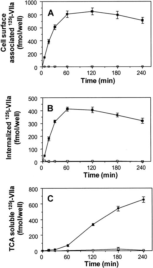
BHK cells were exposed to 10 nM 125I-VIIa in the presence and absence of anti-TF IgG, and the amount of TF-specific cell surface–associated, cell surface–internalized, and cell surface–degraded VIIa was followed with time. A time-dependent binding of 125I-VIIa to the cell surface TF followed by internalization and degradation was observed with BHK(TF) cells. In comparison, only a negligible amount of 125I-VIIa was bound and processed by untransfected BHK cells (Figure1). Association to the surface of BHK(TF) cells was half-maximal after approximately 20 minutes of exposure to125I-VIIa. Fifty percent of the cell surface–associated125I-VIIa, which corresponds to approximately 4000 fmol/106 cells, was internalized within an hour following the treatment. This corresponds to an internalization constant (k1 = 0.076 minutes−1). When 10 nM VIIa was offered to the cells, the maximal degradation rate achieved in this TF-dependent VIIa endocytosis was 2650 fmol/106 cells per hour. The amount of VIIa internalized correlated well with the amount of VIIa offered, up to about 30 nM 125I-VIIa, a concentration that could saturate TF sites on the cell surface (data not shown).
TF-specific binding, internalization, and degradation of VIIa in BHK cells.
125I-VIIa (10 nM) was added to confluent monolayers of BHK cells (■) or BHK cells transfected with full-length TF (BHK(TF)) (▪) that were preincubated for 60 minutes with control buffer or 200 μg/mL rabbit antihuman TF IgG at room temperature. Cell surface association (A), internalization (B), and degradation (C) were determined at various time points as described in “Materials and methods.” TF-specific binding, internalization, and degradation were determined by subtracting the values obtained in the presence of antihuman TF IgG from the values obtained in its absence. Nonspecific binding of 125I-VIIa to BHK(TF) cells in the presence of antihuman TF IgG was 10% to 15% of the total binding. A similar amount of 125I-VIIa was bound to parental untransfected BHK cells in both the absence and presence of antihuman TF IgG. Data are the mean ± SD of 3 independent experiments in triplicate.
Addition of 125I-VIIa in complex with soluble recombinant TF1-219 to untransfected BHK cells did not change the rate of VIIa binding, internalization, or degradation (data not shown). These data document that membrane attachment of TF is needed for the binding and internalization of VIIa. The kinetics of cell surface association, internalization, and degradation of FFR-VIIa were indistinguishable from that of VIIa (data not shown).
Internalization of VIIa demonstrated by immunofluorescence microscopy
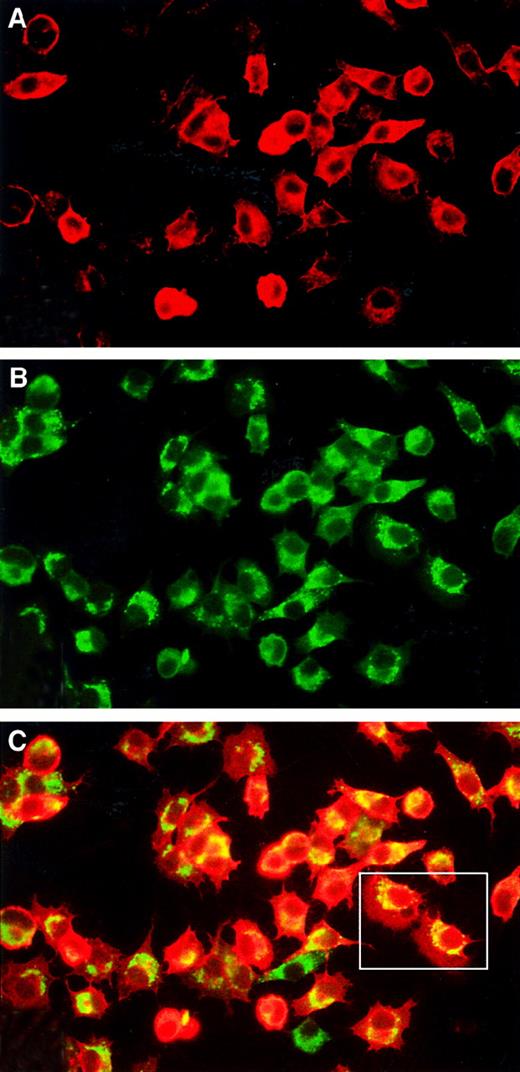
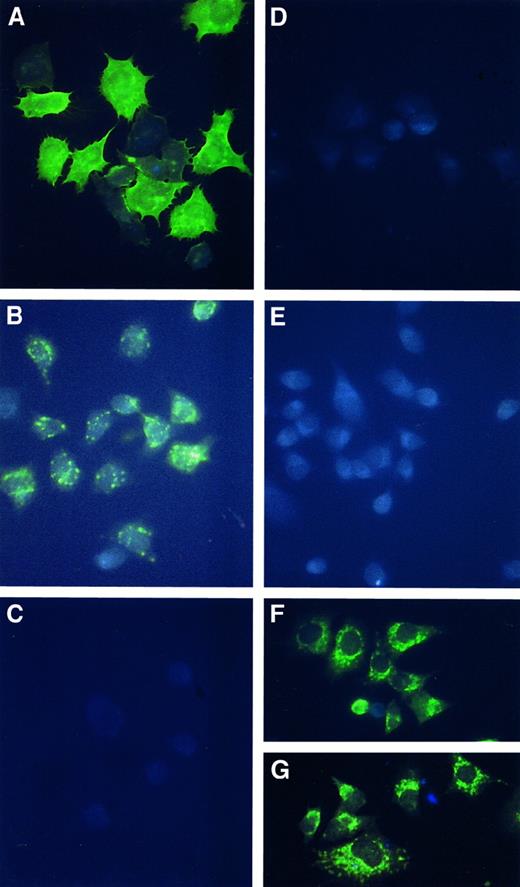
BHK(TF) cells treated with 10 nM VIIa for 1 hour at 37 °C were analyzed by immunofluorescence microscopy (Figure2). TF and VIIa were both immunostained as a diffuse membrane staining and as bright spots in vesicular perinuclear compartments (Figure 2A,B). Double-labeling experiments revealed that TF and VIIa were colocalized in most cells (Figure 2C). As expected, cells incubated with medium alone were negative for VIIa staining (Figure 3D). Staining was localized primarily to the perinuclear compartments when the cells were incubated with VIIa at 37°C for 1 hour and subsequently treated with a low-pH glycine wash (Figure 3B), indicating that cell surface–associated VIIa, but not internalized VIIa, was removed by this treatment. As shown in Figure 3C, perinuclear staining, as well as cell surface–associated staining, was absent when the cells were exposed to VIIa at 4°C to prevent internalization and subsequently treated with a glycine wash to remove cell surface–associated VIIa. This finding was consistent with the observation that more than 90%125I-VIIa associated to the cell surface of BHK(TF) cells at 4°C could be displaced by the glycine wash. Immunostaining for TF revealed no significant difference in staining pattern between cells treated with buffer (Figure 3F) and those treated with VIIa (Figure3G). One should note that very bright staining of intracellular TF masked the fluorescence of cell surface–stained TF (Figure 3F), thus giving the false impression that the cells apparently lack TF on their cell surface.
Double-immunofluorescence staining for TF and VIIa.
BHK(TF) cells were incubated with 10 nM VIIa for 1 hour at 37°C and then stained for VIIa (A) and TF (B) as described in “Materials and methods.” (C) Pictures of panels A and B have been merged to highlight areas with colocalization (yellow). Note that most of the cells show a high degree of colocalization of TF and VIIa. In a subset of these, colocalization in perinuclear vesicles is quite evident.
Immunofluorescence staining for VIIa or TF.
BHK(TF) cells treated with 10 nM VIIa (A-C, G) or buffer (D-F) were immunofluorescence stained with VII antibodies (A-E) or TF antibodies (F, G). (A) Cells were treated with 10 nM VIIa for 1 hour at 37°C, and the unbound VIIa was removed before fixing the cells. (B) Cells were treated with 10 nM VIIa for 1 hour at 37°C, the unbound VIIa was removed, and the cell surface–associated VIIa was eluted with low-pH buffer to illustrate the cell surface–internalized VIIa. (C) Cells were treated with 10 nM VIIa for 1 hour at 4°C, the unbound VIIa was removed, and the cell surface–associated VIIa was eluted with low-pH buffer. (D, E) Cells were treated the same as in panels A and B, respectively, except that VIIa was not included in the incubation medium. (F, G) Immunostaining of TF after 1-hour exposure of BHK(TF) cells at 37°C to the control medium and 10 nM VIIa, respectively.
Determination of bulk membrane turnover
To determine whether the internalization of VIIa was an active process or represented a bulk membrane turnover, internalization of VIIa was compared to the internalization of ricin. Ricin binds to terminal galactose residues on membrane glycoproteins and glycolipids and is internalized by any structure that pinches off from the membrane. Thus ricin internalization has been used as a general marker for bulk membrane turnover.28 29 The initial VIIa internalization rate (32% ± 5% within the first 5 minutes) was approximately 2-fold higher than that of ricin (17% ± 7%). The finding that endocytosis of VIIa is significantly faster than that of ricin might indicate that internalization of VIIa is facilitated and does not just represent bulk membrane turnover.
VIIa is not internalized via LRP in a TFPI-dependent mechanism
Previous studies revealed an endocytosis mechanism in which VIIa was internalized by LRP in a TFPI-dependent process involving localization of the TF/TFPI/VIIa complex to clathrin-coated pits.17 To test whether this process was also operative for VIIa endocytosis by BHK(TF) cells, we first studied the effect of cytosolic acidification. This treatment impedes clathrin-dependent endocytosis, and as expected, pre-exposure of BHK(TF) cells to 10 mM acetic acid impeded the internalization of transferrin, which is known to proceed via this pathway. Cytosolic acidification decreased transferrin internalization by 50% in contrast to VIIa internalization, which was unaffected by this treatment. This observation suggests that VIIa endocytosis by BHK(TF) cells proceeds via a clathrin-independent mechanism. In further experiments to rule out the involvement of LRP and TFPI in VIIa internalization, we studied the effect of RAP and heparin on VIIa internalization. RAP is known to block the binding of all established ligands to LRP receptors,30 and heparin is expected to displace TFPI from cell surface heparan sulfate proteoglycans31 and thereby impede TFPI-dependent LRP internalization of the TF/VIIa complex. Neither RAP nor heparin affected the VIIa endocytosis (Table1). In further experiments, TFPI/Xa was exogenously added to the cells to test the effect of TFPI/Xa on VIIa cell surface association and internalization. As expected, 10 nM TFPI/Xa markedly inhibited the cell surface TF/VIIa activity (data not shown). However, as shown in Table 1, TFPI/Xa had no effect on VIIa binding and internalization.
Effect of receptor-associated protein, heparin, and tissue factor pathway inhibitor/factor Xa on tissue factor–specific binding and internalization of coagulation factor VIIa
| . | Cell surface–associated VIIa, % of control . | Internalized VIIa, % of control . |
|---|---|---|
| RAP | 104 ± 5 | 102 ± 2 |
| Heparin | 96 ± 5 | 106 ± 12 |
| TFPI/Xa | 112 ± 8 | 105 ± 7 |
| . | Cell surface–associated VIIa, % of control . | Internalized VIIa, % of control . |
|---|---|---|
| RAP | 104 ± 5 | 102 ± 2 |
| Heparin | 96 ± 5 | 106 ± 12 |
| TFPI/Xa | 112 ± 8 | 105 ± 7 |
Confluent monolayers of BHK(TF) cells were incubated with 10 nM125I-VIIa for 1 hour in the absence or presence of 100 nM RAP, 1 U/mL heparin, or 10 nM TFPI/Xa. Complexes of TFPI/Xa were prepared by incubating equimolar amounts of recombinant full-length TFPI and Xa for 30 minutes at 37°C. Data obtained with the cells in the absence of TFPI/Xa, heparin, or RAP were set to 100%. Data are the mean ± SD of 3 independent experiments in triplicate.
VIIa indicates coagulation factor VIIa; RAP, receptor-associated protein; TFPI/Xa, tissue factor pathway inhibitor/factor Xa; BHK(TF), baby hamster kidney stably transfected with full-length tissue factor.
TF cytoplasmic domain or its potential acylation at C245 is not required for TF-dependent endocytosis of VIIa
BHK cell lines were stably transfected with a cytoplasmic-deleted construct of TF, des(248-263)TF, and with a C245S substitution construct of TF. The resulting cell lines, BHK(TFΔcyto) and BHK(TFC245S), were used to elucidate the role of the cytoplasmic domain in TF-dependent internalization of VIIa. As previously described by Sorensen et al,25 the Xa generation activity of the BHK(TFΔcyto) and BHK(TFC245S) cell lines was comparable to that of the BHK cell line transfected with full-length TF, BHK(TF), whereas the VIIa binding capacity of the BHK(TFC245S) cell line was approximately 50% of that observed with the 2 other TF-expressing cell lines.
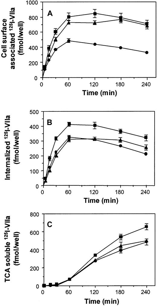
Figure 4 shows VIIa cell surface association, internalization, and degradation observed with BHK(TF), BHK(TFΔcyto), and BHK(TFC245S). As expected, less VIIa was bound to the cell surface of BHK(TFC245S) cells. Still, the rate of the subsequent internalization and degradation reactions observed with the BHK(TFΔcyto) and BHK(TFC245S) cell lines was similar to those of BHK(TF) cells. This suggested that VIIa endocytosis takes place independently of the cytoplasmic domain and also independently of possible acylation of C245.
TF-specific binding, internalization, and degradation of VIIa in BHK(TF), BHK(TFΔcyto), and BHK(TFC245S) cells.
Experiments were performed as described in Figure 1 with BHK(TF) (■), BHK(TFΔcyto) (▴), and BHK(TFC245S) (●). The data represent the cell surface–associated VIIa (A), internalized VIIa (B), and degraded VIIa (C). Data are the mean ± SD of 3 independent experiments in triplicate.
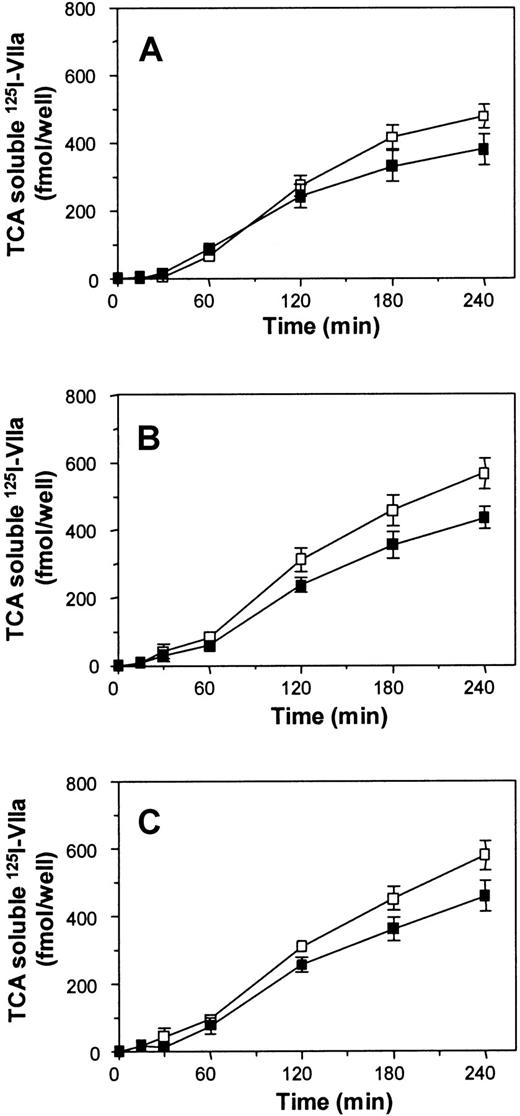
In contrast to association and internalization of VIIa, which were not affected by addition of TFPI/Xa, TFPI/Xa induced a small but consistent increase in VIIa-degradation in BHK(TF) cells (Figure5). A slightly enhanced VIIa degradation was also evident with BHK(TFΔcyto) and BHK(TFC245S) cell lines, indicating that the TF cytoplasmic domain and its potential modifications played no role in the TFPI/Xa-dependent enhancement of VIIa degradation.
Effect of TFPI/Xa on TF-specific VIIa degradation in BHK(TF), BHK(TFΔcyto), and BHK(TFC245S) cells.
Confluent monolayers of BHK(TF) (A), BHK(TFΔcyto) (B), or BHK(TFC245S) (C) cells were incubated with 10 nM125I-VIIa for 1 hour in the absence (▪) or presence (■) of 10 nM TFPI/Xa, and the degradation of VIIa was determined as described in “Materials and methods.” Data are the mean ± SD of 3 independent experiments in triplicate.
Recycling of internalized TF/VIIa
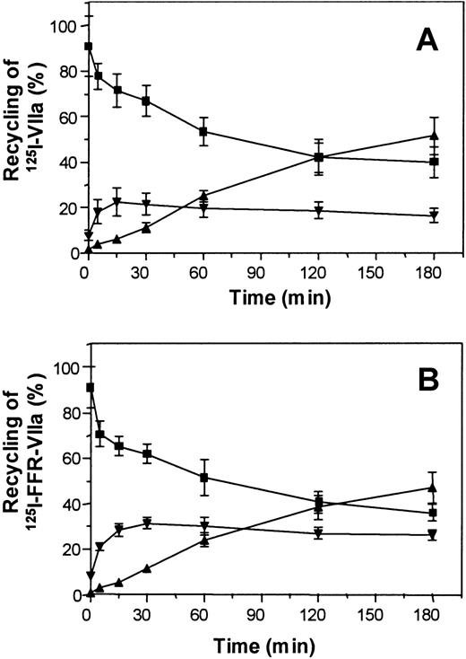
Next we examined whether the internalized VIIa or FFR-VIIa could be recycled back to the cell surface. Monolayers of BHK(TF) cells were allowed to internalize 125I-VIIa for 1 hour. The cell surface–associated radioactivity was completely removed by treating the cells with low-pH glycine, and the cells were allowed to stand at 37°C for recycling of VIIa. The amount of intracellular VIIa and the total amount of excreted, intact, or degraded VIIa were then determined at various time points as described in “Material and methods.” As shown in Figure 6A, a significant portion (about 20%) of the internalized VIIa pool was excreted as intact (TCA-precipitable) VIIa. Experiments run in parallel suggested that the recycled TCA-precipitable VIIa was functionally fully active. Thus, the pooled samples from the overlaying medium and the low-pH glycine eluate contained an amount of VIIa that, when quantified by a Xa generation assay with lipidated TF, corresponded well with the amount of TCA-precipitable VIIa (data not shown). Recycling of TCA-precipitable protein was even more pronounced with125I-FFR-VIIa, accounting for 25% to 30% of the internalized FFR-VIIa (Figure 6B).
Recycling of internalized VIIa.
Confluent monolayers of BHK(TF) cells were allowed to internalize 10 nM 125I-VIIa or 125I-FFR-VIIa for 1 hour. Thereafter, the cell surface–associated radioactivity was removed by a low-pH glycine wash. After the glycine wash, the monolayers were washed 3 times with buffer B and then incubated with buffer B for various times at 37°C for recycling of (A) 125I-VIIa or (B)125I-FFR-VIIa. Total radioactivity in the internalized pool measured after the first glycine wash was set to 100%. Subsequent measurement of radioactivity at various time points of the residual internalized pool (▪), recycled intact protein (▾), and recycled degraded protein (▴) was performed as described in “Materials and methods.” Data are the mean ± SD of 3 independent experiments in triplicate.
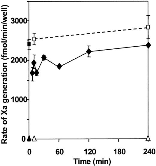
Next, we studied the appearance on the cell surface of functionally active VIIa from the internalized pool by measuring the ability of monolayers to support X activation. In these experiments, BHK(TF) cells were treated with 10 nM VIIa for 1 hour at 37°C, cell surface–associated VIIa was removed by glycine wash, and the appearance of recycled VIIa was monitored directly by the ability of monolayers to support X activation in the absence of exogenously added VIIa. The Xa-generating activity was compared to that of monolayers without VIIa preincubation, which exhibited a negligible activity, and to that of monolayers with 10 nM VIIa exogenously added, which was set to 100% activity. As shown in Figure 7, the recycling of VIIa appears to be a rapid process because functionally active VIIa reappeared on the cell surface as soon as the cell surface–associated VIIa was removed. It is interesting to note that the recycled VIIa was capable of generating as much as 60% to 65% of maximal Xa generation capacity. Similar experiments were performed with BHK(TFΔcyto) and BHK(TFC245S) cell lines, where VIIa recycling was evaluated by the ability of recycled VIIa on the cell surface to support X activation. Data from these experiments revealed a recycling pattern similar to that described in Figure 7 for BHK(TF) cells (data not shown), suggesting that the cytoplasmic domain of TF was not obligatory for recycling of VIIa.
Effect of VIIa recycling on X activation.
BHK(TF) cells were treated with 10 nM VIIa at 37°C for 1 hour. Cell surface–associated VIIa was then removed by glycin wash, and the monolayers were washed 3 times with buffer B. Measurement of subsequent recycling of TF/VIIa activity at different time points was performed by incubating the cells with 100 nM X for 5 minutes and measuring the amount of Xa generated (♦). The activity of cells not preincubated with VIIa and not subjected to glycine wash (▴, ▪) and the activity of cells not preincubated with VIIa but washed with glycine (▵, ■) were measured for comparison at 10 and 240 minutes in the presence of 100 nM X (▴, ▵) or in the presence of 100 nM X and 10 nM exogenous VIIa (▪, ■).
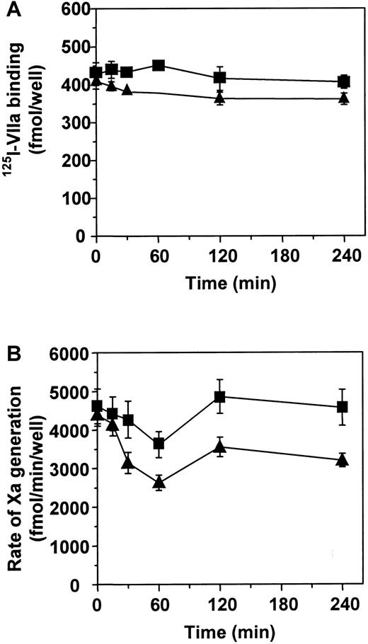
The above experiments demonstrated that VIIa was internalized and recycled to the cell surface. However, the fate of TF in this process was not evident. Therefore we next examined the amount of TF exposed to the surface during TF-dependent internalization of VIIa. Monolayers of BHK(TF) cells were allowed to internalize VIIa or FFR-VIIa for varying time intervals from 15 minutes to 4 hour before the cell surface–associated VIIa or FFR-VIIa was removed by treating the cells with low-pH glycine. Thereafter, the amount of cell surface–exposed TF was measured either by VIIa binding or by TF/VIIa functional activity. As shown in Figure 8A, the total amount of VIIa binding sites on BHK monolayers was unchanged during internalization of VIIa or FFR-VIIa. In contrast, however, we observed a transient down-regulation of the amount of functionally active TF sites during the internalization cycle (Figure 8B).
Internalization and recycling of TF.
Monolayers of BHK(TF) cells were allowed to internalize VIIa (▪) or FFR-VIIa (▴) for various times, from 15 minutes to 4 hours. The cell surface–associated VIIa or FFR-VIIa was subsequently removed by low-pH buffer wash, and the exposure of TF on the cell surface was measured by either 125I-VIIa binding (A) or the cell surface–associated TF/VIIa functional activity (B) as described in “Materials and methods.” Data are the mean ± SD of 3 independent experiments in triplicate.
Discussion
The binding of VIIa to a number of cells in TF-dependent and TF-independent reactions has been studied in great detail.1,2 In comparison, little is known about the fate of cell surface–associated VIIa and the possible catabolism of cell surface TF/VIIa complexes. Recent studies have described 3 different mechanisms for VIIa and TF/VIIa internalization. Chang and Kisiel32 described a TF-independent internalization and degradation of VIIa in human hepatoma cells. Hamik et al17found that TF was down-regulated on monocytes in a reaction involving VIIa, TF, TFPI, and LRP. Iakhiaev et al18 have provided suggestive evidence that VIIa complexed with TF on fibroblasts was endocytosed by 2 different pathways: LRP-independent pathway in the absence of TFPI/Xa and LRP-dependent pathway in the presence of TFPI/Xa. None of the above studies addressed the role of cytoplasmic domain of TF in TF-mediated VIIa endocytosis. In the present study we investigated VIIa-endocytosis in further detail using BHK cells transfected with TF and TF variants.
The data presented in the current study show a significant internalization and degradation of VIIa in BHK(TF) cells, whereas there was no detectable internalization and degradation of VIIa observed in wild-type BHK cells. These data clearly document that VIIa internalization and degradation in the kidney cell (or BHK cell) is strictly dependent upon its binding to cell surface TF. Cytosolic acidification experiments suggested that VIIa was catabolized by clathrin-independent endocytosis. Further experiments showed that neither RAP nor heparin affected the TF-mediated endocytosis of VIIa. This excludes the possible involvement of LRP and endogenous TFPI in VIIa endocytosis and provides further support for clathrin-independent endocytosis of VIIa. Addition of the TFPI/Xa complex resulted in only a slight increase (approximately 25%) in VIIa degradation.
These data differ from the earlier published data in monocytes17 and fibroblasts,18 which showed a more pronounced effect of TFPI on TF and VIIa degradation. A possible reason could be that compared to TF, BHK cells may have only a limited number of LRP and/or cell surface heparan sulfate proteoglycans which are needed for internalization of TF/TFPI/VIIa/Xa complexes via clathrin-dependent mechanism.17,18 In this context it should be noted that although kidney cells mainly express megalin (another member of the low-density lipoprotein (LDL) receptor family) and not LRP, it has been reported that some kidney cells also express LRP.33 Further, because RAP inhibits the endocytosis mediated by both LRP and megalin,34 the failure of RAP to inhibit endocytosis of VIIa in the present study suggests that members of the LDL receptor family are not involved in TF-mediated endocytosis of VIIa in kidney cells.
Although the earlier studies17,18 described above suggest that TF/VIIa is actively internalized, these studies have not eliminated the possibility that the observed internalization of TF and VIIa is a result of and follows normal membrane turnover. The data presented herein provide evidence that TF-mediated endocytosis of VIIa is an active receptor-mediated process and not the result of a general turnover of bulk membrane components. The endocytosis rate of k1 = 0.076 minutes−1, which corresponds to approximately 50% of cell surface–associated VIIa internalized in 20 minutes, is substantially higher compared to the turnover rate of bulk membrane components, which in general corresponds to 15% to 25% internalization per hour.35-38 A comparison of internalization of VIIa and ricin, a marker for membrane turnover,30 provides further evidence that the rate of VIIa internalization was much higher compared to the rate of general membrane turnover.
Internalization of receptors for endocytosis via clathrin-coated pits is coded by specific amino acid sequences in the cytoplasmic domain.39 Such sequences are not present in the TF cytoplasmic domain. The sorting sequences for clathrin-independent internalization are not known. Because the cytoplasmic domain of TF comprises a cysteine and a few serines susceptible to posttranslational modifications, it is possible that these potential modifications may affect the affinity of transmembrane receptors for membranes, subcellular location, and interaction with other proteins, which in turn could affect the internalization and degradation of receptor-associated ligands. However, the data of the present studies show that the cytoplasmic domain of TF does not play a role in the TF-dependent endocytosis of VIIa. In this respect, endocytosis of TF/VIIa complexes is similar to the endocytosis of thrombin/thrombomodulin (TM) complexes. Internalization of thrombin/TM complexes was reported to occur predominantly through a clathrin-independent mechanism, and the deletion of the cytoplasmic domain has no effect on the constitutive internalization of TM as well as thrombin/TM complexes.40-42
The data on VIIa recycling with BHK(TF) cells (Figure 6) confirm and extend recent observations in fibroblasts.18 Our data show that a proportion (approximately 20%) of internalized VIIa recycles to the cell surface as an intact protein which is fully capable of activating factor X. It is interesting to note that although VIIa occupies only about 10% of available TF sites on the cell surface, recycled VIIa confers the cell with approximately 60% to 70% of its total X-activating capacity (Figure 7). This agrees well with previous reports showing that the maximal procoagulant activity of TF-expressing cells is reached at a much lower VIIa concentration than needed for saturation of an additional pool of coagulant inactive or “cryptic” TF sites available for binding of VIIa on the surface.26 43 Although direct evidence for such a mechanism is missing, these data are consistent with the hypothesis that VIIa is internalized primarily from the pool of functionally active TF sites and then recycled back to the same pool representing a preferred residence for VIIa. This model may also explain other observations, such as a transient decrease in functionally active TF (Figure 8) and similar internalization rates in BHK(TFC245S) and BHK(TFΔcyto) cells (Figures4 and 5), which differ substantially in VIIa binding capacity but are equally active in their capacity to activate X. It is clear, however, that the above model cannot in itself account for the total endocytotic process of VIIa including the extent of VIIa internalized and subsequently degraded. It is possible that uptake and rapid cycling of VIIa are 2 distinct events, where VIIa bound to an active pool of TF follows rapid recycling of TF without going to a degradation pathway, and VIIa bound to nonfunctional TF is internalized and directed to a degradation pathway.
The present data, along with recent reports,17,18 suggest that the fate of cell surface TF/VIIa complexes might differ with different cell types. VIIa bound to TF on kidney cells (as suggested by the present data) is, for example, internalized predominantly through a clathrin-independent mechanism, and the formation of the inhibitor complex on the cell surface with TFPI/Xa has no significant effect on the internalization of VIIa. In contrast, formation of the inhibited complex (with either TFPI or TFPI/Xa) appears to be a prerequisite for the internalization of TF in monocytes, and the internalization and degradation of TF proceeds via a LRP/clathrin-dependent mechanism.17 Both the above mechanisms are apparently operative in fibroblasts, ie, a substantial amount of VIIa is internalized in the absence of TFPI/Xa via a LRP/clathrin-independent mechanism, whereas the presence of TFPI/Xa induces an additional internalization of VIIa by a LRP/clathrin-dependent pathway.18 Further, the present data obtained with kidney cells suggest a rapid and preferential recycling of functional TF/VIIa complexes, whereas no such apparent evidence for preferential recycling for functional TF/VIIa complexes was found in fibroblasts.18
In conclusion, our data show that TF-dependent internalization and degradation of VIIa in kidney cells do not depend on interactions involving the cytoplasmic domain of TF, TFPI/Xa does not affect the internalization, and the majority of internalized functional TF/VIIa complexes rapidly recycle back to the cell surface.
From Novo Nordisk A/S, Maalov, Denmark, Elke Gottfriedsen and Berit Lassen, Department of TF/VIIa Research, and Steen Kryger, Department of Pharmacological Research, provided excellent technical assistance; Jesper Bøggild Kristensen, Department of Isotope Chemistry, prepared the iodine-labeled ligands. Prof Bo van Deurs, Structural Cell Biology Unit, Department of Medical Anatomy, The Panum Institute, University of Copenhagen, Denmark, and Dr Mirella Ezban, Novo Nordisk, are gratefully acknowledged for their discussion and helpful suggestions.
Supported in part by grant HL 58869 (L.V.M.R.) from the National Heart, Lung, and Blood Institute, National Institutes of Health, Bethesda, MD, and a grant (C.B.H.) from the Danish Academy of Technical Sciences (EF639), Copenhagen, Denmark.
Submitted May 3, 2000; accepted November 13, 2000.
The publication costs of this article were defrayed in part by page charge payment. Therefore, and solely to indicate this fact, this article is hereby marked “advertisement” in accordance with 18 U.S.C. section 1734.
References
Author notes
Lars C. Petersen, Department of TF/VIIa Research, Novo Nordisk A/S, Novo Nordisk Park, 2760 Maalov, Denmark; e-mail: lcp@novo.dk.

This feature is available to Subscribers Only
Sign In or Create an Account Close Modal