Vascular endothelial growth factor (VEGF) is a potent angiogenic peptide with biologic effects that include regulation of hematopoietic stem cell development, extracellular matrix remodeling, and inflammatory cytokine generation. To delineate the potential role of VEGF in patients with myelodysplastic syndrome (MDS), VEGF protein and receptor expression and its functional significance in MDS bone marrow (BM) were evaluated. In BM clot sections from normal donors, low-intensity cytoplasmic VEGF expression was detected infrequently in isolated myeloid elements. However, monocytoid precursors in chronic myelomonocytic leukemia (CMML) expressed VEGF in an intense cytoplasmic pattern with membranous co-expression of the Flt-1 or KDR receptors, or both. In situ hybridization confirmed the presence of VEGF mRNA in the neoplastic monocytes. In acute myelogenous leukemia (AML) and other MDS subtypes, intense co-expression of VEGF and one or both receptors was detected in myeloblasts and immature myeloid elements, whereas erythroid precursors and lymphoid cells lacked VEGF and receptor expression. Foci of abnormal localized immature myeloid precursors (ALIP) co-expressed VEGF and Flt-1 receptor, suggesting autocrine cytokine interaction. Antibody neutralization of VEGF inhibited colony-forming unit (CFU)-leukemia formation in 9 of 15 CMML and RAEB-t patient specimens, whereas VEGF stimulated leukemia colony formation in 12 patients. Neutralization of VEGF activity suppressed the generation of tumor necrosis factor-α and interleukin-1β from MDS BM–mononuclear cells and BM–stroma and promoted the formation of CFU-GEMM and burst-forming unit-erythroid in methylcellulose cultures. These findings indicate that autocrine production of VEGF may contribute to leukemia progenitor self-renewal and inflammatory cytokine elaboration in CMML and MDS and thus provide a biologic rationale for ALIP and its adverse prognostic relevance in high-risk MDS.
Introduction
The ineffective hematopoiesis and heightened risk of leukemia transformation that distinguish the myelodysplastic syndromes (MDS) derive from features inherent in the malignant clone and its interaction with the micro-environment.1-4Excessive production of growth-inhibitory cytokines such as tumor necrosis factor (TNF)-α is demonstrable in trephine biopsies and in bone marrow plasma of patients with MDS,4-6 whereas in vitro neutralization of TNF-α activity enhances the outgrowth of hematopoietic progenitors.7 Similarly, plasma TNF-α concentration directly correlates with the magnitude of nucleotide pyrimidine oxidation and the depletion of cellular glutathione in myelodysplastic CD34+ bone marrow mononuclear cells.8
Although cellular elaboration of angiogenic peptides represents an important feature of the malignant phenotype in solid tumors and multiple myeloma,9-11 the pattern of expression and relevance in other hematologic malignancies remains undefined. Vascular endothelial growth factor (VEGF) is a potent angiogenic peptide with diverse biologic activities that include the regulation of embryonic stem cell development, extracellular matrix remodeling, and local generation of inflammatory cytokines.12-19 VEGF exerts its biologic effects by interaction with either of 2 high-affinity tyrosine kinase receptors, the 160-kd c-fms–like tyrosine kinase (Flt-1 or VEGFR-1) and the 180- to 210-kd fetal liver kinase-1 (KDR, Flk-1, or VEGFR-2).20,21 Cellular expression of these VEGF receptors is not restricted to proliferating endothelial cells but is also demonstrable in macrophages, megakaryocytes, and primitive hematopoietic stem cells.11,16,22,23 Indeed, homozygous gene knockout studies have shown that expression of the KDR receptor is essential for hematopoietic development.15 VEGF exerts both growth-enhancing and suppressive effects on the in vitro formation of hematopoietic progenitor that is lineage- and maturation-dependent. Recombinant human (rhu-)VEGF promotes the expansion of granulocyte-macrophage progenitors but inhibits the formation of erythroid bursts and multipotent progenitors.24 Prolonged systemic administration of rhu-VEGF in murine models results in intense medullary hyperplasia of immature myeloid elements and arrest of erythroid maturation.25
Recent investigations suggest that cellular expression of VEGF and other angiogenic peptides may contribute to the pathobiology of hematopoietic malignancies. We reported that hematopoietic cell lines of diverse histogenic origin produce and secrete VEGF and commonly co-express one or both VEGF receptors.11 Similarly, Fiedler et al26 reported the overexpression and secretion of VEGF in 72% of acute myelogenous leukemia (AML) specimens and the corresponding expression of Flt-1 or KDR gene message in 52% and 19% of patients, respectively. Pruneri et al27 recently reported that bone marrow microvessel density is increased in MDS and AML and that it directly correlates with myeloblast percentage. To delineate the role of VEGF in MDS, we investigated the pattern of VEGF and receptor expression in bone marrow clot sections and evaluated the relation to cytokine elaboration and the effects of VEGF neutralization and stimulation on leukemia and committed progenitor formation.
Materials and methods
Blood and marrow specimens
Heparinized bone marrow specimens were obtained from patients and normal allogeneic bone marrow donors as excess pathologic material after obtaining written, informed consent. Low-density mononuclear cell (MNC) fractions were isolated by Ficoll-Hypaque (Sigma, St Louis, MO) density gradient centrifugation. After they were washed with phosphate-buffered saline (Gibco Life Technologies, Gaithersburg, MD), cells were resuspended in Iscove modified Dulbecco medium (IMDM; Gibco) supplemented with 20% fetal bovine serum (FBS; Hyclone, Logan, UT). Bone marrow stromal layers were established from MNC fractions as previously described.28 Stroma was incubated in culture for 5 weeks with 0.5-vol media replacement weekly. Stromal cultures were visually inspected using an inverted microscope and, when confluent, subjected to treatment with VEGF or anti-VEGF antibody in fresh IMDM with 10% FBS, and supernates were taken after a 24-hour incubation at 37°C with 5% CO2. Heparinized peripheral blood was obtained from patients and healthy volunteers, and the plasma component was isolated by centrifugation at 900g for 20 minutes and cryopreserved at −20°C. Two independent observers (T.M.G., A.F.L.) confirmed each specific diagnosis.
Cell lines
The KG1 acute myeloid leukemia cell line was obtained from the American Type Culture Collection (ATCC, Rockville, MD). KG1 cells were maintained in suspension cultures containing 20% FBS and IMDM.
Cytokines and reagents
Recombinant human VEGF was purchased from R & D Systems (Minneapolis, MN). The recombinant human monoclonal VEGF-neutralizing antibody A.4.6.1,29 which recognizes all isoforms of human VEGF, was kindly provided by Dr Napeoleone Ferrara (Genentech, San Francisco, CA). Recombinant human erythropoietin was provided as a gift from Ortho Biotech (Raritan, NJ; manufactured by Amgen, Thousand Oaks, CA).
Liquid suspension cultures
Short-term suspension cultures were performed using 1 × 10⁶ bone marrow MNC incubated in media consisting of IMDM and 10% FBS, in the presence or absence of either the VEGF neutralizing recombinant human antibody A.4.6.1 or rhu-VEGF (R & D Systems) at the concentrations indicated. Supernatants were harvested after a 24-hour incubation and cryopreserved at −20°C for subsequent quantitation of cytokine concentration.
Enzyme-linked immunosorbent assays
Cytokine concentrations in patient plasma and cell culture supernatants were determined using commercially available enzyme-linked immunosorbent assay (ELISA) kits for human interleukin-1β (IL-1β; sensitivity, 0.125 pg/mL), tumor necrosis factor-α (TNF-α; sensitivity, 0.5 pg/mL), VEGF (sensitivity, 31.2 pg/mL), and granulocyte-macrophage colony stimulating factor (GM-CSF; sensitivity, 1.0 pg/mL). All kits were obtained from R & D Systems, and procedures followed the manufacturer's protocol. Analyses were performed in triplicate with calibrations performed in duplicate using recombinant cytokine standards in the appropriate diluent. The color of the chromogenic reaction was evaluated spectrophotometrically at 490 nm for IL-1β and TNF-α and at 450 nm for VEGF using a plate reader (Biotech Laboratories, Laguna Hills, CA). The VEGF ELISA recognizes 2 secreted isoforms of VEGF (121 kd and 165 kd), but it lacks reactivity with the bound forms of the cytokine (189 kd and 206 kd).
Progenitor and leukemia colony-forming assays
The blast colony-forming capacity of leukemia progenitors (CFU-L) was evaluated using a modification of methods previously described.30 Briefly, 1 × 10⁵ patient BM–MNCs were plated in 0.2 mL methylcellulose (0.8%) and 10% FBS with IMDM in 96-well microtiter. Microwells were plated in triplicate in the presence or absence of varied concentrations of the A.4.6.1 antibody or rhu-VEGF and incubated in a moist atmosphere with 5% CO2. Aggregates of more than 20 cells were counted with an inverted microscope after a 7-day culture, and colony recovery was compared to growth in control plates. CFU-L assays using KG1 cells were initiated at a cell density of 5 × 10⁵ cells (n = 8). Additional aliquots of BM–MNC (2 × 10⁵/mL) were plated in cytokine-defined medium containing 30% FBS, 3 U/mL erythropoietin, 100 U/mL GM-CSF, 100 U/mL IL-3, 5 ng/mL stem cell factor (R & D Systems), and various concentrations of the A.4.6.1 antibody or rhu-VEGF for 14 days. In addition, the growth of bone marrow colony-forming unit granulocyte, erythroid, macrophage, megakaryocyte (CFU-GEMM); burst-forming unit-erythroid (BFU-E); and colony-forming unit granulocyte-macrophage (CFU-GM) was assessed. As previously described,31 hematopoietic colonies (more than 40 cells/colony) and clusters (3-40 cells) were scored after 14 days' incubation using an inverted microscope, and results were expressed as mean colony number per 1 × 10⁵ plated and percentage of colony growth in control cultures.
Immunohistochemistry
Immunohistochemical analysis was carried out on formalin-fixed, paraffin-embedded bone marrow clot sections as previously described.32 Slides were stained for expression of VEGF, Flt-1/VEGFR-1 (Santa Cruz Biotechnology, Santa Cruz, CA) and KDR/VEGFR-2 (Santa Cruz Biotechnology; Sigma). The anti-VEGF antibody used in these studies has been demonstrated to be specific for VEGF and does not cross-react with other known VEGF/placental growth factor family members. Its specificity has been established through the use of VEGF-specific blocking peptides. Both the KDR and Flt-1 antibodies are also specific and do not cross-react with each other or with other protein tyrosine kinase membrane receptors. All reactions were performed using an automated immunostainer (GenII; Ventana Medical Systems, Tucson, AZ). Detection of bound antibody was assessed through the use of immunoperoxidase methodologies with diaminobenzidine serving as the color substrate or by alkaline phosphatase methodologies using a biotinylated goat–anti-rabbit antibody (DAKO-Patts, Santa Barbara, CA) in conjunction with alkaline phosphatase-conjugated streptavidin followed by nitroblue tetrazolium/5-bromo-4-chloro-3-indolyl phosphate (NBT/BCIP) as the color substrate. The Ventana Medical Systems antibody diluent was used as a negative control. Nuclei were counterstained with methyl green or hematoxylin, and sections were evaluated by light microscopy. Endogenous peroxidase was inhibited with methyl alcohol containing 0.01% H2O2.
The degree of expression in tumor cells was judged at 400 × magnification as 4+ (very intensely positive), 3+ (moderately intensely positive), 2+ (moderate), 1+ (faint), or 0 (completely negative) throughout the sample. Samples were judged by 2 independent reviewers.
In situ hybridization
Formalin-fixed, paraffin-embedded BM clots or cores were sectioned 3 μm thick and placed on glass slides with a “sausage” control section containing human placenta, liver, spleen, colon, and pancreas. Slides were baked for 1 hour at 60°C and then deparaffinized in 2 changes of xylene for 10 minutes each and 2 changes of 100% ethanol for 2 minutes each, followed by a graded series of alcohols (95%, 80%, 70%) and, finally, 2 changes of diethylpyrocarbamate-treated water. They were then placed in APK wash (Ventana Medical Systems). All further steps were carried out using an automated in situ hybridization instrument (GenII; Ventana Medical Systems). The details of this procedure have been previously published.32-34 A 24-mer VEGF-specific oligonucleotide probe was designed based on published sequences and was synthesized with 6 biotins at the 3′ end (Research Genetics, Huntsville, AL). Before addition to the probe, slides were treated with Protease 1 (Ventana Medical Systems) for 4 minutes. Two hundred microliters of probe diluted to a concentration of 1 ng/μL in a hybridization solution composed of 35% formamide, 5 × Denhardt, 10% dextran sulfate, 100 μg/mL salmon sperm DNA, and 4 × SSC was manually added to each slide. For a negative control, the hybridization solution alone was applied. The slides were then denatured at 65°C for 4 minutes, followed by hybridization at 45°C for 60 minutes. After hybridization, 3 stringency washes—1 × SSC, 0.5 × SSC, and 0.1 × SSC—were performed for 4 minutes each at 50°C to remove unbound probe. Detection was carried out at 40°C by incubating the slides in streptavidin–alkaline phosphatase for 1 hour (Boehringer-Mannheim Biochemicals, Indianapolis, IN) followed by overnight incubation in NBT/BCIP substrate. The slides were counterstained off the instrument with contrast red (Kierkegaard & Perry Laboratories, Gaithersburg, MD). Hybridization with a d(T)30 oligonucleotide probe confirmed the integrity of the mRNA in each sample. To ensure that the oligonucleotide probe recognized mRNA and not genomic DNA, a subset of slides was incubated in either DNAse or RNAse before the addition of the probe. The degree of expression was assessed in a manner identical to that for the immunohistochemistry samples.
Statistical analysis
Statistical analyses were performed using the Studentt test (2-tailed for equal variances). P < .05 was considered significant.
Results
Detection of VEGF and VEGF-receptor expression
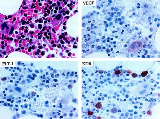
Cellular expression of VEGF and its receptors Flt-1/VEGFR-1 and KDR/VEGFR-2 were evaluated by immunohistochemical staining in bone marrow clot sections from 46 patients with MDS and 17 patients with relapsed (n = 7) or secondary (n = 10) AML. Results were compared to the pattern of protein expression in normal bone marrow specimens (n = 9). Morphologic subtypes of MDS classified according to the French-American British (FAB) criteria35 and the corresponding frequencies of protein detection are summarized in Table1. In normal bone marrow, the detection of VEGF expression in hematopoietic elements was uncommon (Figure1). Using immunohistochemistry, VEGF protein expression was not observed in erythroblasts, lymphocytes, or plasma cells. A faint Flt-1 signal was observed in monocytes–histiocytes and in rare myeloid elements, whereas a strong KDR signal was observed in scattered histiocytes.
Summary of immunohistochemical detection of VEGF and its receptors in MDS and AML
| Diagnosis . | Number . | VEGF . | Flt-1 . | KDR . |
|---|---|---|---|---|
| Normal | 9 | 0* | 0* | 0* |
| AML | 17 | 14 | 13 | 0 |
| Myelodysplasia | ||||
| RA | 8 | 8 | 7 | 2 |
| RARS | 6 | 3 | 2 | 0 |
| CMML | 10 | 8 | 8 | 3 |
| RAEB ± t | 22 | 16 | 15 | 4 |
| Diagnosis . | Number . | VEGF . | Flt-1 . | KDR . |
|---|---|---|---|---|
| Normal | 9 | 0* | 0* | 0* |
| AML | 17 | 14 | 13 | 0 |
| Myelodysplasia | ||||
| RA | 8 | 8 | 7 | 2 |
| RARS | 6 | 3 | 2 | 0 |
| CMML | 10 | 8 | 8 | 3 |
| RAEB ± t | 22 | 16 | 15 | 4 |
Samples were negative with occasional positive cells observed.
Representative pattern of immunohistochemical staining of normal marrow for VEGF, FLT-1/VEGFR-1, and KDR/VEGFR-2.
(upper left) Hematoxylin and eosin staining. (upper right) Faint VEGF signal in megakaryocytes, monocytes, and occasional myelocytes. (lower left) Low-intensity Flt-1/VEGFR-1 signal in macrophages–histiocytes and rare myeloid elements. (lower right) Strong KDR/VEGFR-2 staining in scattered histiocytes (1000 × magnification).
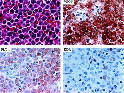
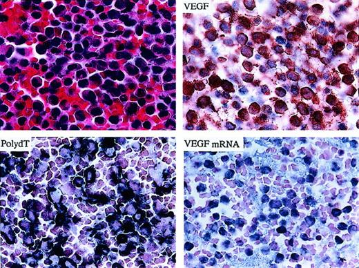
VEGF expression in MDS and AML specimens was detected in a diffuse cytoplasmic pattern in myeloid and monocyte precursors with varied signal intensity (Table 1). In bone marrow specimens from 8 of 10 patients with chronic myelomonocytic leukemia (CMML), cytoplasmic expression of VEGF, ranging from faint (+1) to intensely positive (+4), was detected in myelomonocytic cells, whereas no signal was detected in erythroblasts and plasma cells (Figure 2; Tables 2 and 3). Concordant membranous expression of the Flt-1/VEGFR-1 receptor was observed in monocyte and myeloid precursors in all CMML specimens (Table 1). Using 2 separate antibodies, a similar pattern of cellular expression, albeit of lower intensity, was detected for the KDR/VEGFR-2 receptor in 33% (3 of 10) of the CMML specimens examined. To confirm that the high-intensity VEGF protein signal detected in CMML resulted from overexpression of the VEGF gene transcript, VEGF mRNA expression was assessed by in situ hybridization using a VEGF-specific oligonucleotide probe (Figure 3). Indeed, we found that the cellular expression of VEGF mRNA was restricted to neoplastic myelomonocytic cells in a pattern analogous to that observed for the VEGF protein.
Demonstration of co-expression of VEGF and Flt-1 in neoplastic myelomonocytic leukemia cells in a patient with CMML.
(upper left) Hematoxylin and eosin staining. (upper right) IHC for VEGF demonstrating strong protein expression. (lower left) Same patient demonstrating co-expression of the Flt-1/VEGFR-1 receptor. (lower right) Rare KDR/VEGFR-2 positive cells were observed (1000 × magnification).
Effect of rhu-VEGF and VEGF neutralization (rhu-mAb-VEGF) on leukemia colony formation in advanced MDS
| UPN . | FAB type . | Immunohistochemistry . | CFU-L/105cells ± SD . | ||||
|---|---|---|---|---|---|---|---|
| VEGF . | Flt-1 . | KDR . | Control . | rhu-mAb-VEGF . | rhu-VEGF . | ||
| UAZ006 | RAEB | 0 | 0 | 0 | 25 ± 1 | 10 ± 6.6* | 140 ± 19* |
| UAZ007 | RAEB | 3 | 1 | 0 | 64 ± 24 | 34 ± 24* | 164 ± 39* |
| UAZ016 | RAEB | ND | ND | ND | 19 ± 14 | 22 ± 19 | 63 ± 31* |
| UAZ017 | RAEB | 0 | 0 | 0 | 20 ± 13 | 12 ± 7.6 | 39 ± 6.2 |
| UAZ018 | RAEB | 3 | 3 | 1 | 111 ± 57 | 106 ± 79 | 181 ± 51 |
| UAZ019 | RAEB | ND | ND | ND | 41 ± 22 | 9.3 ± 2.0* | 94 ± 71 |
| UAZ013 | RAEB-t | 4 | 3 | 3 | 182 ± 71 | 114 ± 76 | 384 ± 85* |
| UAZ020 | RAEB-t | 4 | 2 | 0 | 11.3 ± 0.5 | 0* | 21.7 ± 2* |
| UAZ021 | RAEB-t | 3 | 3 | 0 | 20 ± 5.5 | 3 ± 2.6* | 28 ± 8 |
| UAZ022 | RAEB-t | 1 | 4 | 0 | 6 ± 2 | 1.7 ± 2* | 17 ± 11* |
| UAZ023 | RAEB-t | 3 | 2 | 0 | 103 ± 23 | 63 ± 25 | 158 ± 80 |
| UAZ024 | CMML | 0 | 0 | 0 | 238 ± 8.5 | 195 ± 7.7* | 247 ± 1.1 |
| UAZ025 | CMML | 3 | 1 | 0 | 65 ± 30 | 30 ± 7.5 | 128 ± 21* |
| UAZ026 | CMML | 3 | 0 | 0 | 53 ± 23 | 52 ± 5.9 | 83 ± 12* |
| UAZ027 | CMML | 4 | 1 | 0 | 234 ± 50 | 266 ± 17 | 281 ± 24 |
P < .05 versus controls.
ND, not done.
Effect of VEGF neutralization with rhu-mAb-VEGF on bone marrow progenitor growth
| UPN . | FAB type . | Immunohistochemistry . | Control (colony no. ± SD) . | rhu-mAb-VEGF (colony no. ± SD) . | ||||||
|---|---|---|---|---|---|---|---|---|---|---|
| VEGF . | Flt-1 . | KDR . | CFU-GEMM . | BFU-E . | CFU-GM . | CFU-GEMM . | BFU-E . | CFU-GM . | ||
| UAZ001 | N1 Donor | 0 | 0 | 0 | 301 ± 16.2 | 692 ± 99 | 1257 ± 164 | 290 ± 13.6 | 718 ± 135 | 1361 ± 109 |
| UAZ002 | N1 Donor | 0 | 0 | 0 | 89 ± 8 | 570 ± 161 | 745 ± 57 | 107 ± 17.5 | 676 ± 44 | 839 ± 41 |
| UAZ003 | RA | 3 | 3 | 0 | 1.7 ± 1.5 | 2.7 ± 0.6 | 123 ± 33 | 7 ± 4.3 | 11.3 ± 44-150 | 190 ± 70 |
| UAZ004 | RA | 1 | 2 | 0 | 6.3 ± 4.2 | 47 ± 11 | 224 ± 37 | 19.3 ± 5.04-150 | 67 ± 23 | 222 ± 15 |
| UAZ005 | RA | ND | ND | ND | 1.3 ± 0.6 | 11 ± 8 | 47 ± 19 | 2.7 ± 1.5 | 14 ± 4.5 | 49 ± 17.6 |
| UAZ006 | RAEB | 0 | 0 | 0 | 1 ± 1 | 0.3 ± 0.6 | 723 ± 173 | 1 ± 0.6 | 1 ± 0 | 940 ± 450 |
| UAZ007 | RAEB | 3 | 1 | 0 | 13 ± 6.5 | 6.7 ± 3 | 1122 ± 244 | 15 ± 4.6 | 22 ± 9.14-150 | 1376 ± 369 |
| UAZ008 | RAEB | 2 | 2 | 0 | 0.3 ± 0.5 | 0.7 ± 1.1 | 93 | 1.6 ± 1.6 | 6 ± 5.3 | 441 |
| UAZ009 | RAEB | 2 | 2 | 1 | 6.7 ± 4.6 | 41 ± 11 | 1380 | 10 ± 2 | 77 ± 134-150 | 1345 |
| UAZ010 | RAEB | 3 | 1 | 0 | 8.3 ± 4.1 | 54 ± 13 | 730 ± 54 | 16.7 ± 2.34-150 | 82 ± 114-150 | 727 ± 36 |
| UAZ011 | RAEB-t | 1 | 1 | 0 | 0 | 1.7 ± 2.8 | 79 ± 14.7 | 0 | 1.7 ± 1.5 | 121 ± 12 |
| UAZ012 | RAEB-t | 4 | 3 | 3 | 18.7 ± 8 | 15.3 ± 26 | 193 ± 124 | 18.7 ± 3 | 39 ± 4 | 179 ± 46 |
| UAZ013 | RAEB-t | 4 | 3 | 3 | 70 ± 18.7 | 88 ± 39 | 1759 ± 300 | 105 ± 134-150 | 141 ± 304-150 | 2072 ± 375 |
| UAZ014 | RARS | 1 | 2 | 0 | 36.7 ± 6.4 | 53 ± 12 | 801 ± 136 | 62 ± 9.14-150 | 54 ± 20 | 791 ± 87 |
| UAZ015 | RARS | 4 | 2 | 0 | 84.7 ± 12 | 543 ± 83 | 1178 ± 250 | 175.3 ± 394-150 | 776 ± 614-150 | 1313 ± 113 |
P < .05 versus controls.
ND, not done.
Demonstration of abundant VEGF mRNA and protein signals in neoplastic myelomonocytic leukemia cells (CMML).
(upper left) Hematoxylin and eosin staining. (upper right) IHC demonstrating strong VEGF protein expression. (lower left) Poly d(T) staining confirming integrity of total mRNA. (lower right) ISH demonstrating strong expression of the VEGF message in a patient with CMML (1000 × magnification).
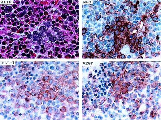
A similar pattern of cellular expression was detected in morphologic subtypes of MDS other than CMML and in AML specimens. Of particular importance, foci of myeloid precursors clustered in the central marrow space, abnormal localized immature precursors (ALIP), consistently displayed intense expression of VEGF, myeloperoxidase, and the Flt-1/VEGFR-1 receptor (Figure 4). Overall, the expression of VEGF was observed in myeloid and monocyte precursors from 76% (35 of 46) of patients with MDS and in myeloblasts from 82% (14 of 17) of patients with AMLs. Co-expression of the Flt-1 receptor was demonstrated in 69% (32 of 46) of patients with MDS and 76% (13 of 17) of patients with AML. Membranous expression of the KDR receptor in blasts and myelomonocytic precursors was detected in 19% (9 of 46) of patients with MDS, but it was not found in any of the patients with AML. If KDR was detected in patients with MDS, it was always found in conjunction with Flt-1 receptor expression. Neither the intensity of cellular VEGF expression nor the distribution or frequency of receptor expression varied among relapsed or secondary forms of AML.
Co-expression of VEGF, Flt-1/VEGFR-1, and myeloperoxidase in a cluster of leukemic blasts (ALIP) in a patient with RAEB.
(upper left) Hematoxylin and eosin staining demonstrating a cluster of ALIP. (upper right) IHC staining for myeloperoxidase (MPO) demonstrating strong expression in ALIP clusters. (lower left) IHC staining demonstrating expression of the Flt-1/VEGFR-1 receptor in a cluster of ALIP cells. (lower right) IHC demonstration of co-expression of VEGF protein in the same cluster of ALIP cells (1000 × magnification).
Leukemia progenitor formation
To determine whether VEGF serves as a trophic stimulant of leukemia progenitor self-renewal in MDS, we evaluated the effects of VEGF neutralization and stimulation on leukemia colony formation (CFU-L) in bone marrow specimens from a group of 15 patients with CMML (n = 4) or RAEB-t (n = 11) with which there was sufficient viably frozen material to perform functional assays. BM-MNCs were plated in cytokine-deficient medium with and without 0.1 to 50 μg/mL of the A.4.6.1 antibody or 1 to 100 ng/mL rhu-VEGF, and leukemia colony formation was assessed after a 7-day incubation. Antibody neutralization of VEGF activity inhibited CFU-L formation in a concentration-dependent fashion in 47% (7 of 15) of the patient specimens, with the magnitude of inhibition ranging from 20% to 100% compared to control cultures (mean, 68% inhibition). Incubation with rhu-VEGF yielded concentration-dependent stimulation of CFU-L formation in 8 patients, including each of the antibody-inhibitable specimens, with maximal stimulation ranging from 1.5-fold to 5.6-fold of control colony growth (mean, 2.44-fold). No significant changes were observed in myeloid cluster growth in response to VEGF neutralization or stimulation. Table 2 illustrates maximal changes in mean leukemia colony number according to culture condition in individual patient specimens.
Immunocytochemical stains of cytospin preparations of the KG1 AML cell line showed that these cells co-express VEGF protein and the Flt-1/VEGFR-1 and KDR/VEGFR-2 receptors. To determine whether VEGF receptor ligation triggers a proliferative growth response in a purified leukemia cell population, CFU-L formation was assessed in methylcellulose cultures containing 10% FBS after a 24-hour incubation of KG-1 cells with and without rhu-VEGF supplementation of 50 μg/mL or IL-1 100 U/mL. Cultures were grown in quadruplicate and were regrown on 3 separate occasions. VEGF exposure before culture initiation stimulated leukemia colony formation 2.4-fold compared to control cultures—mean ± SD colony number, 53 ± 20 (controls) versus 127 ± 52 (VEGF), whereas supplementation with optimal IL-1 concentrations yielded a 52% increase in colony recovery (81 ± 10). These data indicate that VEGF directly stimulates clonogenicity of receptor-competent AML cells.
ELISA quantitation of VEGF, TNF-α, and IL-1β
To quantitate VEGF elaboration by BM-MNC in MDS and the relation to inflammatory cytokine concentrations, BM plasma concentrations of VEGF, TNF-α, and IL-1β by ELISA in specimens from patients with MDS (n = 24) were evaluated, and the results were compared to those from a group of healthy allogeneic BM donors (n = 15). Mean plasma concentrations of IL-1β (3.42 ± 2.94 vs 1.61 ± 0.79 pg/mL;P = .013) and TNF-α (3.14 ± 2.22 vs 1.35 ± 0.83 pg/mL; P = .004) were significantly higher in MDS BM plasma than in the plasma of healthy donors. However, concentrations of immunoreactive VEGF were significantly lower (13.8 ± 21.6 vs 58.6 ± 9.7 pg/mL; P < .001) in MDS BM plasma than in the plasma of healthy allogeneic BM donors, and they were often below the limits of assay detection. To determine whether the lower concentrations of VEGF detected in MDS BM plasma reflected excessive local receptor binding of the peptide or impaired secretion by myeloid precursors, we evaluated VEGF elaboration from BM-MNC in 24-hour suspension cultures. Among the 12 MDS patients studied, we demonstrated elaboration of VEGF from BM-MNC, with corresponding cytokine concentrations in 24-hour culture supernatants ranging from 7.5 to 34 pg/mL. Mean VEGF concentrations in culture supernatants increased with BM myeloblast percentage (RA/RARS, 9.2 ± 7.5 pg/mL; RAEB/RAEB-t, 17.4 ± 21.3 pg/mL) and monocyte count (CMML, 22.3 ± 2.8 pg/mL).
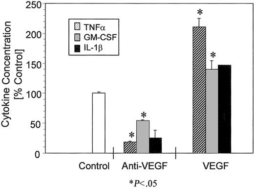
To determine whether VEGF elaboration from BM precursors impacted local generations of TNF-α and IL-1β, we evaluated the effect of VEGF neutralization on inflammatory cytokine generation in 24-hour liquid cultures of MDS BM-MNC by ELISA assay. Neutralization of VEGF activity with 0.1 to 50 μg/mL A.4.6.1 antibody was evaluated in 19 patients and resulted in concentration-dependent suppression of both TNF-α (n = 10) and IL-1β (n = 9) generation (Table4). VEGF neutralization suppressed the concentration of inflammatory cytokines 25% to 100%, yielding mean concentrations of TNF-α and IL-1β that were 44% and 39% of concentrations in supernatants from control cultures. In a separate series of experiments, the effects of VEGF on cytokine elaboration from bone marrow stroma (n = 3; UPN023) were evaluated (Figure5). Exposure to the A.4.6.1 antibody (1 μg/mL) for 24 hours decreased TNF-α and GM-CSF concentrations in stroma supernatants 81% (10.49 ± 0.23 pg/mL vs 1.97 ± 0.17 pg/mL; P < .001) and 54% (39.06 ± 2.45 vs 21.10 ± 1.82; P < .001, respectively, whereas rhu-VEGF (50 ng/mL) increased the TNF-α concentration 2.05-fold (P = .006) and GM-CSF 1.4-fold (P = .038). Supernatant concentrations of IL-1β decreased 77% with VEGF neutralization (0.81 ± 0.44 vs 0.18 ± 0.06;P = .139) and increased 1.5-fold with VEGF exposure (P = .076); however, changes were not statistically significant.
Effect of VEGF neutralization on TNF-α and IL-1β generation from MDS BM-MNC in 24-hour suspension culture
| UPN . | TNF-α (pg/mL) . | IL-1β (pg/mL) . | ||
|---|---|---|---|---|
| Control . | Anti-VEGF . | Control . | Anti-VEGF . | |
| UAZ006 | 1.096 ± 0.17 | 0.336 ± 0.0323-150 | < 0.125 | < 0.125 |
| UAZ009 | 9.534 ± 1.2 | 2.973 ± 0.213-150 | 5.217 ± 0.12 | 0.903 ± 0.193-150 |
| UAZ012 | 1.157 ± 0.9 | 0.518 ± 0.39 | 1.781 ± 0.20 | 0.748 ± 0.043-150 |
| UAZ015 | 37.79 ± 0.47 | 18.41 ± 1.493-150 | 8.796 ± 1.4 | 5.675 ± 0.173-150 |
| UAZ016 | 6.731 ± 1.45 | 6.813 ± 0.14 | 8.619 ± 0.41 | 5.418 ± 0.413-150 |
| UAZ017 | 3.46 ± 0.79 | 3.101 ± 0.11 | 0.912 ± 0.50 | 0.482 ± 0.33 |
| UAZ018 | 4.69 ± 0.26 | 4.91 ± 0.81 | 8.951 ± 0.24 | 6.980 ± 0.553-150 |
| UAZ019 | 3.083 ± 0.13 | 0.767 ± 0.183-150 | 0.905 ± 0.09 | 0.02 ± 0.013-150 |
| UAZ020 | 4.933 ± 1.49 | 2.003 ± 0.193-150 | 4.394 ± 0.18 | 1.754 ± 0.053-150 |
| UAZ021 | 3.683 ± 0.13 | 1.897 ± 1.03-150 | 1.92 ± 0.03 | 0.091 ± 0.063-150 |
| UAZ022 | 9.699 ± 2.92 | 5.030 ± 0.483-150 | 7.272 ± 1.08 | 7.360 ± 0.12 |
| UAZ023 | 5.492 ± 0.55 | 3.034 ± 0.023-150 | 3.497 ± 0.29 | 3.204 ± 0.11 |
| UAZ024 | 3.937 ± 0.02 | 2.94 ± 0.463-150 | 4.568 ± 0.07 | 3.27 ± 0.713-150 |
| UAZ025 | 17.925 ± 8.94 | 20.422 ± 0.62 | 13.126 ± 0.40 | 13.314 ± 0.18 |
| UAZ028 | 11.617 ± 1.37 | 12.321 ± 0.64 | 6.93 ± 0.99 | 4.79 ± 0.44 |
| UAZ029 | 59.039 ± 2.03 | 53.834 ± 1.85 | 8.355 ± 0.41 | 7.656 ± 0.30 |
| UAZ030 | 53.543 ± 1.23 | 51.607 ± 0.98 | 8.089 ± 0.14 | 7.791 ± 0.55 |
| UAZ031 | 66.66 ± 1.10 | 15.791 ± 0.393-150 | 8.012 ± 0.31 | 7.778 ± 0.41 |
| UAZ032 | 50.435 ± 5.61 | 49.471 ± 0.56 | 8.037 ± 0.27 | 7.755 ± 0.49 |
P < .05 versus controls.
Effect of VEGF on cytokine elaboration from bone marrow stroma.
Bone marrow stroma from a patient with RAEB were exposed to the A.4.6.1 VEGF-neutralizing antibody (1 μg/mL)—ie, anti-VEGF or rhu-VEGF (50 ng/mL)—for 24 hours, and cytokine concentrations in stroma supernatants were analyzed by ELISA. Changes in cytokine concentration are expressed as percentage value in control supernatants ± SD. *Statistically significant changes in cytokine concentration (P ≤ .05).
Effect of VEGF neutralization on committed progenitor formation
To determine whether neutralization of VEGF activity improved the formation of committed hematopoietic progenitors in MDS, BM-MNC from 13 patients (RA/RARS, 6; RAEB, 3; RAEB-t, 4) and 2 healthy donors were plated in cytokine-defined medium with and without 0.1 to 50 μg/mL A.4.6.1 antibody, and colony formation was scored after a 14-day incubation. Antibody neutralization of VEGF improved the mean recovery of CFU-GEMM approximately 3-fold (range, 1.3- to 7.5-fold) in 38% (5 of 13) of patient samples, whereas BFU-E was increased 3.7-fold in 46% (6 of 13) of samples (range, 1.5- to 10.7-fold). There was no consistent improvement in mean CFU-GM colony recovery (1.17-fold; range, 0.9- to 1.9-fold) (Table 3). Significant changes in progenitor recovery were not observed with VEGF neutralization in bone marrows from normal donors.
Discussion
Although it is well established that the growth of solid tumors is dependent on the formation of neovasculature, the role of angiogenic factors in hematopoietic malignancies has only recently been investigated. Fiedler et al26 demonstrated the expression of VEGF gene message in 72% of AML specimens; Aguayo et al36 reported that myeloblast VEGF protein content is an independent prognostic variable inversely correlated with disease-free and overall survival. Pruneri et al27 and Hussong et al37 described increased microvessel density in bone marrow trephine biopsies from patients with AML and MDS, the magnitude of which directly correlated with myeloblast percentage. The current studies demonstrate that cellular expression and elaboration of VEGF by malignant monocytes and myeloblasts in MDS and AML may have potent biologic effects that contribute to both leukemia progenitor self-renewal and ineffective hematopoiesis in these diseases. Cellular expression of VEGF was restricted to monocyte and myeloid precursors, and was associated with membranous co-expression of either the Flt-1/VEGFR-1 or the KDR/VEGFR-2 receptor, or both, in 69% (32 of 46) and 19% (9 of 46) of patients with MDS, respectively (Table 1). Our findings of VEGF and FLT-1/VEGFR-1 expression in AML are consistent with the molecular studies of Fiedler et al.26 Although KDR/VEGFR-2 was detected infrequently by these investigators, we did not detect its expression in a group of 17 patients with AML. Possible explanations for this discrepancy may lie in the patient selection or in the method for detection. Fiedler et al26 used RT-PCR to detect KDR/VEGFR-2 messages whereas we used immunohistochemistry to examine the protein. Although solution-phase RT-PCR is an extremely sensitive technique, it lacks the specificity to distinguish expression in individual cells. It is possible that contamination by endothelial cells or macrophages may have contributed to the KDR signal in the study by Fiedler et al.26 Although we did not use antigen retrieval methodologies for immunohistochemical detection of VEGF and its receptors, 2 MDS cases that were negative by IHC demonstrated a significant inhibition of CFU-L and a decrease in both TNF-α and IL-1β, suggesting that the receptors were present and functional. It is possible that using antigen retrieval methodologies would have allowed us to detect VEGF receptor expression in these samples.
Antibody neutralization of VEGF activity suppressed leukemia progenitor formation in 46% of patients, whereas rhu-VEGF promoted the growth of leukemia colonies in 56% of CMML and RAEB-t patient specimens. These findings suggest that VEGF serves as a trophic factor that supports leukemia progenitor self-renewal by either autocrine interaction or paracrine induction of myeloid growth factors from stromal elements. Indeed, rhu-VEGF stimulated the in vitro growth of KG1 AML cells, indicating that VEGF has direct trophic effects in receptor-competent leukemias. We cannot exclude indirect paracrine stimulation of leukemic cells by myeloid growth factors elaborated by VEGF-receptor competent cells in the patient specimens studied. We11 and others26 previously reported that VEGF is a potent stimulant of M-CSF, G-CSF, IL-6, stem cell factor, and GM-CSF production from human umbilical cord endothelial cells. Indeed, our findings of VEGF stimulation of GM-CSF elaboration from bone marrow stroma and the clonogenic response observed in selected VEGF-receptor–naive cases support this notion (Table 2). These findings indicate that the production of VEGF by malignant myeloid precursors serves as both an autocrine growth stimulus and a diffusible paracrine signal mediating the local generation of growth factors that foster leukemia survival and self-renewal. Additionally, these data may explain the adverse prognostic relevance of myeloblast VEGF content in AML described by Aguayo et al.36
Although plasma concentrations of VEGF are elevated in patients with solid tumors, bone marrow plasma concentrations from MDS patients were significantly lower than corresponding levels from normal allogeneic donors, whereas corresponding concentrations of TNF-α and IL-1β were significantly elevated. Secretion of the 121-kd and 165-kd VEGF isoforms by MDS BM-MNC was confirmed in suspension cultures, and increased with myeloblast and monocyte burden. These apparent contrasting observations may be explained by excessive local binding of VEGF within the marrow micro-environment.13 Malignant cells from few, if any, epithelial malignancies express receptors capable of binding VEGF. The co-expression of one or both tyrosine kinase receptors by monocyte and myeloid precursors in MDS may facilitate autocrine receptor saturation. In addition, VEGF, unlike TNF-α and IL-1β, is a heparin-binding protein.38,39The 165-kd VEGF monomer in particular, which represents the most common of the 5 isoforms of the cytokine, is retained by binding to heparan sulfate proteoglycan, which is abundant in the bone marrow extracellular matrix and on the cell surface of hematopoietic progenitors.40 Although the predominant isoform of VEGF produced by myelodysplastic BM-MNC has not as yet been investigated, VEGF165 is recognized as a more potent and bioavailable endothelial cell mitogen.41 42
Cellular expression of VEGF and its receptors in normal marrow was detected in megakaryocytes and tissue macrophages, but rarely in myeloid cells. Tordjman et al43 recently described the expression of VEGF165 mRNA in the more primitive cell population of CD34+/CD38− hematopoietic progenitors. Although primitive stem cells capable of hematopoietic and endothelial cell differentiation express the KDR/VEGFR-2 receptor,16,44 cellular expression of Flt-1/VEGFR-1 mRNA within the hematopoietic compartment has been described in monocytes, where it functions to trigger rolling or migration on endothelial cells22,23 and in CD34+progenitors.45,46 The frequent and often intense co-expression of VEGF and Flt-1/VEGFR-1 by myelodysplastic myeloid precursors we observed suggests a physiologically conserved pattern of gene expression that is up-regulated in malignant progenitors. Of particular interest, monocytoid cells in CMML were distinguished by greater VEGF protein signal intensity and cellular elaboration than myeloid precursors in other morphologic subtypes of MDS. This finding may reflect the high frequency of RAS proto-oncogene activation in CMML, which is recognized to induce transcriptional activation of VEGF gene expression.1 47
Investigations by Broxmeyer et al24 have shown that rhu-VEGF exerts growth-enhancing and -suppressive effects on in vitro colony formation of hematopoietic progenitors mediated by direct cellular interaction and indirect effects mediated by other molecules. VEGF stimulated the proliferation of mature subsets of CFU-GM but inhibited formation of the more primitive progenitors, CFU-GEMM and BFU-E. In single-cell assays, VEGF had no effect on primitive progenitor formation from CD34+ cells but retained enhancing effects on myeloid progenitors. In our studies, neutralization of VEGF activity with the A.4.6.1 antibody suppressed the generation of TNF-α and IL-1β from MDS BM-MNC and BM-stroma supernatants and enhanced the recovery of CFU-GEMM and BFU-E in methylcellulose cultures. The relation between the enhancement of primitive progenitor recovery and the suppression of TNF-α and IL-1β elaboration with antibody neutralization was not evaluated because of insufficient sample size. Nevertheless, improvement in progenitor recovery with VEGF neutralization supports the findings of Broxmeyer et al24 that VEGF exerts an inhibitory effect on progenitor growth mediated, at least in part, by a direct effect of the cytokine. In addition, VEGF stimulates the local generation of inflammatory cytokines by the induction of matrix metalloproteinase cleavage of membrane-bound TNF-α and Fas ligand from effector cells to yield biologically active, soluble forms of the molecules.17-19 In this manner, VEGF elaboration by monocyte and myeloid precursors in MDS may act to sustain leukemia progenitor self-renewal, but they also contribute to the premature death of VEGF-nonresponsive erythroid progenitors.
Perhaps the most compelling observation from these investigations is the uniform co-expression of VEGF and one or both of its receptors by myeloblasts clustered in the central marrow space, ALIP.48This pattern of dislocation of myeloid precursors from a para-trabecular locale represents an adverse prognostic feature in MDS associated with imminent risk for leukemia transformation. Our findings suggest that such foci of myeloblasts emerge as a result of autocrine or paracrine VEGF stimulation, or both, supplanting physiologic sources of cytokine production. Alternatively, such foci may represent sites of localization of proliferating myeloblasts adjacent to medullary neovasculature. Autocrine VEGF stimulation may promote the homotypic adhesion of myeloid precursors through the up-regulation or activation of cell adhesion molecules, analogous to its effects on endothelial cell targets.49 Additional studies are warranted to delineate the relation between ALIP–bone marrow microvessel distribution and integrin expression–activation status. Nevertheless, these observations suggest that VEGF is a potentially important peptide signal reinforcing leukemia cell survival and contributing to ineffective hematopoiesis in MDS. These studies provide a biologic rationale for the clinical investigation of anti-angiogenic agents in patients with MDS.
Supported in part by National Cancer Institute grant CA-32102, National Institute of Environmental Health Sciences grant ESO6694, and the Jeffrey Anderson Memorial Leukemia Research Fund.
The publication costs of this article were defrayed in part by page charge payment. Therefore, and solely to indicate this fact, this article is hereby marked “advertisement” in accordance with 18 U.S.C. section 1734.
References
Author notes
William T. Bellamy, Department of Pathology, University of Arizona, 1501 N Campbell Avenue, Tucson, AZ 85724; e-mail: wbellamy@u.arizona.edu.

This feature is available to Subscribers Only
Sign In or Create an Account Close Modal