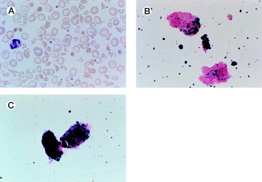
Renal Hemosiderosis Due to Intravascular Hemolysis. A 54-year-old man presented with jaundice, dark urine, and a worsening anemia. The patient has thalassemia minor and had a splenectomy 30 years ago. He had a porcine mitral valve prosthesis placed in 1979 for rheumatic heart disease; he subsequently had a St. Jude mitral valve prosthesis placed in 1990 after developing endocarditis, and later that year required a revision and a larger St. Jude prosthesis. He has had a normal hematocrit in the past and is on chronic anticoagulation therapy with warfarin. At the time of referral in June 1998, his hematocrit was 26%, with an mean corpuscular volume of 59. His peripheral smear is shown in (A). His iron studies were consistent with iron-deficiency, and his haptoglobin was low. The patient’s urine iron stain showed hemosiderin-laden renal tubular epithelial cells as well as free hemosiderin (B and C). Interestingly, an echocardiogram showed normal functioning of the prosthesis with no evidence for a perivalvular leak. His anemia improved on oral iron therapy, and his hemolysis has remained brisk. (Courtesy of Vincent E. Herrin, MD, Fellow in Hematology/Oncology, and Joe C. Files, MD, Chief of Hematology, Associate Chairman of Medicine, Division of Hematology, Department of Medicine, University of Mississippi School of Medicine, University of Mississippi Medical Center, 2500 N State St, Jackson, MS 39216.)
Renal Hemosiderosis Due to Intravascular Hemolysis. A 54-year-old man presented with jaundice, dark urine, and a worsening anemia. The patient has thalassemia minor and had a splenectomy 30 years ago. He had a porcine mitral valve prosthesis placed in 1979 for rheumatic heart disease; he subsequently had a St. Jude mitral valve prosthesis placed in 1990 after developing endocarditis, and later that year required a revision and a larger St. Jude prosthesis. He has had a normal hematocrit in the past and is on chronic anticoagulation therapy with warfarin. At the time of referral in June 1998, his hematocrit was 26%, with an mean corpuscular volume of 59. His peripheral smear is shown in (A). His iron studies were consistent with iron-deficiency, and his haptoglobin was low. The patient’s urine iron stain showed hemosiderin-laden renal tubular epithelial cells as well as free hemosiderin (B and C). Interestingly, an echocardiogram showed normal functioning of the prosthesis with no evidence for a perivalvular leak. His anemia improved on oral iron therapy, and his hemolysis has remained brisk. (Courtesy of Vincent E. Herrin, MD, Fellow in Hematology/Oncology, and Joe C. Files, MD, Chief of Hematology, Associate Chairman of Medicine, Division of Hematology, Department of Medicine, University of Mississippi School of Medicine, University of Mississippi Medical Center, 2500 N State St, Jackson, MS 39216.)


This feature is available to Subscribers Only
Sign In or Create an Account Close Modal