Human tonsil germinal center (GC) B cells rapidly undergo apoptosis in culture. Annexin-V binding shows an early event in this process. In the present study, this method has been used to label apoptotic GC B cells and to analyze additional surface molecules. The expression of all of the molecules studied was reduced in apoptotic (annexin-V+) GC B cells, and the reduction was more marked for CD11a, CD21, CD22, CD49d, and CD54, molecules that participate in survival interaction for GC B cells. The analysis of CD54, one of the molecules that was more drastically reduced, showed that GC, but not mantle zone, B cells actively secrete CD54 to the culture supernatant (SN). The secreted CD54 was partly released from the GC B cells in a particulate form as demonstrated by centrifugation. Further experiments using filtration, fluorescence microscopy, electron microscopy, and flow cytometry analysis showed that GC B cells released to the culture SN a population of spherical membranous vesicles of about 0.18 μm in size, similar to the blebs described in other apoptosis systems. Bleb formation depended on active metabolism, Ca2+, and, in part, on microfilament integrity. GC B-cell–derived blebs were clearly associated with apoptosis, as antiapoptotic stimuli prevented their formation. In addition, GC B-cell–derived blebs contained the adhesion molecules previously studied. Consequently, bleb formation might contribute to the surface molecule loss occurring in apoptotic GC B cells. Finally, a chemotaxis assay showed that GC B-cell blebs were chemotactic for human monocytes, suggesting that this mechanism might operate in vivo.
PROGRAMMED CELL DEATH (PCD) or apoptosis is a physiological process leading to the elimination of useless and harmful cells, which is very important for preserving tissue homeostasis in multicellular organisms.1 Recent reports indicate that apoptosis is regulated by the complex interaction of several families of proteins that have been conserved throughout evolution.2,3 One of these protein families consists of cysteine-containing, aspartate-specific proteases termed caspases, and appears to play a key role in the effector phase of apoptosis.4 PCD causes characteristic morphological changes in the cells, which include the release of small vesicles derived from the cytoplasmic membrane (the phenomenon known as blebbing), shrinkage of the cell and the detachment from the surrounding structures, chromatin condensation, and nuclear and cellular fragmentation.5 The process ends with the phagocytosis of dead cells and apoptotic bodies either by neighboring cells or by specialized phagocytes.6
Germinal centers (GC) are globular microstructures that consist mainly of B lymphocytes, which arise within the follicles of secondary lymphoid tissues on the entrance of T-dependent and some T-independent Ag.7-9 B cells undergoing cell death by apoptosis are a main feature of functional GC. Elimination of apoptotic GC B lymphocytes is carried out by cells of the monocyte/macrophage lineage that, in this location, are called tingible body macrophage.10 Increasing evidence supports the view that the apoptotic pathway is involved in several aspects of the B-cell physiology within the GC. Thus, a basal GC B-cell propensity to apoptosis11,12 allows the rescue of those cells that, after accumulating somatic mutations in their Ig V genes, bear an antibody (Ab) with high affinity for the Ag exposed on the follicular dendritic cell (FDC) surface, a process that leads to memory B-cell selection.13-16 Together with the process of Ig V gene somatic hypermutation, it has recently been documented that a fraction of GC B cells goes through a new phase of V(D)J Ig gene recombination and B-cell receptor (BCR) editing of as yet unknown significance.17-19 It is thought that PCD also operates to assure the deletion of B cells bearing defective or autoreactive Ab generated by these two genetic processes. In this regard, it has been shown that an important proportion of GC B cells undergoes apoptosis by the specific recognition of Ag in soluble form.20-22 This finding has been related to a possible mechanism for maintaining self-tolerance by the elimination of self-reactive B-cell clones generated by the Ig diversification processes occurring during the GC response.20-22 The propensity of GC B lymphocytes to die is further evidenced by the observation that they rapidly undergo massive apoptosis in culture.13,23 In addition, human GC founder B cells have been shown to trigger the apoptosis program even before the onset of somatic mutations.24 Therefore, GC are sites where B cells exhibit an enhanced tendency to PCD, which is critical for the action of appropriate selective mechanisms.
Several surface molecules have been demonstrated to participate in the rescue of GC B cells from apoptosis. Besides the above-mentioned role for the BCR, the ligation of CD40 appears to deliver survival signals to these cells.13,25 Moreover, the adhesion molecules CD11a, CD29-CD49d, and CD54 have been shown to contribute to the FDC-GC B-cell interaction, which appears to be required as well for maintaining the latter cells alive.26-29 In addition, CD21, the complement receptor 2 (CR2), which is expressed by GC B cells and which can interact with C3d and CD23 expressed on the FDC surface, also plays a relevant role in GC B-cell survival.30 31
The initial aim of this study was to examine the fate of several human GC B-cell surface molecules during their spontaneous apoptosis in vitro. To this end, use was made of an early event occurring in apoptotic cells, including GC B cells; this event is the translocation of phosphatydilserine residues from the inner to the outer leaflet of the cell membrane, a phenomenon that can be shown by binding with labeled annexin-V.32 The results show that apoptotic GC B cells rapidly lose a variety of surface molecules, mainly those involved in their adhesion to FDC and surrounding estractures. Further experiments showed that, at least in part, these molecules were released from the cell surface as membranous vesicles similar to those described as apoptotic blebs in many cell systems. Finally, these GC B-cell–derived blebs exhibited chemoattractive activity on monocytes, suggesting that this mechanism might be operative in vivo.
MATERIALS AND METHODS
Materials.
Cycloheximide (Cx), Cytochalasin B (CkB), phorbol 12-myristate 13-acetate (PMA), zymosan, and PKH26-GL were purchased from Sigma (St Louis, MO). Z-Val-Ala-DL-Asp-fluoromethylketone (VAD-fmk) was obtained from Bachem Feinchemikalien AG (Budendorf, Switzerland). EDTA was purchased from Pharmacia Biotech (Uppsala, Sweden). Agar was provided by Difco Laboratories (Detroit, MI). Interleukin-4 (IL-4) was provided by Peprotech Inc (Rocky Hill, NJ). The 24-well flat-bottomed plate used for cell culture was from Nunc (Roskilde, Denmark). Anti-CD40 mouse monoclonal antibody (MoAb) (MAB 89), fluorescein isothiocyanate (FITC)-labeled mouse MoAb against CD21 and CD54 and phycoerythrin (PE)-labeled mouse MoAb against CD11a, and CD40 were obtained from Immunotech (Luminy, France). FITC-labeled mouse MoAb against CD20, CD22, HLA DR, and CD11a, and PE-labeled mouse MoAb against CD20, CD21, CD22, HLA DR, CD38, CD45, CD49d, CD54, and CD58, and control FITC-labeled and PE-labeled MoAb of appropriate isotypes were provided by Becton Dickinson (San Jose, CA). FITC-labeled annexin-V was obtained from Bender Medsystems (Vienna, Austria). Human CD54 enzyme-linked immunosorbent assay (ELISA) kit was purchased from R&D System Inc (Minneapolis, MN). Isopore filter (0.1-μm pore) used to filter cell culture supernatants was provided by Millipore Corp (Bedford, MA). Polyvinylpyrrolidone-free polycarbonate membrane disc (5-μm pore) used in chemotaxis assays was obtained from BioRad Laboratories (Richmond, CA).
Tonsil cell and blood monocyte preparation.
Tonsillar tissue was obtained from subjects undergoing tonsillectomy for chronic tonsillitis. Single-cell suspensions were prepared and the cells were separated into T- and non-T (B)-cell populations by a previously reported rosette technique. B cells were fractionated further into GC and mantle zone (MZ) B cells on a discontinuous Percoll gradient as detailed elsewhere.33 Purity of GC B cells was determined by immunofluorescence and flow cytometry, as CD38+ CD20high cells, and exhibited a viability higher than 95%, as determined by trypan blue exclusion test. Monocytes for chemotaxis assays were obtained from healthy volunteers’ blood. Briefly, blood samples were drawn in sterile tubes, containing 14.3 IU of sodium heparin/mL. Mononuclear cells were isolated by bouyant density centrifugation on Lymphoprep (Nyergaard, Oslo, Norway) at 450g for 35 minutes. The cells were recovered from the interface, washed twice, and resuspended at 1 × 10⁶monocytes/mL in RPMI 1640 medium supplemented with L-glutamine (10 mmol/L). Monocytes were distinguished by differential cell count performed on smears using a nonspecific esterase stain as previously described.34 The monocyte proportion in 13 cell preparations ranged from 15.5% to 24% (17.6% ± 2.5%; mean ± standard error of mean [SEM]).
Cell culture.
GC and MZ B cells and T cells (106 cells/mL) were incubated at 37°C with 5% CO2 in a culture medium consisting of RPMI 1640 supplemented with 10% fetal calf serum (FCS), L-glutamine (10 mmol/L), and gentamycin (0.05 mg/mL) for 2 hours unless indicated otherwise. GC B cells were also cultured in the presence of anti-CD40 MoAb (1 μg/mL) + IL-4 (2 ng/mL) and the peptide VAD-fmk (200 μmol/L) for the indicated times. In some experiments, cells were stained with PKH26-GL alone or combined with FITC-conjugated MoAb before the culture.
Preparation of cell-free supernatant (SN) and subcellular particles derived from cultured GC B and T cells.
Tonsil GC and MZ B cells and T cells (106/mL) were incubated for 2 hours or the indicated times in medium, and then the culture SN was centrifuged at 103g for 10 minutes at 4°C to remove cells and large cellular fragments (cell-free SN). Cell-free SN obtained from cultured GC B cells was then centrifuged at 105g for 18 hours at 4°C, and the soluble (GC SN SOL) and the precipitated (GC SN PREC) fractions were recovered, and adjusted to the initial volume in medium. In some experiments, cell-free SN obtained from GC B-cell cultures was filtered on a 0.1-μm pore membrane to remove subcellular particles from the cell-free SN (GC SN FILT).
Cell and subcellular membranous particle staining.
Two-color staining of GC B cells with fluorochrome-conjugated MoAbs and annexin-V binding assay was performed as previously described.35 In brief, GC B cells (106/mL) cultured for 2 hours, or the indicated time, were incubated with FITC-conjugated annexin-V, either alone or in combination with optimal concentrations of PE-labeled MoAbs against CD11a, CD20, CD21, CD22, CD38, CD40, CD45, CD49d, CD54, CD58, and HLA DR in 1 mL of Ca2+ HEPES buffer (10 mmol/L HEPES, 1.8 mmol/L CaCl2, 150 mmol/L NaCl) for 30 minutes in the dark at 4°C. After two washes with the same buffer, the cells were analyzed by flow cytometry and by fluorescence microscopy. To explore the presence of membranous cellular particles in culture SN, tonsillar GC B and T cells (106 cells/mL) were stained with the lipid intercalating dye PKH26-GL (4 μmol/L) for 5 minutes at room temperature. Staining reaction was stopped by adding a similar volume of FCS, and the cells were washed twice in culture medium. Stained cells were cultured (106 cells/mL) for 2 hours, unless indicated otherwise. At the end of the culture period, the cell-free SN was obtained and the presence of red fluorescent cellular particles was analyzed by flow cytometry. To investigate the requirements for the generation of membranous particles, GC B cells were stained with PKH26-GL as above and cultured for 12 hours in the presence and in the absence of cycloheximide (10 μg/mL), cytokalasin B (5 μg/mL), anti-CD40 MoAb (1 μg/mL) + IL-4 (2 ng/mL), EDTA (1 mmol/L), PMA (1 ng/mL), and the peptide VAD-fmk (200 μmol/L). At the end of the culture period, cell-free SN was recovered and analyzed by flow cytometry. In some experiments, cell-free SN obtained from cultured GC B cells was passed through a 0.1-μm pore filter, and the presence of stained particles in the elutant was analyzed as above. To test the presence of certain GC B-cell surface molecules in the membranous particle, PKH26-GL–stained GC B cells were additionally labeled with appropriate FITC-conjugated MoAbs, washed, and cultured for 2 hours at 106 cells/mL in culture medium. The cell-free SN was analyzed by flow cytometry, assessing the expression of these molecules on the PKH26-GL+ particle fraction.
Flow cytometry.
Fluorescence-activated cell sorting (FACS) analysis was performed on a FACScalibur instrument (Becton Dickinson) equipped with an air-cooled argon ion laser emitting 15 mW at 488 nm. The instrument was equipped with three fluorescence detector photomultiplier tubes, with green fluorescence (FITC, FL1) being collected through a 585/42-nm bandpass, orange/red (PE, FL2) through a 585/42-nm bandpass, and red (PerCP, FL3) through a 650-nm longpass filter. Two-color cell analysis was performed as previously reported.35 In the analysis of cellular-derived vesicles contained in the cell-free SN, light scatter and fluorescence signals were recorded in logarithmic mode. In experiments to test requirements for cell-derived vesicle generation, cell-free SN was acquired for 2 minutes at medium flow rate, and the event numbers were recorded. Negative controls were established as previously described.36
Electron microscopy (EM).
GC B cells (2 × 10⁶ cells/mL) were cultured for 2 hours either in a semisolid medium consisting of agar (0.5% wt/vol in culture medium) or on a 0.1-μm Isopore membrane placed at the bottom of a well of a 24-well microculture plate, and these preparations were used for transmission and scanning EM studies, respectively. In addition, cell-free SN obtained from these cultures was passed through a 0.1-μm pore membrane, and the presence of subcellular vesicles deposited on these membranes was also investigated by both transmission and scanning EM. The different samples were treated with glutaraldehyde (2.5%) overnight at 4°C, with osmium tetroxide (1%) for 30 minutes and then dehydrated. Samples for scanning EM were critical-point dried, mounted in specimen stubs, gold coated, and analyzed.
Chemotaxis assay.
Monocyte chemotaxis was evaluated by a modified Boyden chamber technique as previously described.37 The chambers were separated by polyvinylpyrrolidone-free polycarbonate membrane discs and incubated at 37°C in 5% CO2 in air for 90 minutes. Different preparations of the SN of cultured GC B and T cells were used. Culture medium and zymosan activated serum (ZAS) were used as negative and positive controls, respectively. Two independent observers counted the migrated cells within a square reticle in 10 high-power immersion oil fields (hpf). Each experiment was performed in triplicate chambers and the mean calculated.
RESULTS
Loss of surface molecules by CG B cells undergoing apoptosis.
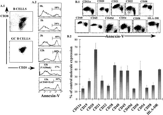
It is well known that GC B cells rapidly undergo spontaneous apoptosis in culture. Annexin-V binding has been described as a good marker for early stages of apoptosis.32 Accordingly, highly purified GC B cells were obtained from human tonsils (Fig 1A.1), and their capacity to bind FITC-annexin-V was tested after different periods of culture. As can be seen in Fig 1A.2, the proportion of annexin-V+ GC B cells increased with time and, after 12 hours, most of the cells (88% ± 5%; mean ± standard error of mean [SEM]; n = 6) showed the annexin-V+ phenotype. This phenomenon could be partly reversed by adding to the culture either anti-CD40 MoAb + IL-4, a well-established antiapoptotic stimulus in this cell system,13 or the broad-spectrum caspase-inhibitor VAD-fmk.38 These results indicate that annexin-V binding can be used as a marker of GC B-cell apoptosis. The method allowed the analysis by flow cytometry of additional surface molecules in apoptotic (annexin-V+) and nonapoptotic (annexin-V−) GC B cells. GC B cells cultured for 2 hours were used in this analysis because this short period of time was sufficient for a substantial proportion of the cells to reach the apoptotic phenotype (from 10% ± 1.5%, at 0 hour, to 33% ± 3%, at 2 hours; mean ± SEM; n = 15). Figure 1B.1 shows an example of this study and Fig 1B.2 summarizes data obtained in five different experiments, representing the percentage of remaining molecule expression, determined as the mean fluorescence intensity (MFI), in apoptotic GC B cells in comparison to live GC B cells. As can be seen, annexin-V+ GC B cells showed reduced expression of all the surface molecules explored. The reduction was not equal for all of the molecules. Thus, CD20 was the least affected; CD38, CD40, CD45, and HLA DR exhibited an intermediate level of reduction; and CD11a, CD21, CD22, CD49d, CD54, and CD58 showed the lowest level of expression. Survival stimulus (CD40 + IL-4 and VAD-fmk) delayed the entrance of GC B cells into the annexin-V+ stage, but apoptotic cells emerging in these cultures showed a low expression of surface molecule similar to that exhibited by untreated cells (data not shown). During the first 6 hours of culture, the surface molecule loss shown by apoptotic GC B cells was not associated with a decrease in cellular size, as detected by changes in the forward scatter values. The level of surface molecule expression by annexin-V+ GC B cells determined as the MFI for each molecule was similar in cells cultured from 0 to 12 hours (data not shown).
Human GC B-cell purification (A.1), annexin-V binding expression kinetics (A.2), and surface molecule expression (B). (A.1) Unfractionated and GC B cells were obtained from human tonsils and the expression of CD20 and CD38 on their surface was monitored by immunofluorescence and flow cytometry. Dot plots corresponding to a representative experiment are shown. (A.2) GC B cells (106cells/mL) were cultured for indicated times in the absence and in the presence of anti-CD40 MoAb (1 μg/mL) + IL-4 (2 ng/mL) or VAD-fmk (200 μmol/L) and the cells were then labeled with FITC-annexin-V and analyzed by flow cytometry. Histograms of annexin-V expression in one representative experiment are shown. (B.1) GC B cells were cultured for 2 hours and simultaneously labeled with FITC-annexin-V and additional MoAb directed to the indicated molecules. Dot plots of one representative experiment are shown. All of the surface molecules examined were positive in more than 90% of the freshly isolated GC B cells, except for CD49d, which was positive in only 39% ± 5% (mean ± SEM) of the cells. Axis scales of dot plots are logarithmic. (B.2) Expression of surface molecules in annexin-V+ GC B cells. The values were obtained as the percentage of the MFI shown for each surface molecule studied on the annexin-V+ cells, with respect to that observed on annexin-V− cells, which was considered the control expression. Results represent the mean ± SEM of five experiments.
Human GC B-cell purification (A.1), annexin-V binding expression kinetics (A.2), and surface molecule expression (B). (A.1) Unfractionated and GC B cells were obtained from human tonsils and the expression of CD20 and CD38 on their surface was monitored by immunofluorescence and flow cytometry. Dot plots corresponding to a representative experiment are shown. (A.2) GC B cells (106cells/mL) were cultured for indicated times in the absence and in the presence of anti-CD40 MoAb (1 μg/mL) + IL-4 (2 ng/mL) or VAD-fmk (200 μmol/L) and the cells were then labeled with FITC-annexin-V and analyzed by flow cytometry. Histograms of annexin-V expression in one representative experiment are shown. (B.1) GC B cells were cultured for 2 hours and simultaneously labeled with FITC-annexin-V and additional MoAb directed to the indicated molecules. Dot plots of one representative experiment are shown. All of the surface molecules examined were positive in more than 90% of the freshly isolated GC B cells, except for CD49d, which was positive in only 39% ± 5% (mean ± SEM) of the cells. Axis scales of dot plots are logarithmic. (B.2) Expression of surface molecules in annexin-V+ GC B cells. The values were obtained as the percentage of the MFI shown for each surface molecule studied on the annexin-V+ cells, with respect to that observed on annexin-V− cells, which was considered the control expression. Results represent the mean ± SEM of five experiments.
Presence of CD54 in the supernatant of GC B-cell culture.
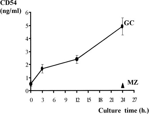
The surface molecule loss observed in apoptotic GC B cells could be explained by either endocytosis or external release. To determine which of these two possible mechanisms operates, the presence of CD54, one of the molecules that was more drastically lost, was determined in the supernatant of GC B-cell cultures by ELISA. Figure 2 shows that CD54 was readily detected in the SN of GC B-cell cultures, and its quantity increased over the time of culture. This phenomenon appeared to be restricted to GC B cells, because purified tonsillar MZ B cells, which express similar levels of CD54 on their surface, did not release detectable quantities of this molecule into the 24-hour culture supernatant (Fig2). To investigate further this phenomenon, cell-free SN obtained after 2 hours of GC B-cell culture was ultracentrifuged, and the recovery of CD54 was additionally determined in both the precipitate and the ultrasupernatant fraction. The results showed that 42.2% ± 9.6% (mean ± SEM; n = 3) of the total CD54 contained in the cell-free supernatant (3.09 ng/mL ± 0.36; n = 3) was recovered in the precipitate fraction. This finding suggested that, at least in part, CD54 was released by the GC B cells in a particulate form.
CD54 detection in culture supernatant. Tonsillar GC and MZ B cells were cultured, and the cell-free SN was recovered after the indicated times and tested for their CD54 content by an ELISA technique. Results (ng/mL) represent the mean ± SEM of eight experiments.
CD54 detection in culture supernatant. Tonsillar GC and MZ B cells were cultured, and the cell-free SN was recovered after the indicated times and tested for their CD54 content by an ELISA technique. Results (ng/mL) represent the mean ± SEM of eight experiments.
Identification of membranous particles released into the GC B-cell supernatant.
Previous reports have established that apoptotic cells produce plasma membrane rounded vesicles called blebs.5 Therefore, experiments were designed to test whether the GC B-cell SN contained particles that could be identified as blebs. Cell-free SN obtained from 2-hour GC B-cell cultures was passed through a 0.1-μm pore filter, and the filter was examined by EM. Figure3A shows the presence of spherical vesicles retained on the filter as detected by either scanning or transmission EM (Fig 3A.1 and A.2, respectively). These particles appeared as rounded plasma membrane vesicles, with an average diameter of 0.180 μm ± 0.05 (mean ± standard deviation [SD]). Later in the culture, particles of larger size were also isolated from the SN of GC B-cell cultures, some of them containing chromatin-condensed material, corresponding to apoptotic bodies (data not shown). In additional experiments, GC B cells were incubated for 2 hours in semisolid cultures to observe bleb formation. Figure 3B shows the process of blebbing by apoptotic GC B cells as detected by fluorescence microscopy (Fig 3B.1) and by transmission and scanning EM (Fig 3B.2 and B.3, respectively).
Microscopy analysis of released vesicles (A) and their cellular generation (B). (A) Cell-free SN obtained from GC B cells cultured for 2 hours was passed through a filter (0.1-μm pore), and the filter was processed for scanning (A.1) and transmission (A.2) EM. Filter pores are seen as black holes in A.1. (B) GC B cells were stained with PKH26-GL and, after 2 hours, were analyzed under fluorescence microscopy (B.1). GC B cells cultured for 2 hours were studied by transmission EM (B.2) and scanning EM (B.3).
Microscopy analysis of released vesicles (A) and their cellular generation (B). (A) Cell-free SN obtained from GC B cells cultured for 2 hours was passed through a filter (0.1-μm pore), and the filter was processed for scanning (A.1) and transmission (A.2) EM. Filter pores are seen as black holes in A.1. (B) GC B cells were stained with PKH26-GL and, after 2 hours, were analyzed under fluorescence microscopy (B.1). GC B cells cultured for 2 hours were studied by transmission EM (B.2) and scanning EM (B.3).
Flow cytometry analysis of apoptotic GC B-cell–derived blebs.
PKH26-GL is a fluorescent probe specific for the cell membrane that can be used as a stable and nontoxic staining of viable cells39 and cellular membrane particles.36 In an attempt to track apoptotic cell-derived blebs, GC B cells, after labeling with PKH26-GL, were cultured for 2 hours and the presence of fluorescent particles was examined in the cell-free SN by flow cytometry. Figure 4A shows that the forward versus side scatter dot plot analysis of this cell-free SN did not show a clear distinction of the blebs, as their size was close to the detection limit of the cytometer. Nevertheless, the use of the fluorochrome allowed identification of the presence of a population of membranous particles (Fig 4B), which disappeared when the GC B-cell supernatant was filtered through a 0.1 μm pore filter (Fig 4C). In addition, these particles were not present in the supernatant of PKH26-GL–labeled tonsillar T lymphocytes cultured for 2 hours (Fig4D). These results indicate that the method could be useful for detecting GC B-cell–derived blebs and, accordingly, it was used to investigate the kinetics of and the requirements for bleb formation in the present culture system. Figure 5A shows that the kinetics of bleb accumulation in the SN of GC B-cell cultures was linear during the first 12 hours. The generation of blebs markedly decreased when cells were cultured at 4°C and when cells were treated with EDTA (1 mmol/L) and with blockers of GC B-cell apoptosis such as anti-CD40 MoAb + IL-4 and VAD-fmk (Fig 5B). Treatment with cytochalasin B, which disrupts microfilaments, provoked only a partial decrease (P < .03). The inclusion of cycloheximide (an inhibitor of protein synthesis) and the PKC-activator, PMA, did not significantly modify GC B-cell bleb generation (Fig 5B).
Analysis of GC B-cell–derived vesicles by flow cytometry. Tonsillar GC B cells and T cells were stained with the cell membrane-specific probe, PKH26-GL, and then cultured for 2 hours. After this period, cell-free SN was collected and the presence of membranous vesicles was monitored by flow cytometry. (A) Dot plot representation of the forward scatter (FS) versus side scatter (SS) values of the SN containing GC B-cell–derived particles is shown. FL2 histogram obtained from the analysis of the same sample as in (A), either before (B) or after (C) filtering through a 0.1-μm pore membrane and of SN obtained from T-cell culture (D).
Analysis of GC B-cell–derived vesicles by flow cytometry. Tonsillar GC B cells and T cells were stained with the cell membrane-specific probe, PKH26-GL, and then cultured for 2 hours. After this period, cell-free SN was collected and the presence of membranous vesicles was monitored by flow cytometry. (A) Dot plot representation of the forward scatter (FS) versus side scatter (SS) values of the SN containing GC B-cell–derived particles is shown. FL2 histogram obtained from the analysis of the same sample as in (A), either before (B) or after (C) filtering through a 0.1-μm pore membrane and of SN obtained from T-cell culture (D).
Kinetics of and requirements for the generation of vesicles derived from cultured GC B cells. GC B cells were stained with PKH26-GL, cultured for 2 hours, and the cell-free SN was analyzed by flow cytometry. (A) A dot plot of SS versus FL2 parameters was used to define GC B-cell–derived membranous particles. (B) The count of PKH26-GL+ particles detected in the SN obtained for indicated times was recorded. Results of one experiment representative of three are shown. (C) PKH26-GL–stained GC B cells were cultured for 12 hours at 4°C and at 37°C in the absence (control culture) and in the presence of cycloheximide (Cx, 10 μg/mL), cytochalasin B (CkB, 5 μg/mL), EDTA (1 nmol/L), anti-CD40 MoAb (1 μg/mL) + IL-4 (2 ng/mL), PMA (10 ng/mL), and VAD-fmk (200 μmol/L). Values of the counts of PKH26-GL+ particles were recorded and expressed as percentages of the control figures. Results represent the mean ± SEM of four experiments.
Kinetics of and requirements for the generation of vesicles derived from cultured GC B cells. GC B cells were stained with PKH26-GL, cultured for 2 hours, and the cell-free SN was analyzed by flow cytometry. (A) A dot plot of SS versus FL2 parameters was used to define GC B-cell–derived membranous particles. (B) The count of PKH26-GL+ particles detected in the SN obtained for indicated times was recorded. Results of one experiment representative of three are shown. (C) PKH26-GL–stained GC B cells were cultured for 12 hours at 4°C and at 37°C in the absence (control culture) and in the presence of cycloheximide (Cx, 10 μg/mL), cytochalasin B (CkB, 5 μg/mL), EDTA (1 nmol/L), anti-CD40 MoAb (1 μg/mL) + IL-4 (2 ng/mL), PMA (10 ng/mL), and VAD-fmk (200 μmol/L). Values of the counts of PKH26-GL+ particles were recorded and expressed as percentages of the control figures. Results represent the mean ± SEM of four experiments.
Presence of surface molecules in the blebs derived from apoptotic GC B cells.
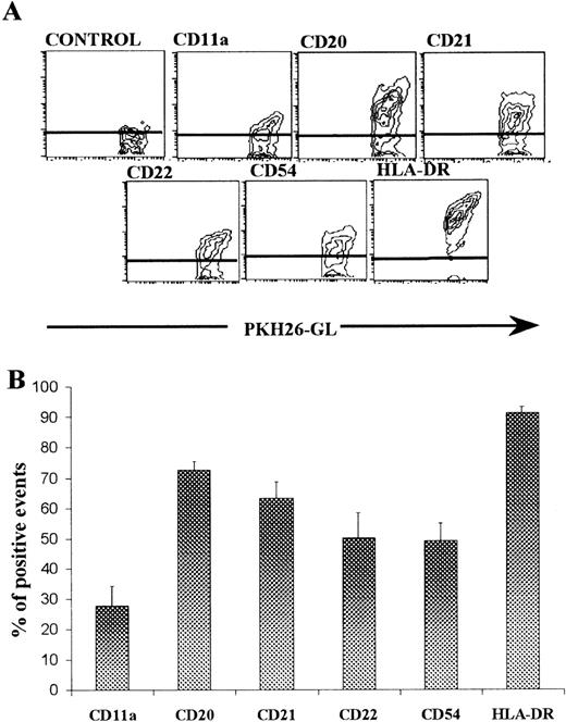
The labeling of GC B-cell membrane with PKH26-GL neither modified annexin-V binding nor affected the detection of surface molecules by immunofluorescence and flow cytometry (data not shown). Therefore, this method was used to investigate the presence of the previously studied surface molecules in the GC B-cell–derived blebs. As can be seen in Fig 6A, PKH26-GL–stained vesicles, at least in part, expressed CD11a, CD20, CD21, CD22, HLA DR, and CD54. Figure 6B shows that, apart from CD11a, all of the molecules studied were detected on most of the PHH26-GL+ particles.
Expression of surface molecules by vesicles derived from cultured GC B cells. GC B cells were stained with PKH26-GL and FITC-conjugated MoAb against several surface molecules present on GC B cells and against an irrelevant antigen (control). After 2 hours, cell-free SN was obtained and analyzed by flow cytometry. (A) Contour plots of FL2 (PKH26-GL staining) versus FL1 (indicated molecule) from one representative example are shown. (B) Values were expressed as the percentage of particles positive for each molecule. Results represent the mean ± SEM of three experiments.
Expression of surface molecules by vesicles derived from cultured GC B cells. GC B cells were stained with PKH26-GL and FITC-conjugated MoAb against several surface molecules present on GC B cells and against an irrelevant antigen (control). After 2 hours, cell-free SN was obtained and analyzed by flow cytometry. (A) Contour plots of FL2 (PKH26-GL staining) versus FL1 (indicated molecule) from one representative example are shown. (B) Values were expressed as the percentage of particles positive for each molecule. Results represent the mean ± SEM of three experiments.
Role of the blebs derived from apoptotic GC B cells as monocyte chemoattractant.
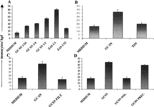
In the final set of experiments, the possibility that blebs derived from apoptotic GC B cells could be chemotactic for monocytes was tested. To this end, the activity of cell-free SN obtained from 2-hour GC B-cell cultures was determined in a chemotaxis assay that used normal blood monocytes. Figure 7A shows that GC B-cell SN exhibited detectable chemotactic activity that increased in a concentration-dependent manner. This activity was not a nonspecific phenomenon, as similar SN obtained from cultured T lymphocytes isolated from the same tonsils did not produce any effect in the assay (Fig 7B). The monocyte-chemotactic activity shown by GC B-cell SN was lost when it was passed through a 0.1-μm pore filter (Fig 7C), suggesting that the activity was associated with cellular particles released into the SN. In addition, Fig 7D shows that the monocyte-chemotactic activity shown by the GC B-cell culture SN was almost exclusively restricted to the membranous precipitate (GC SN PREC), but not in the soluble (GC SN SOL), fraction obtained by ultracentrifugation.
Chemoattractant activity on human monocytes by vesicles derived from GC B cells cultured for 2 hours. (A) Monocyte chemotaxis was assessed using as stimulus culture medium (negative control), ZAS (positive control), and different dilutions of GC B-cell culture SN (GC SN). (B) The chemoattractant activity of GC SN was compared with that of a similar supernatant obtained from T-cell cultures (T SN). (C) Comparison of the chemoattractant effect of GC SN before and after being passed through a 0.1-μm pore membrane (GC SN FILT). (D) GC SN was centrifuged at 105g, and the precipitated material (GC SN PREC) and the soluble fraction (GC SN SOL) were obtained and tested in the chemotaxis assay. The values were expressed as the mean count of migrated monocytes per high-power field. Results represent the mean ± SEM of four experiments.
Chemoattractant activity on human monocytes by vesicles derived from GC B cells cultured for 2 hours. (A) Monocyte chemotaxis was assessed using as stimulus culture medium (negative control), ZAS (positive control), and different dilutions of GC B-cell culture SN (GC SN). (B) The chemoattractant activity of GC SN was compared with that of a similar supernatant obtained from T-cell cultures (T SN). (C) Comparison of the chemoattractant effect of GC SN before and after being passed through a 0.1-μm pore membrane (GC SN FILT). (D) GC SN was centrifuged at 105g, and the precipitated material (GC SN PREC) and the soluble fraction (GC SN SOL) were obtained and tested in the chemotaxis assay. The values were expressed as the mean count of migrated monocytes per high-power field. Results represent the mean ± SEM of four experiments.
DISCUSSION
It is well established that GC B cells are prone to undergo PCD in vivo, as well as in vitro, and this capacity appears to be essential for the selection of memory B cells and probably for the deletion of defective and autoreactive B-cell clones generated during the GC response.40,41 In the present study, annexin-V binding, which shows the phosphatidylserine translocation to the membrane external leaflet, was used for detecting spontaneous apoptosis by cultured GC B cells isolated from human tonsils. This method has been demonstrated to define an early stage of apoptosis in many cell systems, including human tonsil GC B cells, where annexin-V positiveness coincides with the appearance of initial DNA condensation and fragmentation.32 Present results reinforce this assumption, as annexin-V binding by GC B cells was clearly delayed by two well-established antiapoptotic signals, such as the ligation of CD40 plus IL-413 and the treatment with VAD-fmk, a peptide that blocks the caspase cascade at several levels.38 The use of annexin-V binding allowed the observation of additional surface molecules on apoptotic GC B cells. Present data indicate that GC B cells undergoing apoptosis exhibit a markedly reduced expression of several surface molecules. The finding that MFI values of surface molecule expression by apoptotic (annexin-V+) GC B cells remained unaltered during at least 12 hours, even in the presence of antiapoptotic stimuli, suggested that the surface molecule loss was a stable phenomenon, apparently occurring during the transition from annexin-V− to annexin-V+ stages. Moreover, both annexin-V binding and surface molecule loss were clearly related to the apoptotic status of the GC B cells, as these phenomena were equally delayed by the antiapoptotic treatments. Therefore, loss of surface molecules seemed to be a part of the GC B-cell apoptosis program.
Several mechanisms can be invoked to explain the reduced expression of apoptotic GC B-cell surface molecules. The detection and analysis in the culture SN of CD54, one of the molecules more drastically lost in the present system, showed that GC B, but not MZ B, cells released considerable quantities of this molecule to the culture medium. In addition, a significant proportion of these molecules was released from the GC B cells in a particulate form, as they could be precipitated by ultracentrifugation. This finding was more relevant in light of the fact that CD54 is a well-known example of a potentially soluble molecule.42 Cellular blebbing has been described as the formation and release of small membranous vesicles that occurs as an initial event of PCD.43 Consequently, the possible existence of this process during the apoptosis of GC B cells was examined. Present data indicate that apoptotic GC B cells released small spherical membranous vesicles of about 0.2 μm in diameter, as demonstrated by EM and flow cytometry. This latter method revealed that the molecules that showed low expression on apoptotic GC B cells could be detected in most of the membranous vesicles derived from these cells, and allowed a more detailed study of the kinetics of release and the metabolic requirement for the generation of these vesicles. Thus, the formation of vesicles derived from apoptotic GC B cells depended on active metabolism and Ca2+ presence and was largely independent on protein synthesis, microfilament integrity, and PKC activation. These characteristics are compatible with those described for the generation of cellular blebs in several models of apoptotic systems.44-48 Further experiments indicated that apoptotic GC B cells also developed surface blebbing, as determined by fluorescence microscopy and EM. Therefore, it is reasonable to think that membranous vesicles found in the SN of GC B-cell cultures are similar to the blebs described during PCD. This idea was also supported by the finding that bleb formation was prevented by stimuli that delayed the appearance of apoptosis in these cells. Taken together, these observations suggest that, at least in part, the blebbing process can contribute to the explanation of the reduction of surface molecule expression by apoptotic GC B cells.
Reduced expression of surface proteins by apoptotic GC B cells was not similar for all of the molecules studied, the adhesion molecules (CD11a, CD21, CD22, CD49d, CD54, and CD58) being those most affected. Many of these molecules are involved in the adhesion of GC B cells to FDC, and possibly also to other surrounding structures, a mechanism that contributes to preserving GC B-cell survival.26-31Loss of contact and detachment is a common feature of apoptotic cells.5,43 In fact, disruption of intercellular contact through integrins and other adhesion molecules causes apoptosis in many cell systems.49 50 Thus, early in the apoptotic program, GC B cells appeared to lose those molecules that allowed their attachment, which probably resulted in the irreversibility of the process.
Apoptotic cells are rapidly eliminated by phagocytosis, which is important to prevent inflammatory reactions. This process is carried out either by neighbor or by “professional” phagocytes depending on the tissue. In the GC, cells of the monocyte/macrophage lineage are specialized in this task, and apoptotic GC B cells are commonly observed to be engulfed by cells of this kind, named for this reason tingible body macrophages.10 Some of the molecular events involved in the recognition and phagocytosis of apoptotic cells by macrophages have been clarified.6 51-53 However, little is known about the mechanisms that locally recruit the macrophage into the apoptotic focus. Monocytes migrate toward particular sites by following the gradient of certain chemotactic factors. Therefore, the possibility that SN derived from cultured GC B cells is chemoattractant for monocytes was assessed. The data demonstrate that SN from cultured GC B, but not T, cells exhibited detectable chemotactic activity for monocytes. In addition, this activity was present in the particulate fraction of the GC B cell SN, as it was eliminated either by filtering the SN on membranes with 0.1 μm exclusion pore, or by centrifugation at 105g. These findings indicate that blebs released by GC B cells undergoing PCD are capable of attracting monocytes in vitro. Accordingly, it can be hypothesized that a gradient of apoptotic blebs released around dying GC B cells attracts local macrophages in vivo. As membrane blebbing is a rather common event in apoptosis, it could also operate to attract macrophages into the apoptotic focus in other cellular systems.
AKNOWLEDGMENT
The authors thank O. Aliseda and J.M. Geraldı́a (EM Division, UCA) and M. Beltrán (Servicio de Anatomı́a Patológica, Hospital Puerta del Mar) for technical assistance in sample preparation and EM analysis.
Supported by Grants No. 96/2116 and 97/1119 from the Fondo de Investigaciones Sanitarias of Spain.
The publication costs of this article were defrayed in part by page charge payment. This article must therefore be hereby marked “advertisement” in accordance with 18 U.S.C. section 1734 solely to indicate this fact.
REFERENCES
Author notes
Address reprint requests to José A. Brieva, MD, Servicio de Inmunologı́a, Hospital Universitario Puerta del Mar, Avenida Ana de Viya 21, 11009 Cádiz, Spain; e-mail:jabrieva@mar.hpm.sas.cica.es.


This feature is available to Subscribers Only
Sign In or Create an Account Close Modal