Dendritic cells (DCs) are highly specialized antigen-presenting cells that distribute widely in all organs. DCs initiate the primary immune response and activate naive T cells and B cells responsible for the acquired immunity. In this study, CCR7 mRNA was proved to be expressed in DCs and their precursors derived from murine bone marrow-derived hematopoietic progenitor cells (HPCs), whereas CCR1 mRNA was expressed in both CD11b−/dullCD11c+ and CD11b+hiCD11c+ DC precursors. CCR6 mRNA was not detected in any murine DC populations. In agreement with the chemokine receptor mRNA expression by each population in the DC differentiation pathway, SLC (also termed as MIP-3β), one of the ligands for CCR7, strongly and selectively chemoattracted both CD11b−/dullCD11c+ and CD11b+hiCD11c+ DC precursors (days 6 to 7) and more mature DCs (days 13 to 14). We have recently found that transforming growth factor-β1 (TGF-β1), a cytokine that is essential for the appearance of Langerhans cells in the skin, polarizes murine HPCs to generate Langerhans-like cells through monocyte/macrophage differentiation pathway. We observed here that TGF-β1 not only inhibited the expression of CCR7 in DCs and DC precursors derived from HPCs, but also inhibited the migration of these cells in response to SLC. This is the first report describing the chemokine and chemokine receptors responsible for murine DC migration and downregulation of DC migration by TGF-β1.
DENDRITIC CELLS (DCs) are characterized by their unique ability to take up, process, and present antigens to T lymphocytes, which control the primary immune response. A variety of DCs with distinct differences in phenotypes and functions are widely distributed in peripheral blood, skin, lymphoid organs, liver, mucoid organs, and various other tissues. This suggests that the origin and development of DCs might be diverse.1 2
Accumulating evidence suggests that DCs can be generated in vitro when human blood monocytes, human CD34+, and murine Lin−c-kit+ hematopoietic progenitor cells (HPCs) are cultured with appropriate combinations of cytokines.3-6 According to our previous study,6,7 murine Lin−c-kit+HPCs differentiated in response to granulocyte-macrophage colony-stimulating factor (GM-CSF) + stem cell factor (SCF) + tumor necrosis factor-α (TNF-α) into two distinct DC precursors with the immunophenotype of CD11b−/dullCD11c+ and CD11b+hiCD11c+ at days 4 to 6. These two populations could differentiate into CD11b−/dullCD11c+ mature DCs at days 10 to 14 with the characteristic phenotype.7 On the other hand, transforming growth factor-β1 (TGF-β1) differentiated these bone marrow-derived HPCs to generate Langerhans cell-like DCs with high expression of E-cadherin through macrophage differentiation pathway8,9 (Fig 1). During development and maturation of DCs, bone marrow-derived progenitors of DCs are destined to distribute to the nonlymphoid tissues, where they acquire the considerable ability to capture antigens. After inciting stimuli that activate the host defense system, DCs at the immature stage will be mobilized from the periphery to T-cell area of the regional lymph nodes or spleen to present antigens to naive T cells.10 11
DC differentiation pathway. (Modified and reprinted with permission from Zhang et al8.)
DC differentiation pathway. (Modified and reprinted with permission from Zhang et al8.)
The molecular mechanism accounting for leukocyte trafficking is imposed upon seven transmembrane-spanning G-protein–coupled receptors and their ligands.12 Chemokines, a rapid expanding family of 8 to 10 kD, and heparin-binding proteins are the most likely candidates to regulate the migration of DCs. Monocyte chemoattractant protein-1 (MCP-1), MCP-2, MCP-3, macrophage inflammatory protein-1α (MIP-1α), MIP-1β, and RANTES have been shown to induce migration of human CD34+ umbilical vein-derived DCs.13 fMLP, C5a, MCP-3, MIP-1α, RANTES,14 and macrophage-derived chemokine (MDC)13 have also been shown to chemoattract human peripheral blood mononuclear cells (PBMC)- or monocyte-derived DCs that express chemokine receptors, such as CXCR1, CXCR2, CXCR4, CCR1, CCR2, and CCR5.14 Recently, Dieu et al15 have reported that CCR7 mRNA expression was upregulated in human CD34+ cord blood-derived DC during maturation, whereas CCR6 mRNA expression was concomitantly downregulated. But, in human studies, the HPC for DCs has not yet been precisely identified, or the differentiation pathway of DCs has not yet been explored in detail. In this study, we have investigated not only the expression pattern of chemokine receptors by murine DCs, but also the migration capacity of DCs in the three differentiation pathways. Furthermore, we also studied the effect of TGF-β1 on the expression of chemokine receptors and on the migration of DCs and their precursors in vitro.
MATERIALS AND METHODS
Cytokines and antibodies.
Recombinant murine SCF and GM-CSF were kindly provided by Kirin Brewery Co (Tokyo, Japan) and Dr T. Sudo (Basic Research Institute of Toray Co, Kanagawa, Japan), respectively. Morinaga Milk Industry Cooperation (Kanagawa, Japan) kindly provided macrophage colony-stimulating factor (M-CSF). Mouse TNF-α was produced as described previously.16 Endotoxin was not detected in these cytokine preparations using a Toxicolor assay kit (Seikagaku-Kogyo, Tokyo, Japan). These cytokines were used at the optimal concentrations as previously described.6 An anti–c-kit antibody (ACK-2) was kindly provided by Dr Sudo and conjugated with biotin by using a NHS-biotin kit (Amersham Pharmacia Biotech, Uppsala, Sweden) according to the manufacturer’s instructions.17 A rat monoclonal antibody (MoAb) to murine DC marker, DEC-205 (NLDC145), was a generous gift of Dr R.M. Steinman (Rockefeller University, New York, NY).18 19 MoAb to mouse E-cadherin was purchased from Dainippon Pharmaceutical Co (Osaka, Japan). Other MoAbs and reagents used for immunostaining were obtained from PharMingen (SanDiego, CA), unless otherwise indicated.
For migration assay, MIP-1α, MIP-1β, MCP-3 and TGF-β1 were purchased from R&D Systems Inc (Minneapolis, MN). RANTES was obtained from PeproTech Inc (Rocky Hill, NJ). Recombinant SLC was prepared as follows. An expression vector pSCFV-1 (a generous gift from Chugai Pharmaceutical Co, Tokyo, Japan) that contained a tryptophane promoter and a leader sequence of the bacterial pelB gene was chosen to express murine SLC. Complementary DNA for SLC was kindly provided by T. Imai (Shionogi Institute for Medical Science, Osaka, Japan) and cloned into pSCFV-1 and used to transform Escherichia coli BL21. The transformant was precultured in a 40-mL LB medium for 12 hours, and the culture fluid was centrifuged at 3,000g for 10 minutes. The pellet was then cultured in 200 mL modified M9 medium for 4 hours at 30°C and cultured with 3-indole acrylic acid for 4 hours. The culture fluids were centrifuged at 3,000gfor 10 minutes, and the pellet was washed with TES buffer (0.2 mol/L Tris, 0.5 mmol/L EDTA, 0.5 mol/L sucrose, pH 8.0) and subsequently washed again with 5 mmol/L MgSO4.20 All of the supernatants during these procedures were pooled and applied to heparin column (Hi-Trap, Heparin; Amersham Pharmacia Biotech) equipped with GradiFrac system (Amersham Pharmacia Biotech). The sample was eluted by a linear gradient of sodium chloride in the range of 0 to 1 mol/L in 0.02 mol/L sodium phosphate buffer (pH 7.0). The fractions containing murine SLC were monitored by sodium dodecyl sulfate-polyacrylamide gel electrophoresis (SDS-PAGE) analysis. The final product showed a single band on SDS-PAGE stained with Coomassie brilliant blue dye, and its amino acid sequence of amino terminus was identical to the mature murine SLC. The concentration was finally determined by BCA kit (Pierce, Rockford, IL).
Mice.
C57BL/6 mice were obtained from Clea Animal Co (Tokyo, Japan) and maintained under pathogen-free conditions in the Animal Facility of Department of Molecular Preventive Medicine, School of Medicine, The University of Tokyo (Tokyo, Japan). All animal experiments complied with the standards set out in the Guidelines for Care and Use of Laboratory Animals of The University of Tokyo.
Suspension culture of Lin−c-kit+HPCs.
Bone marrow cells were obtained by aspirating femurs and tibiae of 8- to 10-week-old female mice. Lin−c-kit+HPCs were isolated from nonadherent bone marrow mononuclear cells (MNCs) using an EPICS ELITE cell sorter (Coulter Electronics, Hialeah, FL), as previously described.7 8 In brief, nonadherent MNCs were stained with an indirect staining process consisting of biotin-conjugated anti–c-kit MoAb and phycoerythrin (PE)-labeled streptavidin followed by a set of fluorescein isothiocyanate (FITC)-labeled MoAbs to CD3 (145-2C11), CD4 (H129.19), CD8 (53-6.7), B220 (RA3-6B2), Gr-1 (Ly-6G), CD11c (2D7), and CD11b (M1/70). The contamination by other types of cells in these preparations was consistently less than 0.5%, as shown by an immunofluorescence analysis.
Purified Lin−c-kit+ HPCs were prepared as previously described.7 8 Briefly, purified HPCs were incubated at 1 × 10⁴ cells/mL in Iscove’s modified Dulbecco medium (IMDM; GIBCO, Rockville, MD) supplemented with 10% fetal bovine serum (FBS), 5 × 10⁻⁵ mol/L 2-mercaptoethanol, penicillin G (100 U/mL), and streptomycin (100 μg/mL) in the presence of GM-CSF + SCF + TNF-α. TGF-β1 was added in the cultures in various combinations, as indicated. Optimal conditions were maintained by splitting these cultures at day 4, and the medium containing fresh cytokines was exchanged every 3 to 4 days.
In some experiments, CD11b−/dullCD11c+and CD11b+hiCD11c+ DC precursor subsets were sorted at day 6 from Lin−c-kit+ HPCs cultures stimulated with GM-CSF + SCF + TNF-α, as previously described.7 8 In other experiments, TGF-β1–treated Lin−c-kit+ HPC cultures stimulated with GM-CSF + SCF were collected at day 13, washed twice, and recultured in the presence of GM-CSF + TNF-α for an additional 3 to 5 days. All of the staining and sorting procedures were performed in the presence of 1 mmol/L EDTA to avoid cell aggregation. Reanalysis of the sorted populations showed purity greater than 98%.
Reverse transcription-polymerase chain reaction (RT-PCR).
Total RNAs were extracted from 2 × 10⁵ indicated cells using RNAzolB (Biotex Laboratories Inc, Houston, TX), according to the manufacturer’s instructions. First-strand cDNA was synthesized at 37°C for 1 hour from 200 ng of total RNA in 25 μL reaction volume using random primers (Promega, Madison, WI). Thereafter, cDNA was amplified for 35 cycles consisting of 94°C for 45 seconds, 60°C for 45 seconds, and 72°C for 2 minutes, with a pair of oligonucleotide primers corresponding to each chemokine receptor. As control, mouse β-actin transcript was amplified in parallel, as previously described.7 The oligonucleotide primers for chemokine receptors were as follows: 5′-GTGTTCATCATTGGAGTGGTG-3′ and 5′-GGTTGAACAGGTAGATGCTGGTC-3′ were designed for murine CCR1, 5′-ACTCTTTGTCCTCACCCTACCG-3′ and 5′-ATCCTGCAGCTCGTATTTCTTG-3′ for murine CCR6, and 5′-CATCAGCATTGACCGCTACGT-3′ and 5′-GGTACGGATGATAATGAGGTAGCA-3′ for murine CCR7. The PCR products were fractionated on 1.5% agarose gel or 5% polyacrylamide gel and visualized either by ethidium bromide staining or Cyber Green staining.
Migration assay.
Cell migration was assessed using a 96-well chemotaxis chamber (Neuroprobe, Pleasanton, CA) with polycarbonate filter (5-μm pore size). Cell suspension (0.5 to 1.0 × 10⁶/mL) was incubated at 37°C for the indicated time. Based on the number measured by Coulter counter, the migration efficiency was calculated by dividing the number of the migrated cells into the lower chamber by that of the initially loaded cells onto the upper chamber. Each experiment was performed in triplicate.
Statistical analysis.
Differences were evaluated using the Student’s t-test.P values of less than .05 were considered to be statistically significant.
RESULTS
Expression of chemokine receptors on distinct DCs and DC precursors.
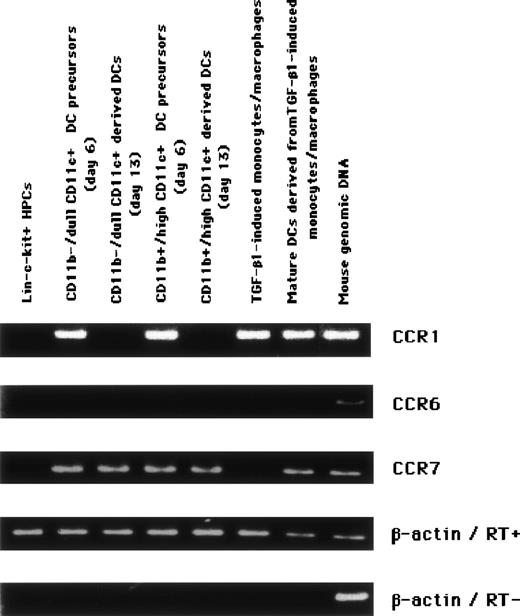
We have recently identified three distinct DC differentiation pathways from murine bone marrow HPCs (Fig 1). These DCs and their precursors could be classified based mostly on the expression pattern of CD11b and CD11c.6 7 To identify the chemokines responsible for the migration of DCs, we first examined the expression of chemokine receptor mRNA in the subpopulations derived from murine Lin−c-kit+ HPCs. Using RT-PCR, the expression of CCR1 mRNA was shown to be expressed in CD11b−/dullCD11c+ and CD11b+hiCD11c+ precursors of DC (days 6 to 7) and was not detected in either mature DCs derived from CD11b−/dullCD11c+ or CD11b+hiCD11c+ DC precursors. To negate the possibility that PCR products of chemokine receptors were attributable to the misamplification from genomic DNA as template instead of mRNA, the expression of a housekeeping gene, β-actin, was visualized exclusively with reverse transcription performed before PCR. In addition, CCR7 mRNA was detected specifically in both CD11b−/dullCD11c+ and CD11b+hiCD11c+ DC precursors and all types of mature DCs, including TGF-β1–induced macrophage-derived ones, whereas CCR6 mRNA was not detected at all in any populations (Fig 2). All of other chemokine receptor mRNAs tested, including CCR2, CCR3, CCR5, CXCR2, CXCR3, and CXCR5, were not detected by RT-PCR in any population.
Differential chemokine receptor expression on DCs and their precursors derived from murine HPCs. Levels of mRNAs in every stage of the differentiation pathway demonstrated in Fig 1 were assessed by RT-PCR. For the positive control, genomic DNA extracted from murine tail was used as the template. PCR product not preceded by reverse transcription to exclude the amplification from genomic DNA was also shown. The results represent three independent experiments.
Differential chemokine receptor expression on DCs and their precursors derived from murine HPCs. Levels of mRNAs in every stage of the differentiation pathway demonstrated in Fig 1 were assessed by RT-PCR. For the positive control, genomic DNA extracted from murine tail was used as the template. PCR product not preceded by reverse transcription to exclude the amplification from genomic DNA was also shown. The results represent three independent experiments.
Identification of chemokines for DCs and DC precursors.
To explore which chemokine receptors are playing a fundamental role in DC migration, we evaluated the migration capacity of DCs and their precursors in response to several corresponding chemokines. The responsiveness to MIP-1α was rather limited to the CD11b−/dullCD11c+ DC precursors (10.8% ± 1.3%, n = 3; Fig 3), and this was no longer active on mature DCs (Fig 4A). Either MIP-1β or RANTES was ineffective on any population of DCs (Figs 3 and 4A and B). Comparing distinct populations with each other, a larger number of CD11b−/dullCD11c+-derived DCs or their precursors were chemoattracted by SLC than were CD11b+hiCD11c+-derived DCs or their precursors. CD11b−/dullCD11c+ precursors acquired higher capacity to migrate in response to SLC in the course of maturation, whereas CD11b+hiCD11c+ DC precursors behaved similarly (Fig 4A and B). However, M-CSF–induced macrophages derived from CD11b+hiCD11c+ did not show any chemotactic response toward SLC because they were lacking in expression of CCR7 (data not shown).
The effect of the various chemokines on the migration of CD11b−/dullCD11c+ and CD11b+hiCD11c+ DC precursors. Optimal concentrations of chemokines (100 ng/mL), TNF- (10 ng/mL), and fMLP (10−7 mol/L) were tested as the ligand for chemotaxis. (▪) CD11b−/dullCD11c+ DC precursors; (▩) CD11b+hiCD11c+ DC precursors. The data represent the mean value ± SD of percentage. The results are representative of more than three experiments.
The effect of the various chemokines on the migration of CD11b−/dullCD11c+ and CD11b+hiCD11c+ DC precursors. Optimal concentrations of chemokines (100 ng/mL), TNF- (10 ng/mL), and fMLP (10−7 mol/L) were tested as the ligand for chemotaxis. (▪) CD11b−/dullCD11c+ DC precursors; (▩) CD11b+hiCD11c+ DC precursors. The data represent the mean value ± SD of percentage. The results are representative of more than three experiments.
The comparison of the migration toward several chemokines between (▪) precursor and (▩) mature DC in (A) CD11b−/dullCD11c+ and in (B) CD11b+hiCD11c+ populations. The data represent the mean value ± SD of percentage. The results are representative of more than three experiments.
The comparison of the migration toward several chemokines between (▪) precursor and (▩) mature DC in (A) CD11b−/dullCD11c+ and in (B) CD11b+hiCD11c+ populations. The data represent the mean value ± SD of percentage. The results are representative of more than three experiments.
Inhibition of CCR7 expression by TGF-β1 on the cytokine-stimulated HPCs.
The differentiating effect of TGF-β1 on macrophages into Langerhans-type cells has been described.8 9 We investigated here whether TGF-β1 affects the expression of chemokine receptors in vitro. GM-CSF+SCF– and GM-CSF+SCF+TNF-α–stimulated HPCs at days 6 to 7 expressed CCR1 and CCR7 (Fig 5), consistent with the results described in Fig 2. Surprisingly, CCR7 induction was inhibited by the addition of TGF-β1 to cytokine-stimulated HPCs during the first 6 to 7 days, although CCR1 expression was not affected by TGF-β1. Downregulation of CCR7 expression by TGF-β1 was no longer seen at days 13 to 14 once TNF-α was added to the culture.
The regulation of CCR7 expression on combined cytokine-stimulated HPCs by TGF-β1.
The regulation of CCR7 expression on combined cytokine-stimulated HPCs by TGF-β1.
Inhibition of chemotactic response of DCs and DC precursors to SLC by TGF-β1.
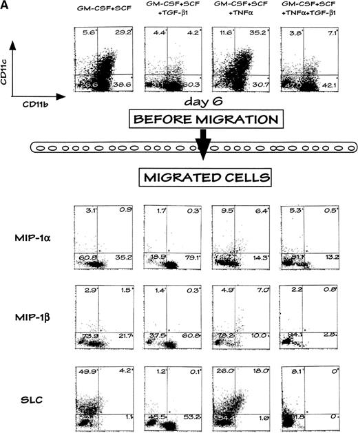
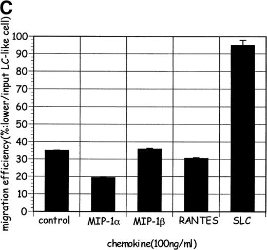
SLC attracted CD11c+ DC precursors in GM-CSF+SCF– and GM-CSF+SCF+TNF-α–stimulated HPCs at days 6 to 7 (25.36% ± 2.87% and 35.27 ± 0.61%, respectively), on which no other chemokines such as MIP-1α or MIP-1β could efficiently act (Table 1 and Fig 6A). However, selective migration by SLC was no longer observed in the presence of TGF-β1, as shown in flow cytometric analysis (Fig 6A). TGF-β1–induced macrophages at day 13 responded weakly to SLC compared with cell population (day 13) derived from GM-CSF + SCF alone, analogous to the other chemokines (Fig 6B). However, mature DCs derived from TGF-β1–induced macrophages completely restored the responsiveness to SLC (Fig 6C) but lost the responsiveness to other chemokines.
Comparison of the Migration Efficiency (Mean Value ± SD) in Response to Chemokines Between Combined Cytokine-Induced DC Precursors (Day 6)
| Chemokine (100 ng/mL) . | Stimulation . | |
|---|---|---|
| GM-CSF + SCF . | GM-CSF + SCF + TGF-β . | |
| Control | 14.15 ± 0.76 | 0 |
| MIP-1α | 25.36 ± 2.87 | 35.27 ± 0.61 |
| SLC | 78.7 ± 7.39* | 10.82 ± 5.4 |
| Chemokine (100 ng/mL) . | Stimulation . | |
|---|---|---|
| GM-CSF + SCF . | GM-CSF + SCF + TGF-β . | |
| Control | 14.15 ± 0.76 | 0 |
| MIP-1α | 25.36 ± 2.87 | 35.27 ± 0.61 |
| SLC | 78.7 ± 7.39* | 10.82 ± 5.4 |
The migration efficiency was calculated as follows. The number of the CD11c+ migrated cells was divided by that in the CD11c+ loaded cells before the migration assay.
The value of the migration efficiency in GM-CSF + SCF–stimulated HPCs is significantly larger than that in GM-SCF + SCF + TGF-β1 (P < .05).
Selective chemoattraction of DC precursors derived from murine HPCs by SLC and its regulation by TGF-β1. (A) Selective migration of DC precursors from GM-CSF+SCF– and GM-CSF+SCF+TNF-–stimulated HPCs was abrogated by TGF-β1. The profiles in the uppermost row indicate the preloaded populations, and those in the rest of the rows indicate the population in the lower chamber after migration assay. (B) TGF-β1–induced macrophages (at day 13; ▩) were less sensitive to SLC than HPC stimulated with GM-CSF+SCF (▪). (C) LC-like mature DCs successively generated with GM-CSF+TNF- from TGF-β1–induced macrophages (at day 13) exhibited high selective migration toward SLC. The values in (B) and (C) represent the mean value ± SD of percentage. The results are representative of more than three experiments.
Selective chemoattraction of DC precursors derived from murine HPCs by SLC and its regulation by TGF-β1. (A) Selective migration of DC precursors from GM-CSF+SCF– and GM-CSF+SCF+TNF-–stimulated HPCs was abrogated by TGF-β1. The profiles in the uppermost row indicate the preloaded populations, and those in the rest of the rows indicate the population in the lower chamber after migration assay. (B) TGF-β1–induced macrophages (at day 13; ▩) were less sensitive to SLC than HPC stimulated with GM-CSF+SCF (▪). (C) LC-like mature DCs successively generated with GM-CSF+TNF- from TGF-β1–induced macrophages (at day 13) exhibited high selective migration toward SLC. The values in (B) and (C) represent the mean value ± SD of percentage. The results are representative of more than three experiments.
DISCUSSION
We have recently established a culture system to generate DCs through three distinct precursors from murine HPC (Fig 1). Either CD11b−/dullCD11c+ or CD11b+hiCD11c+ DC precursor, which appears 6 to 7 days after stimulation with GM-CSF + SCF + TNF-α, becomes phenotypically distinct mature DCs. TGF-β1–induced macrophages (day 13) also differentiate into mature DCs, which resemble Langerhans cells expressing high levels of Ia, CD86, DEC-205, CD40, and E-cadherin, within another 5 days by culturing with GM-CSF + TNF-α.7We speculated that DCs from bone marrow might migrate to distinct organs, due to different expression of chemokine receptors and their responsiveness to chemokines destined by their ontogeny and differentiation pathway. CD11b−/dullCD11c+ DC precursors seemed to be attracted by SLC more efficiently than CD11b+hiCD11c+ precursors. The migration capacity might decrease as CD11b−/dullCD11c+ DC precursors acquire the characteristics of macrophages, represented by the expression of markers such as CD11b and c-fms. In this context, M-CSF–induced macrophages derived from CD11b+hi CD11c+precursors never migrated toward SLC.
The expression of CCR7 on human DCs was induced during the maturation elicited by various stimuli, eg, CD40L, lipopolysaccharide (LPS), interleukin-1 (IL-1), and TNF-α.15,21 Although we could not observe the difference in the expression of CCR7 mRNA between murine DCs and their precursors, the responsiveness to SLC, a ligand for CCR7, increased as CD11b−/dullCD11c+and CD11b+hiCD11c+DC precursors became mature DCs. Furthermore, SLC had chemotactic activity for both DCs and their precursors. Surprisingly, CCR6, one of the candidates mediating the chemotaxis of DC,22 was not expressed on any stage of the differentiation pathway of murine DC tested so far, in line with the results that LARC/MIP-3α never caused the chemotaxis in our system. In contrast, in humans, a peculiar lineage of DCs such as CD34+ cord blood cells-derived premature DCs, but not DC progenitors in peripheral blood or monocytes, preferentially expresses CCR6 and responds to LARC/MIP-3α.10,15 This discrepancy might be due to the species difference. CCR1, which is preferentially expressed on DC precursors, might function instead of CCR6 in mice. A recent study indicated that CXCR3 had the binding affinity to SLC23; however, no populations in our DC differentiation pathway expressed CXCR3. Human macrophage progenitors that form granulocyte-macrophage–colony-forming unit (CFU-GM) have been demonstrated to express CCR7 and also to migrate to ELC/MIP-3β,24 unlike murine HPCs used in this study. Murine Lin−c-kit+ HPCs used in our study could be more undifferentiated cells than CFU-GM forming cells in humans.
In our previous work,8 TGF-β1 could suppress DC maturation from murine Lin−c-kit+ HPCs based on the decreased expression of MHC class II and CD86 and on the reduced capacity of enhancing allogenic MLR. TGF-β1–induced DCs in vitro could prolong murine cardiac allograft survival by inhibiting cellular immunity.25 In addition, infiltrating DCs in colon and basal cell skin cancers25 and the progressing melanoma metastases26 are lacking in CD86 and in the capacity to stimulate T cells because of massive production of TGF-β1 by these tumors.26,27 Collectively, TGF-β1 may regulate immune response negatively through modulating DCs’ development and function. In addition, to examine the effect of TGF-β1 on the migration of DCs was of great interest in acquired immunity and the tumor immunity, besides the immunophenotype and the function of DC. The migration of DC precursors in response to SLC was completely inhibited by TGF-β1. Therefore, it is possible that TGF-β1 not only downregulates the capacity of antigen presentation by DC, but also inhibits the migration of immature DCs that process antigen. Because SLC is expressed on high endothelial venules (HEV) in lymph node and Payer’s patch,28 SLC might recruit DCs at the entry site of the regional lymph node, before the interaction with naive T cell. The situation in which TGF-β1 is produced massively, such as in tumor, may interfere with the establishment of acquired immunity through recruiting less number of sensitized DCs. But, once TNF-α is produced in an inflammatory situation, downregulation of CCR7 expression in immature DCs is deprived. Subsequently, DC matures and is recruited into T-cell area of regional lymph nodes to sensitize naive T lymphocytes.
ACKNOWLEDGMENT
The authors express our sincere gratitude to Dr R.M. Steinman for his kind gift of MoAb to DEC-205 (NLDM145); to Dr T. Sudo for his generous gift of anti–c-kit MoAb, GM-CSF, and SCF; and to Dr T. Imai and Dr O. Yosie (Kinki University, Osaka, Japan) for kindly providing murine SLC cDNA and murine LARC. We also highly appreciate Dr J.J. Oppenheim (NCI-FCRDC, Frederick, MD), Dr K. Kawasaki (Kanazawa University, Kanazawa, Japan), and Dr C. Vestergaard for their critical review of the manuscript.
The publication costs of this article were defrayed in part by page charge payment. This article must therefore be hereby marked “advertisement” in accordance with 18 U.S.C. section 1734 solely to indicate this fact.
REFERENCES
Author notes
Address reprint requests to Kouji Matsushima, MD, PhD, Department of Molecular Preventive Medicine, School of Medicine, The University of Tokyo, 7-3-1, Hongo, Bunkyo-ku, Tokyo 113-0033, Japan; e-mail:koujim@m.u-tokyo.ac.jp.


This feature is available to Subscribers Only
Sign In or Create an Account Close Modal