To the Editor:
Wiskott-Aldrich syndrome (WAS) is an X-linked recessive disorder characterized clinically by the triad of thrombocytopenia, recurrent infections due to defects in the immune system, and severe eczema. The gene responsible for WAS (WASP gene) was recently cloned.1 Since then, large numbers of mutations have been reported in WAS patients with variable clinical phenotypes,2 and it has also been shown that X-linked thrombocytopenia (XLT) resulted from a mutation of the same gene.3
We performed mutation analysis of a boy with WAS and found dual mutations in exon 10 of the WASP gene. The patient died before the present study; thus, only genomic DNA derived from his peripheral blood lymphocytes was available. Genomic DNA was purified, and each exon of the WASP, including flanking introns, was amplified by polymerase chain reaction (PCR). Primers used and each PCR condition were described elsewhere.4 Amplified fragments were purified and directly sequenced using an ABI PRIZM Dye Terminator Cycle Sequencing Ready Reaction Kit (Perkin Elmer, Foster City, CA) and an automated ABI 373A DNA sequencer. The conditions used for the sequencing reaction were also described before.5 To avoid PCR-generated sequence errors, we performed sequencing at least twice using a different set of all PCR products in both strands.
The patient studied had two elder brothers who had been diagnosed as WAS based on their clinical and laboratory findings. They died from severe pneumonia and septicemia with bleeding episodes at 10 months of age and 47 months of age, respectively. At 4 years of age, the patient died from unpredictable intracranial hemorrhage; meanwhile, we obtained his peripheral blood lymphocytes and kept them frozen. After the patient died, the couple had a daughter. Genomic DNAs from his mother and his sister were then obtained to determine their carrier status of the disease after informed consent.
Mutation analysis of the patient's WASP gene showed two mutations in exon 10. One mutation was a one-base insertion (A+) at nucleotide number 1099 or 1100. The other was a one-base deletion (G−) from 5 consecutive Gs of nucleotide number 1127 to 1131. When we made a careful observation of the patient's sequencing results, we found small-tide ambiguous waves after the A+ site in both the directions (data not shown), as if it contained a small amount of the fragments without A+. To confirm this, the fragments of the different PCR including the mutations were cloned into the TA vector PCRII (Invitrogen Corp, Carlsbad, CA), and the sequence of each clone was examined. Most of the clones possessed the dual mutations (A+ and G−); however, we could find some clones with the single mutation, all of which had only the G− mutation (Table1). Sequencing studies of the PCR fragments of the patient's mother and sister showed that both were carriers for the disease. They had the mutant WASP allele together with the normal allele, and the mutant allele included only the G− mutation. The results of the cloning and sequencing experiment of their PCR fragments were shown in Table 1.
Cloning Studies of the Individual WASPAllele
| . | No. of Clones Examined . | Clones With . | |||
|---|---|---|---|---|---|
| A+ and G− . | G− . | A+ . | No Mutation . | ||
| Patient | 51* | 90.2% | 9.8% | 0% | 0% |
| Mother | 15 | 0% | 40% | 0% | 60% |
| Sister | 20 | 0% | 45% | 0% | 55% |
| Control | 10 | 0% | 0% | 0% | 100% |
| . | No. of Clones Examined . | Clones With . | |||
|---|---|---|---|---|---|
| A+ and G− . | G− . | A+ . | No Mutation . | ||
| Patient | 51* | 90.2% | 9.8% | 0% | 0% |
| Mother | 15 | 0% | 40% | 0% | 60% |
| Sister | 20 | 0% | 45% | 0% | 55% |
| Control | 10 | 0% | 0% | 0% | 100% |
*Obtained from 3 independent PCRs.
From these results, we speculated the origin of the patient's dual mutations; the one-base deletion mutation (G−) was derived from his mother, and the other one-base insertion mutation (A+) was generated de novo. The results of the cloning studies suggested that de novo mutation occured after fertilization, possibly at some level of hematological progenitor. It was shown that the patient's clones consisted mostly of the dual mutations, with very minor clones having only the G− mutation. PCR-generated artifacts were very unlikely, because 6 different PCR products showed the same results.
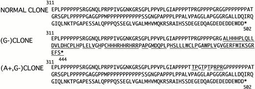
Deduced amino acid sequence in both the mutants were shown in Fig1. In comparison with the single mutation G−, the changes of amino acid in the dual mutation were restricted to the position of 356-365 (Fig 1). The area involved 10 amino acids, and actually 7 amino acids were replaced. Thus, although we could not evaluate the two mutant WASP protein functions, the defect in the case of the dual mutations was considered to be milder than that of the single mutation. A similar case with dual mutations in the WASPgene was recently reported as mild clinical phenotype,6however, the origin of the dual mutations was not discussed.
Comparison of amino acid sequence from Glu311 to the C terminal part of the each WASP allele. The underlined characters indicate substitution of the original sequence. The asterisk denotes the stop codon.
Comparison of amino acid sequence from Glu311 to the C terminal part of the each WASP allele. The underlined characters indicate substitution of the original sequence. The asterisk denotes the stop codon.
Although it is very rarely detected, an additional de novo mutation can reverse severe clinical phenotype in some genetic disorders. Such cases of adenosine deaminase deficiency and atypical X-linked severe combined immunodeficiency were reported, respectively.7 8 In the present case, the additional de novo mutation might change a severe type mutation to a milder one. However, we could hardly confirm the definite difference in clinical severity between the patient and his brothers, who were thought to have only the G− mutation. We thought that the additional mutation was not enough to restore full function of the WASP, or it would take more time to have a definite effect on the phenotype.
The defect of WASP in hematological cells resulted in a growth disadvantage at the stem cell level.9 The present study indicated that there should be a grade of disadvantage depending on the residual WASP functions. The lymphocytes with the dual mutations apparently had an advantage over the cells with the single mutation in terms of cell growth in vivo. These findings encourage future attempts of gene therapy for WAS. Introduction and expression of the normalWASP gene even in small numbers of hematological stem cells could be enough to obtain clinical benefit in gene therapy for WAS patients.
ACKNOWLEDGMENT
We thank Prof K. Kobayashi (Department of Pediatrics, Hokkaido University School of Medicine, Sapporo, Japan) for his critical reading of this manuscript. Supported by a grant from the Ministry of Health and Welfare, Japan.


This feature is available to Subscribers Only
Sign In or Create an Account Close Modal