Abstract
The escape of malignant cells from the immune response against the tumor may result from a defective differentiation or function of professional antigen-presenting cells (APC), ie, dendritic cells (DC). To test this hypothesis, the effect of human renal cell carcinoma cell lines (RCC) on the development of DC from CD34+progenitors was investigated in vitro. RCC cell lines were found to release soluble factors that inhibit the differentiation of CD34+ cells into DC and trigger their commitment towards monocytic cells (CD14+CD64+CD1a−CD86−CD80−HLA-DRlow) with a potent phagocytic capacity but lacking APC function. RCC CM were found to act on the two distinct subpopulations emerging in the culture at day 6 ([CD14+CD1a−] and [CD14−CD1a+]) by inhibiting the differentiation into DC of [CD14+CD1a−] precursors and blocking the acquisition of APC function of the [CD14−CD1a+] derived DC. Interleukin-6 (IL-6) and macrophage colony-stimulating factor (M-CSF) were found to be responsible for this phenomenon: antibodies against IL-6 and M-CSF abrogated the inhibitory effects of RCC CM; and recombinant IL-6 and/or M-CSF inhibited the differentiation of DC similarly to RCC CM. The inhibition of DC differentiation by RCC CM was preceeded by an induction of M-CSF receptor (M-CSFR; CD115) and a loss of granulocyte-macrophage colony-stimulating factor receptor (GM-CSFR; CD116) expression at the surface of CD34+cells, two phenomenon reversed by anti–IL-6/IL-6R and anti–M-CSF antibodies, respectively. Finally, a panel of tumor cell lines producing IL-6 and M-CSF induced similar effects. Taken together, the results suggest that the inhibition of DC development could represent a frequent mechanism by which tumor cells will escape immune recognition.
ALTHOUGH IMMUNOTHERAPY with interleukin-2 (IL-2) and/or interferon α (IFNα) has an established antitumor effect in metastatic renal cell carcinoma (RCC), still a majority of these patients will experience progressive disease after IL-2 treatment.1,2 Tumor-specific antigens have been reported in RCC, but tumor-specific cytotoxic T-cell clones have only been rarely identified,3-5 suggesting a potential impairement of the differentiation of cytotoxic T lymphocytes (CTL) in patients with RCC. The presentation of specific antigen peptides by major histocompatibility complex (MHC) molecules is the initial step for the development of a specific cytotoxic T-cell response. However, tumor cells often express low or undetectable levels of MHC class I and II antigens and most often lack expression of costimulatory molecules (CD80 and CD86), a phenomenon that may result in T-cell anergy.6 Dendritic cells (DC) are professional antigen-presenting cells (APC) required for the initiation of primary T-cell response in vivo.7,8 DC are capable of presenting tumor-specific antigens and of activating a specific antitumor T-cell response in vivo in animal models.8-11 DC are likely to be involved in the development of the spontaneous or therapeutic immune response against RCC tumor in humans, although their precise role in tumor biology in vivo is poorly defined. In vitro, RCC cell lines produce a wide range of cytokines, including transforming growth factor β (TGFβ), IL-6, basic fibroblast growth factor (FGFb), and granulocyte-macrophage colony-stimulating factor (GM-CSF).12-14 Several of these cytokines have been reported to interfere at different steps of the antitumor immune response, in particular antigen presentation and CTL differentiation (for review, see Chouaib et al15).
In the present study, we show that RCC cell lines produce soluble factors that inhibit the differentiation of CD34+progenitors into DC and redirect their differentiation towards the monocyte-macrophage lineage. This phenomenon is shown to be mediated by IL-6 and macrophage colony-stimulating factor (M-CSF).
MATERIALS AND METHODS
Hematopoietic Factors
Recombinant human GM-CSF (rhGM-CSF; specific activity, 2 × 10⁶ U/mg; Schering Plough Research Institute, Kenilworth, NJ) was used at optimal concentration of 100 ng/mL (200 U/mL), recombinant human tumor necrosis factor α (rhTNFα; specific activity, 5 × 10⁶ U/mg; Cetus, Amsterdam, The Netherlands) at 2.5 ng/mL (50 U/mL), recombinant human stem cell factor (rhSCF; specific activity, 4 × 10⁵ U/mg; R&D System, Abingdon, UK) at 25 ng/mL, M-CSF (specific activity, 2 × 10⁶ U/mg; R&D) and IL-6 (specific activity, 106 U/mg; Sandoz, Basel, Switzerland) at 20 ng/mL, and vascular endothelial growth factor165 (VEGF165) (R&D) at 25 ng/mL.
Obtention of RCC Conditioned Medium (CM)
RCC lines obtained from ATCC (CAKI-1 and CAKI-2) or generated in the laboratory (CLB-CAN, CLB-CHA, CLB-GUI, CLB-OTE, CLB-TUG, CLB-TUT, and CLB-VER; Bain C, et al, manuscript submitted) were plated in 100-mm diameter dishes at a density of 5 × 10⁵cells/mL in RPMI 1640 medium supplemented with 2 mmol/L glutamine, 200 UI/mL penicillin, 200 μg/mL streptomycin (GIBCO Laboratories, Grand Island, NY) and 10% fetal calf serum (FCS; Biowittaker, Verviers, Belgium). After 2 days of culture, supernatants were harvested, filtered, aliquoted, and stored at −20°C for further use. Other tumor cell lines conditioned medium were performed in the same conditions using neuroblastoma (SHEP, IMR32, CLB-CA, CLB-ES, SKNAS, and SKNFI), melanoma (CLB-DOR), Burkitt lymphoma (Daudi, Raji, and BJAB), large lung cell carcinoma (H-322), colon carcinoma (HT-29 and SW620), or breast carcinoma cell lines (T47-D, MCF-7, and CLB-SAV) obtained from the ATCC (Rockville, MD) collection or generated in the laboratory.
Preparation of Cells
DC were generated from CD34+ cells isolated from umbilical cord blood samples obtained according to institutional guidelines. Cells bearing CD34 antigen were isolated from mononuclear fractions through positive selection by mini MACS (Miltenyi Biotec, Gmbh, Bergisch Gladbach, Germany), using an anti-CD34 monoclonal antibody (MoAb; Immu 133.3; Immunotech, Marseille, France) and goat antimouse IgG-coated microbeads (Miltenyi Biotec, Gmbh).16 In all experiments, the isolated cells were 80% to 99% CD34+. CD34+ progenitors were seeded for expansion at 5 × 10³ to 104 cells/mL in 24-well plates in complete medium in the presence of GM-CSF (100 ng/mL), TNFα (2.5 ng/mL), SCF (20 ng/mL), and 2% human serum AB+(sAB+) as previously described for 6 days.16 17After 6 days, cells were harvested, numbered, phenotyped, and seeded in absence of sAB+ but in the presence of GM-CSF and TNFα at 5 × 10⁴ cells/mL for 6 additional days, with a last medium change being performed at day 10. Cells were collected at day 12. Eventually, adherent cells were recovered using a 5 mmol/L EDTA solution.
Isolation of CD1a+ and CD14+ Precursors by FACS Sorting
After 6 days of culture in the presence of GM-CSF+TNFα+SCF+sAB+, cells were collected and labeled with fluorescein isothiocyanate (FITC)-conjugated OKT-6 (CD1a; Ortho, Roissy, France) and phycoerythrin (PE)-conjugated Leu-M3 (CD14; Becton Dickinson, Pont de Claix, France).
Cells were separated according to CD1a and CD14 expression into CD1a+CD14− and CD1a−CD14+ fractions using a FACS Starplus (laser setting: power at 250 mw and excitation wavelength at 488 nm; Becton Dickinson). All the procedures of staining and sorting were performed in the presence of 5 mmol/L EDTA to avoid cell aggregation. Reanalysis of the sorted populations showed a purity greater than 98%.
Sorted cells were seeded in the presence of GM-CSF+TNFα with or without 10% RCC CM (5 × 10⁴ cells/mL) for 6 additional days, with a last medium change being performed at day 10. Cells were then recovered at day 12 and adherent cells were removed using a 5 mmol/L EDTA solution.
Mixed Lymphocyte Reaction (MLR) Assay
After 12 days of culture, CD34-derived cells were collected and, after irradiation (30 Gy), used as stimulator cells for allogeneic adult naive T lymphocytes (CD45RA+) purified from healthy volunteer peripheral blood mononuclear cells (PBMC) by immunomagnetic depletion using a cocktail of MoAbs.16 After two rounds of bead depletion, the purity of CD45RA+ was routinely higher than 95%. From 17 to 104 stimulator cells were added to the T cells (2 × 10⁴ cells/well) in 96-well round-bottomed microtest culture plates (Nunc, Rockilde, Denmark). Cultures were performed in RPMI complete medium supplemented with 10% FCS. After 5 days of incubation, cells were pulsed with 0.5 μCi of 3HTdR per well (specific activity, 5 mCi/mmol) for the last 18 hours, harvested, and counted. Tests were performed in triplicates and results were expressed as the mean counts per minute (cpm) ± standard deviation (SD). The levels of 3H-TdR uptake by stimulator cells alone were always less than 100 cpm.
Cell Surface Phenotyping
Cells were processed for double staining at day 6 and day 12 using anti–CD14-PE (Becton Dickinson) and anti–CD1a-FITC (Coulter Clone, Marseille, France). Negative controls were performed with unrelated murine MoAbs. Extensive phenotype were performed at day 12 with MoAbs-PE. The antibodies used were PE labeled (except when specified) and purchased from Becton Dickinson for negative control, CD14, CD15, HLA-DR, CD80, CD16, CD11b; Pharmingen (San Diego, CA) for CD86; Immunotech for CD23, CD83, CDw116, CD40, CD54, and CD58; Caltag Laboratories (Burlingame, CA) for CD32, CD64; Ortho Diagnostic (Raritan, NJ) for CD1a/FITC; and Oncogene Science (Cambridge, MA) for rat control isotype and CD115 (cfms). Fluorescence analysis was performed on a FACScan flow cytometer after acquisition of 5,000 events (Becton Dickinson).
Neutralizing Antibodies
MoAb anti–IL-6 (BE-8) and anti–IL-6R (BR-6) were purchased from Diaclone (Besançon, France). MoAb anti-VEGF and rabbit and mouse Igs used as control antibodies was purchased from R&D. Polyclonal anti–M-CSF was purchased from Genzyme (Paris, France). All antibodies were used at optimal concentrations (10 to 20 μg/mL).
Cytokine Detection
Cytokines were detected in conditioned media and culture supernatants using commercial quantitative sandwich immunoassay kits from Immunotech (IL-6) and R&D System (VEGF and M-CSF).
Phagocytosis
CD34+ cells were cultured in the presence of GM-CSF+TNFα alone or with 10% RCC CM or breast CM for 12 days with or without specific antibodies. During the last 4 hours of the culture, 0.5-μm latex beads coupled to FITC (1/400; Polysciences, Warrington, PA) were added in the culture. To analyze the phagocytic capacity, cells were recovered and washed three times in cold phosphate-buffered saline (PBS), and beads incorporation was evaluated on a FACScan analyzer (histograms) or observed on a fluorescence microscope after cytocentrifugation and MGG coloration.
Statistics
Statistical analyses were performed using Fisher’s exact test and a test of comparison of percentages.
RESULTS
Inhibition of the Differentiation of DC by RCC Medium
Phenotypic modifications.
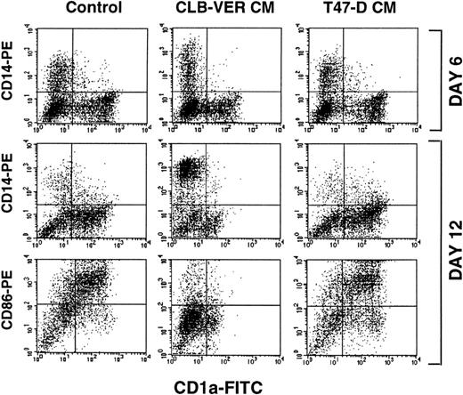
CD34+ progenitors cultured with GM-CSF+TNFα+SCF differentiate into dendritic cells characterized by the acquisition of CD1a and the loss of CD14 antigens at day 1216,18 19(Fig 1). In the presence of 10% final concentration of RCC CM, cultured CD34+ cells displayed an altered phenotype at day 12 with a reduced expression of CD1a antigen (60% CD1a+ with a mean fluorescence intensity [MFI] of 30 in medium alone v 14.6% with an MFI of 5.3 in the presence of RCC CM), whereas breast carcinoma CM (T47-D or MCF-7) had no effect (Fig 1). In 28 different experiments, after a 12-day culture period, 50% to 79% of the cells obtained in the presence of 10% RCC CM expressed CD14 Ag, compared with those cultured in medium or 10% T47-D CM (Fig 1). Among these different experiments, the median reduction of CD1a expression induced by the different RCC CM was 69%, with a range of 32% to 87%. All nine RCC CM tested induced a similar effect (not shown).
Inhibition of the differentiation of DC from CD34+ progenitors by RCC CM. CD34+progenitors were incubated with GM-CSF+TNF alone or in the presence of 10% of RCC CM or breast CM (control). Medium was replaced at days 6 and 10. A double staining with CD14-PE and CD1a-FITC antibodies was performed at day 6. At day 12, cells were labeled with CD14-PE/CD1a-FITC or CD86-PE/CD1a-FITC and analyzed on a FACScan (Becton Dickinson). Results are of one representative experiment performed with different seven CD34+ cells samples.
Inhibition of the differentiation of DC from CD34+ progenitors by RCC CM. CD34+progenitors were incubated with GM-CSF+TNF alone or in the presence of 10% of RCC CM or breast CM (control). Medium was replaced at days 6 and 10. A double staining with CD14-PE and CD1a-FITC antibodies was performed at day 6. At day 12, cells were labeled with CD14-PE/CD1a-FITC or CD86-PE/CD1a-FITC and analyzed on a FACScan (Becton Dickinson). Results are of one representative experiment performed with different seven CD34+ cells samples.
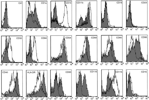
In addition, CD34+ cells cultured in the presence of RCC CM (but not of breast carcinoma CM) displayed an increased expression of IgG receptor molecules (CD32 and CD64), whereas the expression of costimulatory molecules (CD80, CD86, and CD40) and HLA-DR was strongly inhibited (Fig 2). Regarding adhesion molecules, CD58 expression was weakly inhibited, whereas CD54 was not significantly affected. CD83, a specific marker for DC,20and CD116 (GM-CSF receptor α chain [GM-CSFRα]) normally expressed on DC generated from CD34+ were absent when cells were cultured with RCC CM, whereas CD115 (M-CSF receptor [M-CSFR]) was upregulated (Fig 2). In all culture conditions, no CD15 expression was observed on cultured CD34+ cells, demonstrating the absence of granulocytic cells (Fig 2). Altogether, these results demonstrate that RCC CM profoundly affects the characteristic phenotypic differentiation of CD34+ progenitors into DC.16
Phenotypic modifications induced by culture with RCC CM for 12 days. CD34+ cells cultured for 12 days with GM-CSF+TNF (▪) or in the presence of 10% RCC CM (□) were harvested and processed for staining with PE-conjugated MoAbs or uncoupled MoAb shown by PE-conjugated antirat Ig (CD115). Antimouse control antibody was shown in the first histogram. Antirat control antibody overlaps this control (data not shown). Results are representative of four experiments.
Phenotypic modifications induced by culture with RCC CM for 12 days. CD34+ cells cultured for 12 days with GM-CSF+TNF (▪) or in the presence of 10% RCC CM (□) were harvested and processed for staining with PE-conjugated MoAbs or uncoupled MoAb shown by PE-conjugated antirat Ig (CD115). Antimouse control antibody was shown in the first histogram. Antirat control antibody overlaps this control (data not shown). Results are representative of four experiments.
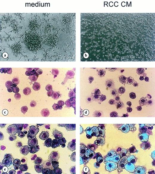
Cells obtained after 12 days of culture in the presence of RCC CM (Fig3B and D) were morphologically distinct from DC (Fig 3A and C) and displayed a macrophage morphology (Fig 3B), a strong adherence to plastic surface (Fig 3B), small nuclei, and numerous intracytoplasmic vacuoles (Fig 3D).
Modulation of cell morphology by RCC medium. Cells cultured from day 6 to day 12 in GM-CSF+TNF alone (a, c, and e) or in the presence of 10% RCC CM (b, d, and f). Cell morphology as evaluated by phase contrast microscopy (a and b) and MGG staining (c and d) and phagocytic capacity (e and f) evaluated by FITC-coupled 0.5 μmol/L latex beads ingestion for the last 4 hours of culture time (counterstained with MGG). Results are representative of 10 different experiments.
Modulation of cell morphology by RCC medium. Cells cultured from day 6 to day 12 in GM-CSF+TNF alone (a, c, and e) or in the presence of 10% RCC CM (b, d, and f). Cell morphology as evaluated by phase contrast microscopy (a and b) and MGG staining (c and d) and phagocytic capacity (e and f) evaluated by FITC-coupled 0.5 μmol/L latex beads ingestion for the last 4 hours of culture time (counterstained with MGG). Results are representative of 10 different experiments.
The inhibitory effect of RCC CM on CD34+ cells differentiation was dose-dependent, being detectable at 1% final concentration and with a maximum inhibitory effect at a 10% final concentration of RCC CM for the VER cell line (Fig 4). It is noteworthy that the magnitude of the effect of RCC CM varied according to the RCC CM tested. Whereas some RCC CM were found to have a major effect at 2% final concentration (CLB-TUT, CLB-GUI, and CLB-OTE), for others (CLB-CAN and CLB-TUG) a 25% final concentration of CM was required to obtain a similar biological effect. In contrast, none of the breast carcinoma CM used (T47-D, M-CF7, and CLB-SAV) was able to block DC differentiation, even when used at high concentration (50% CM; data not shown).
Dose-dependent inhibition of DC differentiation by RCC CM. Cells were cultured from day 6 to day 12 in the presence of an increasing percentage of RCC CM (1% to 20%; ⧫) or breast CM (○). At day 12, cells were harvested and processed for labeling with CD14-PE (—-) and CD1a-FITC (—) antibodies. Results are representative of five experiments.
Dose-dependent inhibition of DC differentiation by RCC CM. Cells were cultured from day 6 to day 12 in the presence of an increasing percentage of RCC CM (1% to 20%; ⧫) or breast CM (○). At day 12, cells were harvested and processed for labeling with CD14-PE (—-) and CD1a-FITC (—) antibodies. Results are representative of five experiments.
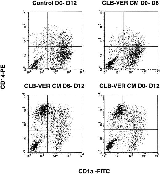
The differential effect of RCC CM during the expansion (day 0 to 6) and the maturation phases (day 6 to 12) on the phenotype of cultured CD34+ cells was then investigated.16 The addition of RCC CM from day 0 to 12 or during the maturation phase only (day 6 to 12) inhibited the acquisition of the DC phenotype (Fig 5). The addition of RCC CM during the proliferative phase only (day 0 to 6) partially affected CD14 and CD1a expression on cultured CD34+ cells at day 6 (Fig 5). However, in this condition, a decrease of CD1a expression intensity was observed (MFImedium = 96 and MFIRCC CM = 53) and was associated with an increase of CD14 intensity (MFImedium = 87 and MFIRCC CM = 179; Fig 1); however, at day 12, the phenotype of cells cultured with RCC CM from day 0 to 6 only was similar to that of cells cultured without RCC CM (Fig 5).
Effect of RCC CM during the different phases of the culture. Double staining (CD14-PE/CD1a-FITC) was performed at day 12 and the results were compared for CD34+ cultured with cytokines alone (control) or in the presence of 10% RCC CM from day 0 to 6 (top right), from day 6 to day 12 (bottom left), or day 0 to day 12 (bottom right). Results are representative of four experiments.
Effect of RCC CM during the different phases of the culture. Double staining (CD14-PE/CD1a-FITC) was performed at day 12 and the results were compared for CD34+ cultured with cytokines alone (control) or in the presence of 10% RCC CM from day 0 to 6 (top right), from day 6 to day 12 (bottom left), or day 0 to day 12 (bottom right). Results are representative of four experiments.
Functional modifications.
In addition to these phenotypic alterations, CD34+ cells cultured with RCC CM displayed important functional alterations. RCC CM induced an increased proliferation rate (1.5- to 2.3-fold increase) during both the proliferation and the maturation phases with all 9 different RCC cell lines supernatants tested (not shown).
In addition, cells generated in RCC CM exhibited a strong phagocytic capacity as evaluated by the ingestion of 0.5-μm latex beads coupled to FITC (Fig 3F) compared with CD34+ cells cultured with cytokines alone (Fig 3E).
The APC function of CD34+ cells cultured with cytokines with or without tumor cell medium (RCC CM and breast carcinoma CM) was evaluated by measuring their capacity to induce the proliferation of purified naive (CD45RA+) allogeneic CD4+ T cells in an MLR. Cells obtained from CD34+ progenitor cells cultured with cytokines in the presence of RCC CM between day 0 and 12 (data not shown) or only during the maturation phase (day 6 to 12) were found to be unable to stimulate naive T-cell proliferation as compared with cells generated without RCC CM (Fig6).
Inhibition of antigen-presenting cell function by RCC CM. CD34+ cells were cultured from day 0 to 6 in GM-CSF+TNF. Medium was then changed and the cells were cultured in the presence of cytokine alone (control; □), RCC CM ([▪] CLB-VER CM; [○] Caki-1 CM), or breast CM (T47-D CM; •) from day 6 to 12. Cells were then harvested, irradiated (30 Gy), and used as stimulator cells for naive T cells (2 × 10⁴ cells/well). The proliferation was evaluated using 3HTdR uptake after 5 days of culture. Results are expressed as the mean ± SD of triplicate culture. These results are representative of five different experiments.
Inhibition of antigen-presenting cell function by RCC CM. CD34+ cells were cultured from day 0 to 6 in GM-CSF+TNF. Medium was then changed and the cells were cultured in the presence of cytokine alone (control; □), RCC CM ([▪] CLB-VER CM; [○] Caki-1 CM), or breast CM (T47-D CM; •) from day 6 to 12. Cells were then harvested, irradiated (30 Gy), and used as stimulator cells for naive T cells (2 × 10⁴ cells/well). The proliferation was evaluated using 3HTdR uptake after 5 days of culture. Results are expressed as the mean ± SD of triplicate culture. These results are representative of five different experiments.
These results indicate that RCC CM inhibits the differentiation of CD34+ cells into DC and triggers their differentiation into macrophages with a poor APC function but a strong phagocytic capacity.
Effect of RCC CM on the two independent subsets of DC development.
At an early time point (day 6) of culture of CD34+ cells in GM-CSF+TNFα+SCF, two subsets of DC precursors identified by the exclusive expression of CD1a and CD14 Ag emerge independently that correspond to two different pathways of DC development: the Langerhans cells (CD1a+) and the CD14-derived DC related to dermal DC or circulating blood DC (CD14+).16
We analyzed the biological effect of RCC CM on these two different subpopulations. [CD1a+CD14−] and [CD1a−CD14+] cells generated from CD34+ cells cultured in GM-CSF+TNFα+SCF were FACS-sorted at day 6. An additional 6 days of culture of these two populations in GM-CSF+TNFα without RCC CM demonstrate that [CD1a+CD14−] as well as [CD1a−CD14+] were able to differentiate into DC in terms of phenotype (Fig 7A) and high APC capacity (Fig 7B), as described previously.16 In contrast, in the presence of 10% RCC CM, CD1a+-derived DC failed to acquire expression of CD86 and exhibited lower levels of HLA-DR expression (Fig 7A). In addition, in an MLR assay, these cells were unable to induce allogeneic naive T-cell proliferation (Fig 7B). In the presence of 10% RCC CM, the day-6 [CD14+CD1a−] subpopulation also failed to differentiate into DC but was driven into macrophages (as described for the nonsorted population) with a specific phenotype (CD14+,CD1a−,HLA-Drlow,CD86−; Fig 7A), a high phagocytic capacity (not shown), and a reduced APC capacity (Fig 7B).
Cord blood CD34+ cells were cultured for 6 days in the presence of GM-CSF+TNF+ SCF+sAB+. Cells were then collected, processed for double staining using anti–CD14-PE and anti–CD1a-FITC, and FACS-sorted into [CD14+CD1a−] and [CD14−CD1a+] populations. Sorted cells were seeded in the presence of GM-CSF+TNF at 5 × 10⁴cells/mL for 6 additional days in the presence or absence of RCC CM (10%), with a last medium change being performed at day 10. (A) Phenotype of purified populations at day 12. At day 12, cells were recovered and analyzed for CD1a, CD14, CD86, and HLA-DR expression by double-color fluorescence. (B) Function of the cells generated. At day 12, cells cultured in the presence of GM-CSF+TNF alone (medium; ▪) or in the presence of RCC CM (□) were recovered, numbered, irradiated (30 Gy), and used as stimulator cells (3.3 × 10³ cells/well) in an MLR with naive CD45RA+ T cells (2 × 10⁴ cells/well).
Cord blood CD34+ cells were cultured for 6 days in the presence of GM-CSF+TNF+ SCF+sAB+. Cells were then collected, processed for double staining using anti–CD14-PE and anti–CD1a-FITC, and FACS-sorted into [CD14+CD1a−] and [CD14−CD1a+] populations. Sorted cells were seeded in the presence of GM-CSF+TNF at 5 × 10⁴cells/mL for 6 additional days in the presence or absence of RCC CM (10%), with a last medium change being performed at day 10. (A) Phenotype of purified populations at day 12. At day 12, cells were recovered and analyzed for CD1a, CD14, CD86, and HLA-DR expression by double-color fluorescence. (B) Function of the cells generated. At day 12, cells cultured in the presence of GM-CSF+TNF alone (medium; ▪) or in the presence of RCC CM (□) were recovered, numbered, irradiated (30 Gy), and used as stimulator cells (3.3 × 10³ cells/well) in an MLR with naive CD45RA+ T cells (2 × 10⁴ cells/well).
These results are consistent with those observed with the bulk population and demonstrate that RCC CM blocks both pathways of DC differentiation.
Effects of RCC CM Is Mediated by IL-6 and M-CSF
RCC cell lines produce a wide range of cytokines, including GM-CSF,14 TGFβ,14 IL-612,13(Table 1), M-CSF (Table 1), and VEGF (range, 2.7 to 6.2 ng/106 cells/48 hours in the 9 RCC cell lines) that could potentially act on DC differentiation. Because GM-CSF and TGFβ are well known inducers of DC differentiation in vitro,21 we examined whether M-CSF, IL-6, or VEGF could be responsible for the inhibitory effects of RCC CM on DC differentiation.22-26 The effects of antibodies against M-CSF, IL-6, or VEGF during the differentiation phase (day 6 to 12) were investigated. CD34+ cells were precultured for 6 days (day 0 to 6) in the presence of GM-CSF+TNFα+SCF+sAB+ and then incubated with GM-CSF+TNFα and 10% RCC CM in presence of neutralizing antibodies anti-VEGF, anti–IL-6/IL-6R (anti–IL-6+anti–gp80/IL-6R), or anti–M-CSF from day 6 to 12. As shown in Fig 8, anti–IL-6/IL-6R and anti–M-CSF partially reversed the phenotypic modifications induced by RCC CM in terms of CD14 expression, whereas control antibodies (mouse IgG or total rabbit Ig) had no effect. Anti–IL-6 or anti–IL-6R alone had similar but weaker effects (not shown). Anti–M-CSF alone partly reversed the effect of RCC CM on CD1a blockade. The combination of anti–IL-6/IL-6R with anti–M-CSF completely reversed the effects of RCC CM on the phenotype of cultured CD34+ cells in terms of CD14 and CD1a expression (Fig 8), CD116 and CD115 expression (Table 2), and CD80, CD86, and HLA-DR expression (not shown).
Correlation Between Cytokine Levels in CM and Blockade of DC Differentiation
| . | IL-6 pg/mL/48 h . | M-CSF pg/mL/48 h . |
|---|---|---|
| Noninhibitory | ||
| Neuroblastoma | ||
| CLB-CA | 165 ± 24 | 109 ± 50 |
| CLB-ES | <15 | 225 ± 31 |
| SKNFI | <15 | 152 ± 32 |
| Burkitt lymphoma | ||
| DAUDI | <15 | <3.9 |
| RAJI | <15 | <3.9 |
| BJAB | <15 | <3.9 |
| Breast carcinoma | ||
| CLB-SA | <15 | 130 ± 21 |
| MCF-7 | <15 | 268 ± 44 |
| T47-D | <15 | 330 ± 36 |
| Small cell lung carcinoma | ||
| H-322 | <15 | <3.9 |
| Colon carcinoma | ||
| SW-620 | <15 | <3.9 |
| Inhibitory | ||
| Neuroblastoma | ||
| IMR-32 | <15 | 222 ± 30 |
| SHEP | 4,035 ± 505 | 2,150 ± 250 |
| SKNAS | 275 ± 10 | 1,032 ± 280 |
| Melanoma | ||
| CLB-DOR | 391 ± 39 | 4,170 ± 348 |
| Renal cell carcinoma | ||
| CLB-VER | 459 ± 67 | 3,190 ± 310 |
| CLB-CHA | 730 ± 45 | 8,530 ± 483 |
| CLB-CAN | <15 | 1,490 ± 231 |
| CLB-GUI | 68,500 ± 1,430 | 14,900 ± 626 |
| CLB-OTE | 68,400 ± 1,037 | 4,330 ± 353 |
| CLB-TUT | 66,800 ± 948 | 2,613 ± 125 |
| CLB-TUG | 420 ± 31 | 763 ± 37 |
| CAKI-1 | 2,010 ± 370 | 9,780 ± 512 |
| CAKI-2 | 3,560 ± 426 | 6,500 ± 731 |
| Colon carcinoma | ||
| HT-29 | 439 ± 59 | <3.9 |
Cells were plated at 5 × 10⁵ cells/mL in 10 mL in petri dishes for 48 hours in complete medium. CM was collected, filtered, aliquoted, and stored at −20°C. IL-6 and M-CSF levels were quantified in CM using commercial sandwich immunoassays, and values were obtained represented mean values ± SD of twice quantifications performed in duplicates.
Inhibition of CD34+ cell differentiation into DC is mediated by IL-6 and M-CSF. These results are expressed as the percentage of CD14+ and CD1a+ cells. CD34+ cells were cultured with GM-CSF+TNF alone () or in the presence of 10% CLB-VER CM (▨) from day 6 to 12. Neutralizing antibodies were added alone or in combination at 10 μg/mL. At day 12, cells were harvested and processed for double labeling with CD14-PE and CD1a-FITC. For all conditions, the SD ranged from 1% to 2%. *P < .01 as compared with RCC CM-treated cells.
Inhibition of CD34+ cell differentiation into DC is mediated by IL-6 and M-CSF. These results are expressed as the percentage of CD14+ and CD1a+ cells. CD34+ cells were cultured with GM-CSF+TNF alone () or in the presence of 10% CLB-VER CM (▨) from day 6 to 12. Neutralizing antibodies were added alone or in combination at 10 μg/mL. At day 12, cells were harvested and processed for double labeling with CD14-PE and CD1a-FITC. For all conditions, the SD ranged from 1% to 2%. *P < .01 as compared with RCC CM-treated cells.
Modulation of CD116 (GM-CSFR) and CD115 Expression by IL-6 and M-CSF
| Cuture Condition . | Antibodies . | CD116 Expression . | CD115 Expression . | ||
|---|---|---|---|---|---|
| % . | MFI . | % . | MFI . | ||
| Medium | 31.5 | 45.8 | 43.3 | 108 | |
| CLB-CHA CM | Control antibodies | 3.3 | 32.5 | 89.9 | 305 |
| Anti–IL-6 + IL-6R | 2.3 | 37.1 | 77.6 | 165 | |
| Anti–M-CSF | 21.9 | 38.2 | 62.7 | 106 | |
| Anti–IL-6 + IL-6R + Anti–M-CSF | 23.7 | 45.1 | 44.3 | 86 | |
| CLB-VER CM | Control antibodies | 4.7 | 30.5 | 83.1 | 232 |
| Anti–IL-6 + IL-6R | 5.9 | 35.1 | 71.7 | 153 | |
| Anti–M-CSF | 18.7 | 36.7 | 40.5 | 92 | |
| Anti–IL-6 + IL-6R + anti–M-CSF | 25.8 | 42.8 | 41.7 | 81 | |
| IL-6 (20 ng/mL) | 15.9 | 37.9 | 79.6 | 309 | |
| M-CSF (20 ng/mL) | 3.3 | 35.6 | 68.0 | 156 | |
| IL-6 + M-CSF (20 ng/mL) | 3.3 | 31.9 | 86.6 | 291 | |
CD34+ cells were cultured in presence of GM-CSF + TNFα alone (medium), with 10% RCC CM (CLB-VER, CLB-CHA), or in presence of recombinant cytokines (IL-6, M-CSF, IL-6 + M-CSF; 20 ng/mL) from day 6 to 12. Control antibodies (MoAb and polyclonal controls; 10 μg/mL) or antibodies against IL-6 + IL-6R (10 μg/mL) and M-CSF (10 μg/mL) were added alone or in combination. At day 12, cells were harvested and processed for staining with mouse and rat control antibodies (not shown), CD116-PE (GM-CSFRα), or CD115 (M-CSF-R) + antirat (IgG1)-PE. Results are expressed as the percentage and MFI of positive cells. Results are representative of three different experiments.
Anti–IL-6/IL-6R and anti–M-CSF abrogated the inhibitory effects of RCC CM on the reversed APC function of cultured CD34+ cells (Table 3). Anti–IL-6/IL-6R and anti–M-CSF also reversed the increase of phagocytic capacity of cultured CD34+ cells induced by RCC CM (Table 4). In contrast, an anti-VEGF antibody did not affect the phenotype (Fig 8) and the function (Table3) of CD34+ cells cultured with RCC CM.
Reversion of the Inhibitory Effect of RCC CM by Neutralizing Antibodies Against (IL-6 + IL-6R) or M-CSF
| Conditions . | Control . | CLB-VER CM (10%) . | IL-6 (20 ng/mL) (cpm × 10−3) . | M-CSF (20 ng/mL) . | IL-6 + M-CSF (20 ng/mL) . |
|---|---|---|---|---|---|
| Control antibodies | 29.7 ± 3.4 | 7.6 ± 1.4 | 12.9 ± 1.3 | 11.5 ± 1.4 | 11.9 ± 0.5 |
| Anti–(IL-6 + IL-6R) | 30.0 ± 2.9 | 13.0 ± 1.4 | 29.1 ± 3.0 | 17.9 ± 1.8 | 17.7 ± 3.0 |
| Anti–M-CSF | 29.6 ± 1.8 | 19.2 ± 3.4 | 15.7 ± 2.0 | 27.0 ± 2.4 | 14.8 ± 1.3 |
| Anti–(IL-6 + IL-6R) + anti–M-CSF | 29.9 ± 1.3 | 25.5 ± 1.5 | 32.1 ± 3.4 | 27.4 ± 1.1 | 25.0 ± 2.6 |
| Anti-VEGF | 30.5 ± 1.8 | 7.7 ± 0.9 | 13.1 ± 1.1 | 11.3 ± 0.7 | 12.3 ± 1.5 |
The CD34+ cells were cultured in presence of GM-CSF + TNFα alone (control) or associated with RCC CM, IL-6, M-CSF, or IL-6 + M-CSF in the presence of neutralizing antibodies against IL6 + IL-6R (10 μg/mL), M-CSF (10 μg/mL), VEGF (10 μg/mL), or control antibodies (10 μg/mL). Cultured CD34+ cells were harvested at day 12, irradiated (30 Gy), and used as stimulator cells (104 cells) in an allogeneic MLR with CD45RA+naive T cells (2 × 10⁴ cells/well) as described in Materials and Methods. Results are expressed as the mean ± SD of triplicate culture. Results are representative of four experiments.
Modulation of Phagocytosis by RCC Medium, IL-6, and M-CSF
| Conditions . | MFI . | |||||
|---|---|---|---|---|---|---|
| Control . | CLB-CHA CM . | Caki-1 CM . | IL-6 . | M-CSF . | IL-6 + M-CSF . | |
| Control antibodies | 8.1 | 41.8 | 28.3 | 75.8 | 56.2 | 94.3 |
| Anti–(IL-6 + IL-6R) | 8.2 | 20.0 | 9.5 | 9.9 | 24.5 | 28.3 |
| Anti–M-CSF | 7.9 | 10.4 | 9.6 | 36.1 | 10.9 | 59.6 |
| Anti–(IL-6 + IL-6R) + anti–M-CSF | 7.8 | 7.1 | 5.7 | 6.8 | 9.7 | 20.6 |
CD34+ cells were cultured with GM-CSF + TNFα alone or in presence of 10% RCC CM (CLB-CHA CM and Caki-1 CM), IL-6 (20 ng/mL), M-CSF (20 ng/mL), or IL-6 + M-CSF from day 6 to 12. Neutralizing antibodies (control antibodies, anti–[IL-6 + IL-6R], and anti–M-CSF) were added only or in combination at 10 μg/mL. At day 12, cells were tested for their capacity to phagocyte FITC-conjugated 0.5-μm latex beads for 4 hours at 37°C in complete medium. Ingestion of FITC-labeled particules was analyzed on a FACS can. Results are of one experiment representative of four.
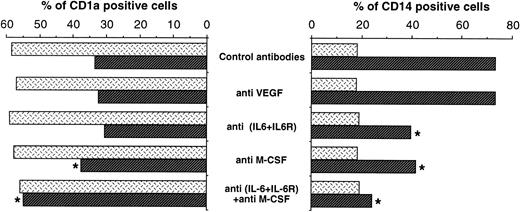
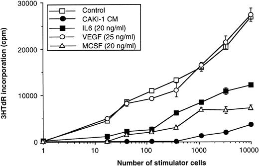
To confirm the role of IL-6 and M-CSF, CD34+ cells were cultured from day 6 to 12 in the presence of recombinant IL-6 (20 ng/mL), M-CSF (20 ng/mL), and VEGF (25 ng/mL) alone or in combination. Exogenous IL-6 and/or M-CSF mimicked the effect of RCC CM in terms of CD14/CD1a modifications (Fig 9) as well as CD80, CD86, HLA-DR, and CD116 expression and cell morphology (not shown), phagocytosis (Table 4), and APC function (Fig 10). Anti–IL-6/IL-6R reversed the effects of both IL-6 and M-CSF in terms of CD14/CD1a phenotype, phagocytic capacity, and APC function. In contrast, anti–M-CSF only slightly interfered with the biological effects of IL-6 in terms of CD14/CD1a phenotype (Fig 9), phagocytic function (Table 4), and APC function (Table 3). Titration performed with recombinant cytokines alone or in combination demonstrates that the minimal concentration of recombinant IL-6 or M-CSF required to induce an effect on DC differentiation was 1 ng/mL and 500 pg/mL for their combination (data not shown).
Inhibition of CD34+ cell differentiation by IL-6 and M-CSF. CD34+ cells were cultured with GM-CSF+TNF alone (control) or in the presence of 10% CLB-VER CM, IL-6 (20 ng/mL), M-CSF (20 ng/mL), or VEGF (25 ng/mL) from day 6 to 12. Neutralizing antibodies ([▪] control antibodies), (░) anti–IL-6+IL-6R, () anti–M-CSF, or (▨) the combination of anti–IL-6+IL-6R and anti–M-CSF were added at 10 μg/mL in the culture. At day 12, cells were harvested and processed for a double staining with CD14-PE and CD1a-FITC. Results are expressed as the percentage of CD14+ and CD1a+ cells. For all conditions, the SD ranged from 1% to 2%.
Inhibition of CD34+ cell differentiation by IL-6 and M-CSF. CD34+ cells were cultured with GM-CSF+TNF alone (control) or in the presence of 10% CLB-VER CM, IL-6 (20 ng/mL), M-CSF (20 ng/mL), or VEGF (25 ng/mL) from day 6 to 12. Neutralizing antibodies ([▪] control antibodies), (░) anti–IL-6+IL-6R, () anti–M-CSF, or (▨) the combination of anti–IL-6+IL-6R and anti–M-CSF were added at 10 μg/mL in the culture. At day 12, cells were harvested and processed for a double staining with CD14-PE and CD1a-FITC. Results are expressed as the percentage of CD14+ and CD1a+ cells. For all conditions, the SD ranged from 1% to 2%.
Inhibitory effect of IL-6 and M-CSF on APC function. CD34+ cells were cultured from day 6 to day 12 with GM-CSF+TNF alone (□) or in the presence of exogenous cytokines (▪) IL-6 (20 ng/mL), (▵) M-CSF (20 ng/mL), (○) VEGF (25 ng/mL), or (•) Caki-1 CM (10%) alone. Cells generated were harvested at day 12, irradiated (30 Gy), and used as stimulator cells (17 to 104 cells) in an allogeneic MLR with CD45RA+naive T cells (2 × 10⁴ cells/well) as described above. Results are representative of five experiments.
Inhibitory effect of IL-6 and M-CSF on APC function. CD34+ cells were cultured from day 6 to day 12 with GM-CSF+TNF alone (□) or in the presence of exogenous cytokines (▪) IL-6 (20 ng/mL), (▵) M-CSF (20 ng/mL), (○) VEGF (25 ng/mL), or (•) Caki-1 CM (10%) alone. Cells generated were harvested at day 12, irradiated (30 Gy), and used as stimulator cells (17 to 104 cells) in an allogeneic MLR with CD45RA+naive T cells (2 × 10⁴ cells/well) as described above. Results are representative of five experiments.
It is noteworthy that recombinant M-CSF (20 ng/mL) induced IL-6 production by cultured CD34+ cells at day 12 (with medium alone, <7.5 pg/mL; with M-CSF, 1.41 ± 0.46 ng/mL in a representative experiment), whereas IL-6 (20 ng/mL) did not affect M-CSF production at day 12, suggesting that M-CSF acts in part by inducing a secondary IL-6 production. These results demonstrate that IL-6 and M-CSF are responsible for the blockade of DC generation from CD34+ cells induced by RCC CM.
Modulation of GM-CSFRα (CD116) and M-CSFR (CD115) Expression on CD34+ Cells by RCC CM, IL-6, and M-CSF
We asked whether the inhibitory effect of RCC CM on CD34+cell differentiation into DC could result from a modulation of CD116 expression. Indeed, RCC CM as well as recombinant M-CSF (Table 2) were found to completely inhibit the expression of CD116 at the surface of CD34+ cells. Recombinant IL-6 also inhibited CD116 expression, although only partially (Table 2). Anti–M-CSF abrogated the inhibitory effect of RCC CM on CD116 expression. Conversely, RCC CM, recombinant IL-6, and recombinant M-CSF were found to induce an upregulation of CD115 expression at the surface of cultured CD34+ cells (Table 2); the combination of recombinant IL-6 and M-CSF exerted an additive effect on CD115 expression (Table 2). Anti–IL-6/IL-6R and anti–M-CSF additively blocked the induction of CD115 expression induced by RCC CM (Table 2). Taken together, these results demonstrate that the inhibition of CD34+ cells differentiation into DC by RCC CM, recombinant M-CSF, and/or recombinant IL-6 is associated by a downregulation of GM-CSFRα expression and the induction of M-CSFR expression.
Correlation Between IL-6 and M-CSF Production and the Inhibition of DC Development in a Panel of Tumor Cell Lines
The supernatants of a panel of tumor cell lines of various histologies (melanoma, lymphoma, breast carcinoma, neuroblastoma, large cell lung carcinoma, and colon carcinoma) were tested for their capacity to block CD34+-derived DC generation and to produce IL-6 and M-CSF. As shown in Table 1, the inhibition of DC differentiation, as evaluated by the modulation of CD1a and CD14 expression, by tumor cell supernatant was highly correlated to IL-6 production (>300 pg/mL; 11 of 14 v 0 of 11; χ2 test, P = 10−4) or M-CSF production (>300 pg/mL; 12 of 14v 1 of 11; χ2 test, P = 2 × 10⁻⁴) by tumor cells. This indicates that a wide range of tumor cell types are capable of blocking the differentiation of DC from CD34+ cells and that this effect is strongly correlated to the production of IL-6 and M-CSF.
DISCUSSION
The results presented here show that RCC cell lines produce soluble factors that inhibit the differentiation of DC from CD34+progenitor cells induced by GM-CSF+TNFα+SCF, in particular the acquisition of the characteristic differentiation Ag of DC (ie, CD1a, CD80, CD83, CD86, HLA-DR, and CD40…) as well as the acquisition of the APC function as evaluated by their capacity to trigger the proliferation of naive allogeneic CD4+ T lymphocytes. Similar inhibitory effects were observed when RCC CM was added in the culture from day 0 to 12 or only during the differentiation period (day 6 to 12).
Two different pathways of DC differentiation from CD34+progenitors have been previously described in this culture system,16 involving the two different populations detectable at day 6 ([CD14+CD1a−] and [CD14−CD1a+]). The inhibitory effect of RCC CM on DC differentiation on the unsorted population at day 6 was also observed on sorted day-6 [CD14+CD1a−] cells. In addition, RCC CM blocked the maturation of [CD1a+CD14−] early differentiated from CD34+ cells in terms of both phenotypic and APC capacity. RCC CM therefore blocks the two pathways of DC differentiation involving the [CD14+CD1a−] and [CD14−CD1a+] subpopulations at day 6.
The magnitude of the effects of RCC CM varied considerably among CD34+ cells obtained from different cord blood donors. As demonstrated previously,16 the respective levels of these two subpopulations, ie, [CD14+CD1a−] and [CD14−CD1a+], emerging at day 6 varied from 12% to 33% for CD14+ subpopulation and 8% to 32% for CD1a+ subpopulation. The variations of the magnitude of the inhibitory effect of RCC CM on DC differentiation are likely to result mostly from the variations relative proportion of [CD14+CD1a−] and [CD14−CD1a+] at day 6, a parameter that is not significantly affected by RCC CM in these experiments.
In the presence of RCC CM, an increased cell yield was observed as compared with that observed in cytokines alone. However, the observed decrease of CD1a+ fraction did not result from the overgrowth of this population by CD14+ cells; indeed, the quantification of the absolute number of CD1a+ and CD14+ cells with or without RCC CM in five different experiments showed a twofold to threefold decrease of the total CD1a cell number in the presence of RCC CM and, conversely, a 20-fold increase of the CD14+ population in the same conditions (data not shown). This could result from the capacity of CD14+ cells to respond to M-CSF signal, whereas CD1a+ cells that do not express M-CSFR are unable to respond to M-CSF.16 Taken together, these results indicate that RCC CM inhibits the differentiation of CD1a cells and induces a functional impairement of the remaining CD1a+ cell population obtained in these conditions.
The results presented here show that tumor cell lines inhibiting DC development produced high IL-6 and/or M-CSF and that the inhibitory effects of RCC CM on the phenotype, APC functions, and phagocytic capacity of cultured cord blood cells were reversed by anti–IL-6/IL-6R and anti–M-CSF antibodies. In addition, recombinant IL-6 with or without recombinant M-CSF was found to mimick the effects of RCC CM. Actually, IL-6 and M-CSF were found to exert distinct and additive inhibitory effects on the phenotypic and functional differentiation of CD34+ progenitors into DC.
Regarding phenotypic modifications, anti–IL-6/IL-6R partially blocked the induction of CD14 antigen expression induced by RCC CM, whereas anti–M-CSF reversed partly the effects of RCC CM on both CD14 and CD1a expression. Importantly, the combination of anti–IL-6/IL-6R and anti–M-CSF additively and completely reversed the phenotypic modifications induced by all RCC CM on cultured CD34+cells. High concentrations of recombinant IL-6 and M-CSF were capable of exerting similar inhibitory effects on the phenotypic differentiation of CD34+ cells, indicating that both cytokines are involved in the phenotypic alterations induced by RCC CM. It is noteworthy that the phenotypic modifications induced by recombinant M-CSF were also inhibited by anti–IL-6/IL-6R antibodies. Because M-CSF induced IL-6 production in these culture conditions, these results suggest that IL-6 is also a second mediator of M-CSF activity in this model.
CD34+ cells cultured in the presence of RCC CM were found to have characteristic features of macrophages in terms of morphology and function, with a high phagocytic capacity and poor APC function. Here also, both M-CSF and IL-6 contributed to this phenomenon. Anti–IL-6/IL-6R antibodies reversed partially the effects of the RCC CM on the phagocytic activity and APC function of cultured CD34+ cells. Anti–M-CSF antibody also partially reversed the inhibitory effect of RCC CM on APC function of cultured CD34+ cells. The combination of anti–IL-6/IL-6R and anti–M-CSF additively reversed the effects of RCC CM on the APC function of cultured CD34+ cells. However, with all cell lines tested, anti–M-CSF alone was found capable of completely inhibiting the increased phagocytic capacity induced by tumor cell medium, indicating that M-CSF plays a predominant role in the acquisition of high phagocytic function by CD34+ cells.
Finally, IL-6 and M-CSF exerted distinct effects on the expansion of cultured CD34+ cells costimulated with GM-CSF and TNFα. The increased proliferation induced by RCC CM was found to be mediated by M-CSF because (1) anti–M-CSF, but not anti–IL-6/IL-6R, reversed this effect and (2) recombinant M-CSF, but not recombinant IL-6, exerted a similar proliferative effect (not shown). This result is in agreement with those reported by Szabolcs et al17demonstrating the capacity of CD14+ subpopulation to proliferate in the presence of M-CSF.
These results are consistent with previous reports showing that IL-6 plays an important role in the monocytopoiesis from myeloid progenitors, that M-CSF induces the differentiation of CD34+ pluripotent cells into macrophages,17,22,23,25,27 and that IL-6 synergizes with M-CSF for the formation of macrophage colonies from purified CD34+ progenitors.24 In addition, anti–IL-6 has been reported to inhibit the generation of colony-forming unit-macrophages (CFU-M) induced by M-CSF from bone marrow precursors.25 However, our results are in contrast with a recent study28 demonstrating that production of high levels of IL-6 (500 to 700 pg/mL; probably by cogenereated monocytes) during DC development from CD34+ progenitors in GM-CSF+TNFα+SCF+sAB+ contributed to increase the yield of DC. Although differences in culture conditions may account for this discrepancy, the most likely explanation may be that the levels of IL-6 described by these investigators are under those required with recombinant IL-6 alone to block DC differentiation. It is noteworthy that the presence of M-CSF in culture medium was not reported in this study.
Taken together, these results demonstrate that IL-6 and M-CSF produced by RCC cells cooperate for the inhibition of the generation of DC from CD34+ cells induced by RCC CM. These results also show that IL-6 and M-CSF redirects the differentiation of these cells towards macrophage lineage. It must be noted that the capacity of anti–IL-6/IL-6R versus anti–M-CSF to reverse the biological effects of RCC CM on CD34+ cell differentiation varied according to the tumor cell line studied and their respective levels of IL-6 and M-CSF production. It is also important to note that the concentrations of exogenous recombinant IL-6 or M-CSF required to block DC differentiation were twofold to 10-fold higher than those present in 10% RCC CM for some RCC cell lines (CLB-CAN and CLB-TUG). This discrepancy suggests that another soluble factor(s) produced by some RCC cell lines may cooperate with IL-6 and/or M-CSF to inhibit DC differentiation.
The results presented here indicate that the molecular mechanisms by which RCC CM antagonize the effects of the GM-CSF+TNFα combination involve the modulation of GM-CSF and M-CSF receptors expression (CD116 and CD115, respectively) at the surface of cultured CD34+cells. RCC CM, recombinant IL-6, and, to a lesser extent, recombinant M-CSF were found to induce an increase of M-CSFR expression at the surface of differentiating CD34+ cells; the induction of CD115 expression by RCC CM is blocked additively by anti–IL-6/IL-6R and anti–M-CSF, a result consistent with previous publications on monocytes.24 Conversely, RCC CM, recombinant M-CSF, and, to a lesser extent, recombinant IL-6 induced a downregulation of GM-CSFRα expression. The loss of CD116 and the acquisition of CD115 expression was found to be an early event during CD34+ cell culture, being detectable at day 8, ie, 2 days after the addition of tumor cell medium to CD34+ cell culture (not shown). The loss of GM-CSFRα expression may render CD34+ cells completely insensitive to the effect of GM-CSF, which is absolutely required for DC differentiation in this model (for review, see Caux and Banchereau29). In contrast, the induction of M-CSFR expression is likely to contribute to support the differentiation of [CD14+CD1a−] cells towards the macrophage lineage.
Preculture of CD34+ cells from day 0 to 6, ie, during the early phase of DC differentiation, induced phenotypic modifications that were completely reversed by an additional 6 days of culture in GM-CSF+TNFα alone. The reversibility of the inhibitory effect of M-CSF on DC differentiation in the early phases of the differentiation process has also been reported by Chapuis et al.30 However, in the present work, it is also clear that the inhibitory effect of IL-6 and/or M-CSF is not reversible after 12 days of culture. We observed that CD34+ cord blood cells cultured from day 0 to 12 or only during the maturation period (day 6 to 12) with RCC CM failed to acquire a DC phenotype or function after additional culture between day 12 and 18 in the presence of GM-CSF+TNFα or GM-CSF+IL-4 (data not shown), demonstrating that macrophages obtained at day 12 were terminally differentiated. This result is also consistent with those described by Szabolcs et al17 demonstrating that day-12 macrophages generated in M-CSF from day-6 CD14+intermediates could not become DC even if recultured in GM-CSF+TNFα.
Several other cytokines have been described to block DC differentiation and functions, in particular VEGF, which is produced at high levels by tumoral cells.26 Although RCC cells produced high levels of VEGF (range, 2.7 to 6.2 ng/mL/106 cells/48 hours in the 9 different cell lines tested), the addition of neutralizing anti-VEGF antibody did not reverse the blocking effects of RCC CM on DC differentiation. Moreover, the addition of exogenous VEGF (25 ng/mL) during the differentiation phase (day 6 to 12) did not affect DC differentiation. This discrepancy could result from the fact that VEGF acts at very early stages of DC differentiation (D. Gabrilovich, personal communication, March 1998), whereas here the effects of VEGF were tested between day 6 and 12.
IL-10 has also been reported to inhibit DC differentiation and function. IL-10 was reported to block the differentiation of monocytes into DC31,32 and to inhibit the APC function of epidermal Langerhans cells33-35 and of monocyte- and CD34+-derived DC.36-39 However, none of the RCC cell lines tested here was found to produce detectable IL-10 levels,40 and IL-10 was not found in the culture supernatants even at day 7 to 9 (not shown). Finally, the use of anti–IL-10 neutralizing antibody did not modify the blocking effect of RCC CM on DC differentiation. This demonstrates that IL-10 is not involved in the inhibitory effects of tumor cell medium described in this study, in contrast with what is observed for monocytes.40
Interestingly, the important role of IL-6 observed in the present report is in agreement with previous clinical observations made in patients treated with IL-2. The presence of high pretreatment serum IL-6 levels has been found inversely correlated to the response to IL-2 and to survival after immunotherapy with IL-2 in patients with RCC41 as well as in melanoma and colorectal carcinoma.42,43 Although the correlation between high serum IL-6 levels and survival could result from the deleterious effects of this cytokine on the general status of these patients,44-46the biological explanation for the inverse correlation with response to IL-2 in several different human tumors was unclear. The present results suggest that these clinical observations may result from the immunosuppressive role of IL-6 on DC differentiation. In the present study, a wide range of tumor cells lines were found capable to produce IL-6 and M-CSF and to block DC differentiation, indicating that the production of IL-6 may be a frequent phenomenon by which tumor cell may protect themselves against the antitumor immune response. Interestingly, the overproduction of IL-6 has been found correlated with a defective immunity in vivo in other diseases, in particular in acquired immunodeficiency syndrome (AIDS) patients.47 The immunosuppressive role of IL-6 described here may account, at least in part, for this observation.
In conclusion, these results indicate that RCC cells inhibit the differentiation of CD34+ cells into DC through an IL-6– and/or M-CSF–dependent mechanism. These results undercover a frequent mechanism by which tumor cell may inhibit the spontaneous and therapeutic antitumor immune response and may account for the inverse correlation between in vivo IL-6 production and immune status in cancer and infectious diseases in humans.
ACKNOWLEDGMENT
The authors are grateful to I. Durand for FACS-sorting; to A. Duc, I. Iacono Di Cacito, and C. Massacrier for their technical assistance; and to the doctors and colleagues from clinics and hospitals in Lyon who provided us with the umbilical cord blood samples.
Supported by grants from Le LIONS Bourg Doyen, Le Comité de Saône et Loire and le comité du Rhône de La Ligue Contre le Cancer, la Ligue Nationale Contre le Cancer and l’ARC.
The publication costs of this article were defrayed in part by page charge payment. This article must therefore be hereby marked “advertisement” in accordance with 18 U.S.C. section 1734 solely to indicate this fact.
REFERENCES
Author notes
Address reprint requests to J.Y. Blay, MD, PhD, Unité“Cytokines et Cancers,” Unité Inserm 453, Centre Léon Bérard, 28 rue Laënnec, 69373 Lyon Cedex 08, France; e-mail: blay@lyon.fnclcc.fr.


![Fig. 6. Inhibition of antigen-presenting cell function by RCC CM. CD34+ cells were cultured from day 0 to 6 in GM-CSF+TNF. Medium was then changed and the cells were cultured in the presence of cytokine alone (control; □), RCC CM ([▪] CLB-VER CM; [○] Caki-1 CM), or breast CM (T47-D CM; •) from day 6 to 12. Cells were then harvested, irradiated (30 Gy), and used as stimulator cells for naive T cells (2 × 10⁴ cells/well). The proliferation was evaluated using 3HTdR uptake after 5 days of culture. Results are expressed as the mean ± SD of triplicate culture. These results are representative of five different experiments.](https://ash.silverchair-cdn.com/ash/content_public/journal/blood/92/12/10.1182_blood.v92.12.4778/5/m_blod42414006x.jpeg?Expires=1771255228&Signature=q8j~gVzqOXIrahF~74cUG7Sbi2~FX7X9OKeuVxVTUTBXNZr1a2OtqIeOL50flOU~6YglW5eipz1A-M0xjv~74FcJY2N8o2nBsF3M~nKydbWaSJ8vIb~8llYUXXwK9-CEOYCsuQsWbE-0VYAN2CfvBWT73Ait5bk1YaHxM1QWOHrk5UydY7FGdcrImqHEuHH0yItsOICTWr~mtTnR~8ZL5ztZe~eb2O6tZOK7rKVV~hSyJa89O4HeVbDfLjOY1bAGKzTUp~MI-yuItLLelvf~6JiEny-a9avVzHruWA~SXVDKctB3dF3WYxNP-Qtw4zDt86srqTwdtyWlDudp0iYn1w__&Key-Pair-Id=APKAIE5G5CRDK6RD3PGA)
![Fig. 7. Cord blood CD34+ cells were cultured for 6 days in the presence of GM-CSF+TNF+ SCF+sAB+. Cells were then collected, processed for double staining using anti–CD14-PE and anti–CD1a-FITC, and FACS-sorted into [CD14+CD1a−] and [CD14−CD1a+] populations. Sorted cells were seeded in the presence of GM-CSF+TNF at 5 × 10⁴cells/mL for 6 additional days in the presence or absence of RCC CM (10%), with a last medium change being performed at day 10. (A) Phenotype of purified populations at day 12. At day 12, cells were recovered and analyzed for CD1a, CD14, CD86, and HLA-DR expression by double-color fluorescence. (B) Function of the cells generated. At day 12, cells cultured in the presence of GM-CSF+TNF alone (medium; ▪) or in the presence of RCC CM (□) were recovered, numbered, irradiated (30 Gy), and used as stimulator cells (3.3 × 10³ cells/well) in an MLR with naive CD45RA+ T cells (2 × 10⁴ cells/well).](https://ash.silverchair-cdn.com/ash/content_public/journal/blood/92/12/10.1182_blood.v92.12.4778/5/m_blod42414007x.jpeg?Expires=1771255228&Signature=ZYpsgfQ-EwJx3GvkBPKMtdytGxFf7MEgqJlj~bdeF1~XRoK5iKYKeYODKmnUbu0AzYtayBWTwRptc9PqQYgssxHLqyc0addh3254d5fKSPspE0pB1ETJwWQD86uMtL2arJ8xh~VY7JE1YFVLLZTY47oHmj4BHNWRMkKnHjGE1agIwaWB-BXSzp4wxbpINAHMQDOKEx62rlTkU~Q4w9REO5ttJXI2afA8447g-ge3Qz0oxozvazTXDH3ClcnsT2rKpWWHsz5sY7TfIAJ1SddLRcKkIPBImmE3a0mJhixZvFMdVtU~WHssWnVN0qCQ0-sObRROy1EjLSyLHqyuQ3QR~Q__&Key-Pair-Id=APKAIE5G5CRDK6RD3PGA)
![Fig. 9. Inhibition of CD34+ cell differentiation by IL-6 and M-CSF. CD34+ cells were cultured with GM-CSF+TNF alone (control) or in the presence of 10% CLB-VER CM, IL-6 (20 ng/mL), M-CSF (20 ng/mL), or VEGF (25 ng/mL) from day 6 to 12. Neutralizing antibodies ([▪] control antibodies), (░) anti–IL-6+IL-6R, () anti–M-CSF, or (▨) the combination of anti–IL-6+IL-6R and anti–M-CSF were added at 10 μg/mL in the culture. At day 12, cells were harvested and processed for a double staining with CD14-PE and CD1a-FITC. Results are expressed as the percentage of CD14+ and CD1a+ cells. For all conditions, the SD ranged from 1% to 2%.](https://ash.silverchair-cdn.com/ash/content_public/journal/blood/92/12/10.1182_blood.v92.12.4778/5/m_blod42414009x.jpeg?Expires=1771255228&Signature=B0VFMuBf9PZl~VazOHpLNoixFZtBF2OqtNdcB-9UMCs-SLMGaeKTQxsCxDYA7FX1At54ldq7MTCicP93DF6fOq0UXQdXjaBAU3JQOMaHrKcweGBKw4E4yG3hLcfKUiLu7KEtX~pE8QiyIO3b44AORotjTDsA4W8X2s5YB1RZJ8ymq2TB7awulD3TukrVmZgLdbPK6z0dTmO3NVoWmyh5R27LutuLPCxzzt7ugCZDLW9BqN2OGyDxPlcujF8BNhrRUGfEWpw2ful5JPm-eWMSHaH3FddAzaNd9qYJzx3S~H-tU285KhrtT~0deRgtelt1uI0EFkUPLXG746iTYYMW6Q__&Key-Pair-Id=APKAIE5G5CRDK6RD3PGA)
This feature is available to Subscribers Only
Sign In or Create an Account Close Modal