Human eosinophils perform several functions dependent on phospholipase A2 (PLA2) activity, most notably the synthesis of platelet-activating factor (PAF) and leukotriene C4 (LTC4). Several forms of PLA2have been identified in mammalian cells. In the present study, the 14-kD, secretory form of PLA2 was detected in human eosinophils by immunocytochemical staining with the specific monoclonal antibody (MoAb) 4A1. In contrast, preparations of neutrophils, monocytes, lymphocytes, and basophils did not show detectable staining. With two MoAbs in a sandwich enzyme-linked immunosorbent assay (ELISA), large amounts of sPLA2 were detected in lysates of eosinophils, that were 20-fold to 100-fold higher than in the other circulating leukocytes (ie, neutrophils, basophils, monocytes, and lymphocytes). In addition, with a commercially available sPLA2 activity assay kit, we were able to show high activity of sPLA2 in human eosinophils relative to neutrophils. Investigations at the ultrastructural level showed that sPLA2 in eosinophils is mainly located in specific granules. Immunoelectron microscopy also visualized sPLA2within phagosomes after addition of opsonized particles to the eosinophils. However, sPLA2 was not detected in the cell-free supernatants of activated eosinophils, in contrast to eosinophil-cationic protein (ECP), which colocalizes with sPLA2 in resting eosinophils. These findings warrant further studies into the role of sPLA2 in eosinophil function.
AN IMPORTANT FEATURE of the pathogenesis of asthma is the accumulation of eosinophils in the lungs, a phenomenon that is correlated with destruction of lung tissue.1,2However, until now it is not clear why eosinophils preferentially accumulate in the lungs and what induces their degranulation and release of the lipid mediators platelet-activating factor (PAF) and leukotriene C4 (LTC4).3-5Eosinophil granule proteins can be cytotoxic for respiratory epithelial cells and pneumocytes,6,7 and PAF and LTC4 can induce bronchoconstriction and bronchial hyperresponsiveness.8 In a previous study, we observed that PAF synthesis by human eosinophils after in vitro activation with STZ (serum-treated zymosan) is completely blocked by the phospholipase A2 (PLA2)-inhibitor mepacrine.9Mepacrine also inhibits LTC4 secretion from eosinophils activated with the tripeptide fMLP (formyl-methionyl-leucyl-phenylalanine).10 Furthermore, the release from eosinophils of preformed mediators, such as eosinophil cationic protein (ECP) and eosinophilic peroxidase (EPO), has been shown to be influenced by the enzymatic activity of PLA2.10 Because PLA2 activity plays such an important role in the release of various eosinophil products, we undertook further studies to characterize PLA2 activity in human eosinophils.
Recently, it has become clear that PLA2 enzymes can be classified in several groups. In man, four proteins have been sequenced: an intracellular or cytosolic 85-kD protein (group IV) and three, so-called secretory, 14-kD PLA2s (belonging to group I, II en V, respectively).11,12 Group I PLA2 is produced by pancreatic acinar cells, functions as a digestive enzyme within the intestinal lumen, and is therefore also referred to as pancreatic PLA2.11 Group II PLA2(sPLA2) can be purified from human synovial fluid,13 platelets, and can be detected in chondrocytes and Paneth cells.14 During septic shock and inflammation, the concentration of sPLA2 in serum is enhanced, but the cellular source of sPLA2 in serum is unknown.15-18 Group V PLA2 is present in several human tissues and in murine P388D1macrophages19 and murine mast cells.20 With antibodies raised against the group II PLA2, we show in this study that human eosinophils express secretory PLA2(sPLA2) at levels much higher than found in other leukocytes.
MATERIALS AND METHODS
Formyl-methionyl-leucyl-phenylalanine (fMLP), cytochalasin B (cyto B), phorbol myristate acetate (PMA), and calcium ionophore A 23187 (Sigma Chemical Co, St Louis, MO) were dissolved in dimethyl sulfoxide (DMSO) at 1,000 times the final concentration for cell incubations. PAF (Sigma) was dissolved in phosphate-buffered saline (PBS) containing human-serum albumin (HSA) (0.5% vol/vol). Interleukin-5 (IL-5) was obtained from Amersham Corp (Amersham, Birmingham, UK). STZ was prepared as described previously.9 Serum-opsonized Sephadex particles (SOS) were prepared by incubating swollen Sephadex G15 beads (150 mg) in 1 mL of human AB serum for 30 minutes at 37°C followed by three washes in 0.9% (wt/vol) NaCl. Washed beads were resuspended in incubation medium (see below) to a concentration of 150 mg/mL. The monoclonal antibodies (MoAbs) 4A1 and 10B2 raised against sPLA2 were kindly provided by Dr F.B. Taylor, Jr (Oklahoma Medical Research Foundation, Oklahoma City, OK). The polyclonal anti-sPLA2 rabbit antiserum was purchased from Cayman Chemical Co (Ann Arbor, MI). All other chemicals were reagent grade. Incubation medium for the cell incubations contained 132 mmol/L NaCl, 6.0 mmol/L KCl, 1.0 mmol/L CaCl2, 1.0 mmol/L MgSO4, 1.2 mmol/L potassium phosphate, 20 mmol/L HEPES, 5.5 mmol/L glucose and 0.5% (wt/vol) human albumin, pH 7.4.
Isolation of granulocytes and neutrophils.
Blood was obtained from healthy volunteers. Granulocytes were isolated from the buffy coat of 500 mL of blood anticoagulated with 13 mmol/L trisodium citrate (pH 7.4) essentially as described before.21 In short, the mononuclear cells were removed by centrifugation of the blood over isotonic Percoll (1.077 g/mL, pH 7.4). After isotonic lysis of the erythrocytes in the pellet with an ice-cold solution containing 155 mmol/L NH4Cl, 10 mmol/L KHCO3, and 0.1 mmol/L EDTA (pH 7.2), the granulocytes were washed twice in PBS and suspended in incubation medium. Where indicated, neutrophils were further purified by depletion of the eosinophils with CD9 MoAbs and magnetic Dynabeads, coated with sheep-antimouse antibodies. The resulting preparations always contained less than 0.2% eosinophils.
Purification of eosinophils.
Eosinophils were purified as previously described.21 In short, granulocytes were incubated for 30 minutes at 37°C to restore the initial density of the cells. After this incubation period, the cells were washed and resuspended in PBS supplemented with HSA (0.5% wt/vol) and trisodium citrate (13 mmol/L). After preincubation of the granulocytes for 5 minutes at 37°C, fMLP (10 nmol/L) was added to the cell suspension, and the incubation was continued for 10 minutes. Subsequently, the eosinophils were purified by centrifugation (20 minutes at 800g) over isotonic Percoll (1.082 g/mL, pH 7.4), washed and resuspended in incubation medium. This suspension contained for >98% eosinophils.
Purification of monocytes, lymphocytes, and basophils.
Monocytes and lymphocytes were isolated from the upper fraction of the Percoll gradient (1.077 g/mL) (the mononuclear cells) by countercurrent centrifugal elutriation as described previously by De Boer and Roos.22 The purities of both cell fractions were > 90%. Basophil purification was performed by depletion of residual contaminating cells (mainly lymphocytes and a few monocytes) from the last elutriation fraction, with antibodies against these cells (CD2, CD14, CD16, CD19) and magnetic Dynabeads as described.23The purity of the basophils in this preparation was > 92%. The mononuclear cell fractions were kept on ice to prevent activation.
Immunocytochemical staining methods.
Human granulocytes (containing about 5% eosinophils), and purified eosinophils, basophils, lymphocytes, and monocytes were centrifuged on microscope slides and fixed in acetone for 10 minutes. All incubation steps were performed at room temperature. The preparations were preincubated with normal goat serum (1:10 diluted in PBS, containing 1% [wt/vol] bovine serum albumin [BSA]) for 15 minutes and were subsequently incubated for 1 hour with the anti-sPLA2 MoAb 4A1 (25 μg/mL). Control preparations were incubated with PBS/BSA without MoAb 4A1 or with irrelevant MoAb of the appropriate subclass. After each incubation step,the preparations were washed with PBS. Next, the preparations were incubated with 0.3% (vol/vol) H2O2, 0.1% (wt/vol) sodium azide in PBS for 20 minutes to block endogenous peroxidase activity. Subsequently, the preparations were incubated for 30 minutes with biotinylated rabbit antimouse-Ig F(ab)2 fragments (Dakopatts, Glostrup, Denmark) 1:200 diluted in PBS containing 10% (vol/vol) normal human serum (NHS). The preparations were washed and incubated for 30 minutes with horseradish peroxidase (HRP)-labeled streptavidin-biotin(AB) Complex (Dakopatts), diluted 1:200 in PBS-NHS. HRP activity was detected by using 3-amino-9-ethylcarbazole (Sigma) in the presence of 0.03% (vol/vol) H2O2 yielding a red color. Preparations were fixed in paraformaldehyde (PFA; 4% vol/vol), counterstained with Mayer's hematoxylin and mounted with glycerolgelatin. The preparations were examined by light microscopy, to determine the localization and intensity of staining of sPLA2. The specificity of this procedure was confirmed by the absence of staining of pancreas tissue preparations (described to contain PLA2 type I14) and positive staining of Paneth cell preparations (described to contain sPLA2,14 results not shown).
Enzyme-linked immunosorbent assay (ELISA) for sPLA2.
Concentrations of sPLA2 in cell lysates or cell supernatants were determined with an ELISA modified from Smith et al.24 MoAb 10B2 and 4A1 against sPLA2 were used as catching and detecting antibody, respectively. In some experiments, the polyclonal anti-sPLA2 rabbit antiserum was used as catching antibody. The amount of sPLA2 was assessed by comparison with a (sigmoidal) calibration curve obtained with purified recombinant human sPLA2 (kindly provided by Dr C. Schalkwijk, Centre for Biomembranes and Lipid Enzymology, University of Utrecht, Utrecht, The Netherlands). The sensitivity of the measurements in this assay amounted to 0.2 to 5 ng/mL. Lysis of the different cell types (2 × 10⁷ cells/mL) was performed for 30 minutes at 4°C in incubation medium with the following additions: 1% NP-40 (vol/vol), phenylmethyl sulfonyl fluoride (PMSF; 100 μg/mL), tosyl-leucyl chloromethylketone (TLCK; 1 mmol/L), leupeptin (0.1 mmol/L), soybean trypsin inhibitor (SBTI; 20 μg/mL) (Sigma), and EDTA (5 mmol/L).
Measurement of sPLA2 activity.
Human eosinophils or neutrophils (depleted for eosinophils) were resuspended (108 cells/mL) in PBS containing 0.34 mol/L sucrose, 10 mmol/L HEPES, 100 μg/mL PMSF, 1 mmol/L TLCK, 0.1 mmol/L leupeptin, and 5 mmol/L EDTA (final pH 7.0), sonicated at 21 kHz and 8 μm peak-to-peak amplitude for 3 × 15-second intervals at 4°C and stored at −80°C until use. sPLA2ELISA measurements of these sonicates show identical amounts of sPLA2 protein compared with lysis in 1% NP-40 as described above (data not shown). sPLA2 activity measurement was performed with a commercially available assay system (Cayman Chemical Company) exactly as recommended by the manufacturer. The assay uses the 1,2-dithio analog of diheptanoyl phosphatidylcholine, which serves as a substrate for most PLA2 with the exception of group IV cytosolic PLA2. On hydrolysis of the thio ester bound at the sn-2 position by PLA2, free thiols are detected by reaction with 5,5′-dithio-2-nitrobenzoate (DTNB). The hydrolysis of DTNB was quantified by measuring the absorbance at 405 nm at different time points and determination of the slope, using 10.0 mmol−1 · L+1 · cm−1 as the adjusted (for pathlength) molar extinction coefficient.
ELISA for ECP.
ECP in cell supernatants was measured with a slightly modified sandwich ELISA previously described by Reimert et al.25 Specific polyclonal rabbit-antihuman ECP antibodies were obtained by immunization of rabbits with highly purified ECP.26 Plates were coated overnight (o/n) with rabbit-antihuman ECP (3 μg IgG/mL). After blocking with BSA (2%, wt/vol), samples and standard (purified ECP) were incubated in various dilutions for 90 minutes at 37°C in a buffer (pH 7.5) containing 0.1 mol/L NaCl, 50 mmol/L NaH2PO4, 0.1% Tween-20, 0.1% CTAB, 0.2% BSA and 20 mmol/L EDTA. Bound ECP was detected after incubation with biotin-conjugated rabbit-antihuman ECP by adding a mixture of avidin and biotinylated alkaline phosphatase according to the manufacturer's description (ABC-Complex Dakopatts). Enzymatic activity was determined with phosphatase substrate tablets (Sigma 104, Sigma). The range of the measurements was 0.10 to 10 ng/mL.
Immunoelectron microscopy.
Eosinophils were prewarmed for 10 minutes, primed with IL-5 (10-10 mol/L) for 30 minutes, and incubated with STZ for 10 minutes. All cells were fixed for 2 hours at room temperature in a graded (2% to 8%) PFA series in PBS to preserve the antigenicity of sPLA2 and were pelleted in 10% gelatin in PBS. This fixation procedure was found to be optimal for staining with antibodies directed against sPLA2 (see below). The gelatin slab was kept at 4°C in PBS containing 1% PFA. Small pieces of gelatin were infiltrated with 2.3 mol/L sucrose in PBS for 4 hours, mounted on copper pins at 4°C, and frozen in liquid nitrogen. About 80 nm cryopreparations were made on an MT-7 ultracryomicrotome (Research and Manufacturing Co, Tucson, AZ) and incubated at room temperature with mouse MoAb anti-sPLA2 10B2 (dilution 1/50) followed by incubation with rabbit antimouse (RAM)-IgG and goat-antirabbit (GAR)-IgG conjugated to 10 nm gold (both at dilution 1/40) (Amersham). As a control, anti-sPLA2 was replaced by a nonrelevant murine IgG. All incubations lasted 1 hour. After immunolabeling, the cryopreparations were embedded in a mixture of methylcellulose and uranyl acetate. All preparations were examined with a Philips CM10 electron microscope (Eindhoven, The Netherlands).
RESULTS
Immunocytochemical staining of leukocytes with MoAbs against sPLA2.
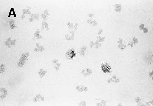
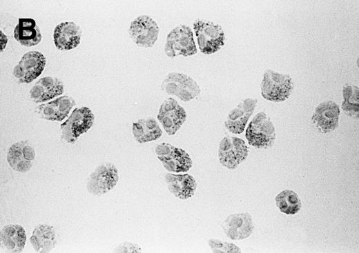
The MoAb 4A1 raised against recombinant human sPLA2 was used to determine the presence of the 14-kD sPLA2 in circulating leukocytes. As positive control for the histochemical staining procedure, HEPG2 cells, which are known to express sPLA2,27 were included in the experiments. The few cells staining positive for sPLA2 in the granulocyte fraction (Fig 1A) had the appearance of eosinophils. Indeed, in cytospins of purified eosinophils, all cells stained positive for sPLA2 (Fig 1B). In Fig 1B, it can be seen that the sPLA2 present in the eosinophils is granular distributed. The same pattern of staining was seen in the HEPG2 cells (data not shown). The immunocytochemical preparations of neutrophils, monocytes, lymphocytes, and basophils did not exhibit a detectable staining (data not shown).
Visualization of sPLA2 in (A) human granulocytes and (B) purified eosinophils. Granulocytes (containing 5% eosinophils) and purified eosinophils were centrifuged on microscope slides, and sPLA2 was detected by immunohistochemical staining with MoAb 4A1 as described in Materials and Methods.
Visualization of sPLA2 in (A) human granulocytes and (B) purified eosinophils. Granulocytes (containing 5% eosinophils) and purified eosinophils were centrifuged on microscope slides, and sPLA2 was detected by immunohistochemical staining with MoAb 4A1 as described in Materials and Methods.
Measurement of the intracellular content and activity of sPLA2.
To quantitate the level of sPLA2 in eosinophils, a sandwich ELISA with MoAbs directed against sPLA2 was applied. Previous studies have shown that this assay is specific for the human sPLA2 enzyme24 and that a strong correlation exists between PLA2 activities and immunoreactive material in plasma samples of patients undergoing IL-2 therapy.28 Table 1 shows that the content of sPLA2, present in the lysates of eosinophils was 20-fold to 100-fold higher than that of the other circulating leukocytes. Although sPLA2 was not visualized in preparations of monocytes and neutrophils immunocytochemically stained for sPLA2, lysates of monocytes and neutrophils (depleted for eosinophils) did contain a relative small amount of sPLA2 (Table 1), whereas the results obtained with lysates of purified basophils were on the borderline of the sensitivity of our assay. The low signal in the neutrophil lysates could not be explained by the presence of contaminating eosinophils (being less than 0.2%). In the lysates of lymphocytes, sPLA2 was not detected.
Detection of sPLA2 in Lysates of Human Leukocytes
| Cell Type . | sPLA2 (ng/107 cells) . |
|---|---|
| Lymphocytes | <0.1 |
| Basophils | 0.11 ± 0.02 |
| Monocytes | 0.15 ± 0.05 |
| Neutrophils | 0.73 ± 0.15 |
| Eosinophils | 19.3 ± 3.30 |
| Cell Type . | sPLA2 (ng/107 cells) . |
|---|---|
| Lymphocytes | <0.1 |
| Basophils | 0.11 ± 0.02 |
| Monocytes | 0.15 ± 0.05 |
| Neutrophils | 0.73 ± 0.15 |
| Eosinophils | 19.3 ± 3.30 |
Human leukocytes were isolated from four different blood donors and lysed (2 × 10⁷/mL) as described in Materials and Methods. Before lysis, neutrophils were depleted of eosinophils by immunodepletion with CD9 antibodies and magnetic beads. The lysates were tested in the sPLA2 ELISA. The results are expressed as means ± SEM of four different donors.
The pronounced difference between neutrophils and eosinophils was confirmed with ELISA measurements in which the monoclonal 10B2 antibody was replaced as catching antibody by a polyclonal anti-sPLA2 rabbit antiserum. The levels of sPLA2 measured in this modified ELISA amounted to 1.4 ± 0.25 and 32.0 ± 5.8 (mean ± standard error of mean [SEM], n = 5) ng sPLA2/107 cells, respectively, for neutrophils and eosinophils. Measurement of sPLA2 activity as described in Materials and Methods showed an activity of 14.6 ± 2.9 (mean ± SEM, n = 3) nmol/min/108 cells in sonicates from human eosinophils and 1.6 ± 0.4 (mean ± SEM, n = 3) nmol/min/108 cells in sonicates from neutrophils, again indicating that eosinophils contain high amounts of sPLA2 relative to neutrophils. It should be noted that for the sPLA2 activity measurement, we have used crude whole cell sonicates. These sonicates might have contained endogenous inhibitors of the sPLA2 enzyme.29
Immunoelectron microscopic studies on the localization of sPLA2 in human eosinophils.
Investigation of the intracellular localization of sPLA2 in unstimulated eosinophils was undertaken by immunoelectron microscopy (immuno-EM). Immunogold-labeled MoAb against sPLA2 was detected in granules that contained the typical eosinophil crystals (Fig 2). It is difficult to ascertain whether the heterogeneity in the labelling in the granules is due to heterogeneity in the localization of sPLA2 or to partial destruction of the epitopes for the antibody due to the fixation procedure used.
Localization of sPLA2 in human eosinophils isolated from peripheral blood. Ultrathin cryosections were incubated with MoAb 10B2, rabbit antimouse IgG and goat antirabbit IgG conjugated to 10-nm gold particles. (A) Overview of an eosinophil showing labeling mainly on the matrix of specific granules, although these granules do show heterogeneity. Nucleus (n); bar = 500 nm. (B) Detail of the cytoplasm showing dense labeling in some granules (closed arrows) and absence of label in others (open arrows). Bar = 200 nm.
Localization of sPLA2 in human eosinophils isolated from peripheral blood. Ultrathin cryosections were incubated with MoAb 10B2, rabbit antimouse IgG and goat antirabbit IgG conjugated to 10-nm gold particles. (A) Overview of an eosinophil showing labeling mainly on the matrix of specific granules, although these granules do show heterogeneity. Nucleus (n); bar = 500 nm. (B) Detail of the cytoplasm showing dense labeling in some granules (closed arrows) and absence of label in others (open arrows). Bar = 200 nm.
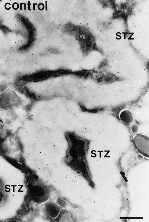
Immunolocalization of sPLA2, after addition of STZ to IL-5–primed eosinophils, was also studied with immuno-EM. After binding, STZ particles were engulfed by the eosinophils with a lag time of about 2 minutes (unpublished data). The cells were fixed 10 minutes after the addition of STZ for the preparation of sections suitable for immuno-EM. Clearly, phagosomes had been formed and immunogold particles depicting sPLA2 were present in these compartments (Fig 3A). Some background staining was noted under these conditions (Fig 3B), but this staining was considerably lower than the staining obtained with the anti-sPLA2 antibody.
Localization of sPLA2 in human eosinophils after incubation of the cells with STZ for 10 minutes. (A) Ultrathin cryosections were incubated as described in the legend to Fig 2. The micrograph shows several STZ particles contained in a phagosome that are strongly labeled for sPLA2. Nucleus (n); bar = 500 nm. (B) Micrograph showing some aspecific labeling of the STZ-treated eosinophils incubated with rabbit antimouse IgG and goat antirabbit IgG conjugated to 10-nm gold particles. Some specific granules (g) are seen near the phagosomes. Bar = 500 nm.
Localization of sPLA2 in human eosinophils after incubation of the cells with STZ for 10 minutes. (A) Ultrathin cryosections were incubated as described in the legend to Fig 2. The micrograph shows several STZ particles contained in a phagosome that are strongly labeled for sPLA2. Nucleus (n); bar = 500 nm. (B) Micrograph showing some aspecific labeling of the STZ-treated eosinophils incubated with rabbit antimouse IgG and goat antirabbit IgG conjugated to 10-nm gold particles. Some specific granules (g) are seen near the phagosomes. Bar = 500 nm.
Detection of sPLA2 in the supernatant of stimulated eosinophils.
Using the sPLA2 ELISA, we measured the sPLA2content in supernatants collected from unstimulated eosinophils and from eosinophils treated with PAF, fMLP, PAF-fMLP, cyto B, or cyto B-fMLP. None of these treatments induced the secretion of sPLA2 into the supernatant. Other stimuli, such as Ca2+-ionophore A23187, PMA, or combinations of these stimuli, also did not result in the release of sPLA2 in the supernatant (data not shown).
In another set of experiments, eosinophils were stimulated with STZ particles or serum-opsonized Sephadex (SOS). As shown in Table 2, ECP was released, especially after stimulation with the much larger SOS particles leading to frustrated phagocytosis (Egesten et al, manuscript in preparation). In agreement with the enhanced percentage of eosinophils able to bind opsonized particles after IL-5 pretreatment,9 the release of ECP was enhanced for both the STZ- and SOS-induced ECP release after IL-5 pretreatment. However, the same supernatants did not contain sPLA2 as determined by the sPLA2 ELISA (Table2).
Release of Granule Proteins From Untreated and IL-5–Primed Eosinophils After Stimulation With Opsonized Particles
| . | ECP % of Total Amount . | sPLA2 . |
|---|---|---|
| Unstimulated | 1.9 ± 0.9 | ND |
| IL-5 | 2.7 ± 1.3 | ND |
| STZ | 1.5 ± 1.2 | ND |
| SOS | 14.1 ± 3.6 | ND |
| IL-5/STZ | 4.6 ± 1.8 | ND |
| IL-5/SOS | 21.8 ± 4.4 | ND |
| . | ECP % of Total Amount . | sPLA2 . |
|---|---|---|
| Unstimulated | 1.9 ± 0.9 | ND |
| IL-5 | 2.7 ± 1.3 | ND |
| STZ | 1.5 ± 1.2 | ND |
| SOS | 14.1 ± 3.6 | ND |
| IL-5/STZ | 4.6 ± 1.8 | ND |
| IL-5/SOS | 21.8 ± 4.4 | ND |
Eosinophils (5 × 10⁶/mL) were incubated with buffer or IL-5 (10−10 mol/L) for 30 minutes at 37°C before STZ (1 mg/mL) or SOS (30 mg/mL) was added. After 15 minutes, the eosinophil suspensions were centrifuged and supernatants were collected for ECP and sPLA2 determination. The results are expressed as means ± SEM of three different experiments.
Abbreviation: ND, not detectable.
DISCUSSION
This study has shown that human eosinophils express high amounts of the secretory 14-kD sPLA2, whereas neutrophils and monocytes express only a small amount. Our data indicate that in future studies on the role of secretory 14-kD sPLA2 in monocytes or neutrophils, contamination by eosinophils has to be excluded because of the very high amounts of this enzyme in eosinophils. In lymphocytes, sPLA2 was not detectable by ELISA or by immunocytochemical staining of cytospin preparations. In basophils, a small amount could be present, the ELISA signal being on the borderline of our assay. A preliminary report has appeared describing a possible role of secretory PLA2 in LTC4 synthesis by human basophils.30
Because we used for this study antibodies raised against the synovial type of sPLA2 (belonging to group II of PLA2s), it is most likely that the enzyme detected by us in human eosinophils is identical to this protein. However, it cannot be completely excluded that another member of the sPLA2 gene family is highly expressed in eosinophils, eg, the recently cloned group V enzyme. Interestingly, this latter protein has recently been shown in murine macrophages19 and mast cells20 to play an important role in arachidonic acid release, a role previously assigned to the group II PLA2 enzyme.
Although eosinophil degranulation and synthesis of PAF and LTC4 are dependent on PLA2 activity, the role of the (secretory) sPLA2 in various eosinophil responses can only be speculated on. The fact that mepacrine has been suggested to specifically inhibit sPLA231 and is able to completely block STZ-induced PAF synthesis in human eosinophils9 might indicate an important role for sPLA2 activity in eosinophil activation. In addition, the presence of sPLA2 in the eosinophil phagosomes may indicate a function for this PLA2 in the killing of microorganisms. Mammalian sPLA2 is able to directly degrade phospholipids of Gram-negative bacteria.32
Despite significant degranulation (as indicated by the release of ECP), sPLA2 did not appear to be released from eosinophils on activation in vitro. sPLA2 is clearly lipolytic and catalyzes a reaction at the water/lipid interface.11 Hence, it is not unlikely that directly after secretion and exposure to mmol/L Ca2+ concentrations, the enzyme rebinds to the membrane. However, immunoelectron microscopy did not yield an indication for the presence of sPLA2 on the outer membrane after eosinophil activation. Likewise, we did not detect sPLA2 on the plasma membrane by indirect immunofluorescence with the MoAbs 4A1 or 10B2 (data not shown). Possibly, insertion of the enzyme into the lipid bilayer prevents binding of the antibodies. Cell fractionation followed by detergent lysis of the various fractions may be helpful to resolve this issue. Bach et al33 have shown that quantification of release of eosinophil granular proteins is hampered by destruction on eosinophil activation. Measurement of remaining eosinophil-derived neurotoxin (EDN) and EPO in activated eosinophils showed a recovery of only 40% in the cell pellet of activated eosinophils, without apparent release of these proteins (<2%). These investigators could not explain this low recovery. Also, we have found a low recovery of sPLA2 protein (as determined by ELISA measurements of sPLA2 in the supernatant and the remaining amount of sPLA2 in the cell pellet of activated eosinophils) of 51% ± 8% and 66% ± 6% (mean ± SEM, n = 3) in PAF/STZ- and PMA-stimulated human eosinophils respectively, compared with a recovery of 95% ± 5% in unstimulated eosinophils. The cause of this poor recovery is currently under investigation.
Previously, Rosenthal et al34 have shown the presence of a 14-kD PLA2 in phagosomes of neutrophils on activation with serum opsonized S. aureus. Under certain conditions, this enzyme can be found in the supernatant of activated neutrophils.34,35 Our study shows that the level of sPLA2 is about 20-fold higher in eosinophils than in neutrophils, but also that under in vitro conditions the enzyme is not readily released from the eosinophils. Although we were not able to show release of sPLA2 protein from eosinophils in vitro, sPLA2 might be released from eosinophils in vivo. Bowton et al36 have recently described enhanced sPLA2 activity in bronchoalveolar lavage (BAL) fluid after antigen challenge in asthmatic patients. These investigators did not examine the cellular source of the sPLA2, although they suggested the mast cell as a possible candidate. However, our results, while not refuting mast cells as an important source of sPLA2, might suggest eosinophils as another cellular source of sPLA2 in BAL fluids. Released sPLA2, eg, in the lungs, may act on other cell types present in the lung: addition of sPLA2 to mast cells results in the release of histamine, one of the agents responsible for the symptoms during an asthmatic attack.37
ACKNOWLEDGMENT
The authors thank Prof. H. van den Bosch (University of Utrecht, The Netherlands) for advice on sPLA2 activity measurements.
Supported by The Netherlands Asthma Foundation (Project No. 14.89.25).
Address reprint requests to A.J. Verhoeven, PhD, Central Laboratory of the Netherlands Red Cross Blood Transfusion Service, Plesmanlaan 125, 1066 CX Amsterdam, The Netherlands.
The publication costs of this article were defrayed in part by page charge payment. This article must therefore be hereby marked “advertisement” in accordance with 18 U.S.C. section 1734 solely to indicate this fact.


This feature is available to Subscribers Only
Sign In or Create an Account Close Modal