Abstract
The receptor-type tyrosine kinase, c-kit is expressed in hematopoietic stem cells (HSC), myeloid, and lymphoid precursors. In c-kit ligand-deficient mice, absolute numbers of HSC are mildly reduced suggesting that c-kit is not essential for HSC development. However, c-kit− HSC cannot form spleen colonies or reconstitute hematopoietic functions in lethally irradiated recipient mice. Based on in in vitro experiments, a critical role of c-kit in B-cell development was suggested. Here we have investigated the B-cell development of c-kitnull mutant (W/W ) mice in vivo. Furthermore, day 13 fetal liver cells from wild type or W/W mice were transferred into immunodeficient RAG-2−/− mice. Surprisingly, transferred c-kit− cells gave rise to all stages of immature B cells in the bone marrow and subsequently to mature conventional B2, as well as B1, type B cells in the recipients to the same extent as transferred wild type cells. Hence, in contrast to important roles of c-kit in the expansion of HSC and the generation of erythroid and myeloid lineages and T-cell precursors, c-kit− HSC can colonize the recipient bone marrow and differentiate into B cells in the absence of c-kit.
THE DOMINANT WHITE spotting locus encodes the receptor-type tyrosine kinase, c-kit,1 and the ligand for c-kit is stem cell factor (SCF )2-7 (for review see Galli et al8 ). Mice bearing mutations at the dominant white spotting locus exhibit developmental abnormalities of several cell lineages including mast cells,9 melanocytes, germ cells, T lymphocytes residing in intestinal epithelium,10 the intestinal cells of Cajal,11,12 lymphoid and myeloid precursors, and hematopoietic stem cells (HSC)13,14 (for review see Russell15 ). The molecular basis of various spontaneous mutations at the dominant white spotting locus has been determined: W, one of these mutant genes, bears a point mutation at the splicing donor site causing the deletion of the transmembrane region; WV bears a point mutation at the kinase domain.16 Mice homozygous for the W gene (W/W mice) thus lack expression of c-kit on the cell surface (null mutant), and die ∼10 days after birth due to severe anemia. In contrast, mast cells from W/WV mice can express c-kit on the cell surface,17 and these mice exhibit only slightly decreased numbers of myeloid and lymphoid precursors of bone marrow.18
The following four observations provide evidence that c-kit has an important role in the proliferation of HSC and myeloid precursors: First, the absolute numbers of HSC are decreased by ∼2.5 fold in SCF-deficient mice.14 Second, intraperitoneal injection of antagonistic anti–c-kit antibody rapidly eliminates HSC and myeloid precursors in the bone marrow.13 Third, a large number of transferred W/WV bone marrow cells cannot reconstitute lethally-irradiated wild type mice (for review see Russell15 ). Fourth, conversely, engraftment of only a single HSC derived from wildtype mice is apparently sufficient to sustain the entire erythropoiesis in recipient W/WV mice.19 Thus, c-kit+ HSC are functionally superior to a much larger number of W/WV HSC. The third and fourth points indicate that in vivo transfer of wildtype or W mutant HSC may provide the most sensitive analysis of c-kit–associated hematopoietic defects.
Based on the following evidence, a crucial role of c-kit –mediated signals in B-cell development had been suggested: First, SCF can synergize with interleukin-7 (IL-7) to promote the proliferation of precursor B cells in vitro20-24; second, anti–c-kit antibody suppresses the growth of B-cell precursors in vitro25; third, in W/WV mice, the number of B precursors in bone marrow is slightly decreased 18; fourth, in vivo early B lymphocyte development of wildtype, immunoglobulin κ chain transgenic,22 as well as RAG-2−/− mice (S.-I. Nishikawa, personal communication, December 1994) is suppressed by intraperitoneal injection of anti-c-kit antibody. However, since infusion of antagonistic anti-c-kit antibody inhibits most HSC in vivo, such analyses could not distinguish whether the antibody directly perturbs development of committed lymphocyte precursors or affects lymphocyte development only indirectly via its effects on HSC.
To investigate the role of c-kit in B lymphocyte development, we have analyzed B-cell development in fetal and 5-day old W/W mice. Furthermore, to amplify potential defects in c-kit− precursors, day 13 W/W or wildtype fetal liver (FL) cells were transferred into lymphocyte-deficient RAG-2−/− mice. Subsequently, B-cell development from donor-type c-kit− or c-kit+ HSC was followed. The data indicate that transferred HSC and/or committed precursors are dependent on c-kit–mediated signals to generate thymocytes, as well as for the expansion of myeloid cells. In marked contrast, HSC can develop normally into precursor B cells in the bone marrow and into mature B1 and B2 type B cells in peripheral lymphoid tissues in the absence of c-kit.
MATERIALS AND METHODS
Mice.Breeding stocks of RAG-2−/−ly5.2/ly5.2 mice26 were kindly provided by Dr F. Alt (The Children's Hospital, Boston, MA). Six- to seven-week old mice were used for transfer experiments. Breeding stocks of WB-W/+ (Ly5.1) mice were obtained from Jackson Laboratory (Bar Harbor, ME). Mice were maintained at the animal facility of the Basel Institute under specific pathogen free conditions. WB-W/+ (Ly5.1) mice were intercrossed to generate day 13 W/W and wildtype fetus. The day on which the vaginal plug was identified was designated as day 0 to isolate fetal liver cells. Homozygous mutant mice were identified by lack of c-kit expression on fetal liver cells by flow cytometry or by the pale appearance of skin of newborn mice. All animal experiments were done complying with standard guidelines of the Basel City.
Monoclonal antibodies (MoAbs).The following primary antibodies were used in this study: phycoerythrin (PE)-coupled and biotinylated ACK-4 (anti–c-kit13; Pharmingen, San Diego, CA), fluorescein isothiocyanate (FITC)-coupled 145-2C11 (anti-CD327), PE-coupled GL3 (anti-γδ T-cell receptor28; Caltag, San Francisco, CA), PE-coupled GK1.5 (anti-CD429; Becton Dickinson, Mountain View, CA), Red613-coupled 53-6.7 (anti-CD830; GIBCO BRL, Gaithersburg, MD), PE-coupled RA3-6B2 (anti-CD45R [B220]31; Caltag), APC-coupled RA3-6B2 (Pharmingen), biotinylated LO-MM (anti-IgM; Caltag), PE-coupled anti-CD4332 (Pharmingen), biotinylated 6C3 (anti-BP-133; Pharmingen), PE-coupled anti-HSA (M1/6934; Pharmingen), PE-conjugated 53-7.3 (anti-Ly-1 [CD5]35; Pharmingen), FITC-conjugated 104-2.1 (anti-Ly5.136) and FITC-labeled and biotinylated A20-1.7 (anti-Ly5.236) (both hybridomas kindly provided by Dr S. Kimura, Sloan-Kettering Cancer Center, New York, NY). Second step reagents were streptavidin-PE, streptavidin-FITC (both from Southern Biotechnology, Birmingham, AL) and streptavidin-APC (Molecular Probes, Eugene, OR).
Transfer of fetal liver cells or bone marrow cells into RAG-2−/− mice.Pregnant females were killed at indicated times of gestation. All subsequent handling was done under sterile conditions. The uterine horns were removed and dissected in phosphate-buffered saline (PBS). Each fetus was separated from uterine decidua and extra embryonic membranes and transferred to a 100 × 15-mm petri dish filled with PBS. Each fetal liver was removed and placed in 1 mL of PBS/5% fetal calf serum (FCS). A cell suspension was prepared by repeated pipetting of the liver through a 23-gauge needle attached to a 1-mL pipette. The nucleated cells were separated by centrifugation at 500g for 10 minutes and filtered through a Nitex filter with a 41-μm pore size (Nitex HC3-41; Tekto, Elmsford, NY.). The absence of c-kit expression in W/W fetal liver was verified by fluorescence-activated cell sorter (FACS) analyses. The final viable cell count (usually greater than 80%) was performed in PBS/5% FCS at a concentration of 8 × 10⁵/0.3 mL. Subsequently, 8 × 10⁵ fetal liver cells (Ly5.1+) were injected intravenously into 4Gy irradiated RAG-2−/− (Ly5.2+) mice. The irradiation was done by Gammacell 40 (Atomic Energy of Canada Limited, Ontario, Canada).
Collection of blood and tissues.Peripheral blood was obtained by incision of the tail tip with a scalpel. Blood was collected into 1.5 mL eppendorf tubes containing 10 μL heparin (Roche, Basel, Switzerland). Nucleated cells were purified using Percoll (Pharmacia, Sweden) following the recommendations of the manufacturer. Bone marrow cells were obtained by flushing femurs and tibias with PBS/5% FCS using a 1-mL disposable syringe; 27- and 22-gauge needles were used for 5-day old and adult mice, respectively. Thymus and spleen were removed using micro scissors and placed in petri dishes with PBS. A cell suspensions were made by grinding tissues between frosted ends of microscope slides. To isolate peritoneal cells, the peritoneal cavity was flushed 5 times with 1 mL of PBS/5% FCS.
Immunofluorence staining and analysis.For phenotypic analysis, single cell suspensions from fetal liver, bone marrow, peritoneum, peripheral blood, splenocytes, or thymocytes were prepared in PBS + 2% FCS. Splenocytes and bone marrow cells were treated with Tris-buffered ammonium chloride at room temperature for 10 minutes to remove red blood cells for staining. A total of 105 to 106 cells were stained with MoAbs as indicated in the figure legends. Flow cytometric analysis was performed on a FACscan (Becton Dickinson) after gating on viable cells. Fluorescence data are displayed as logarithmic contour or dot plots or logarithmic histograms using LYSYS software (Becton Dickinson).
RESULTS
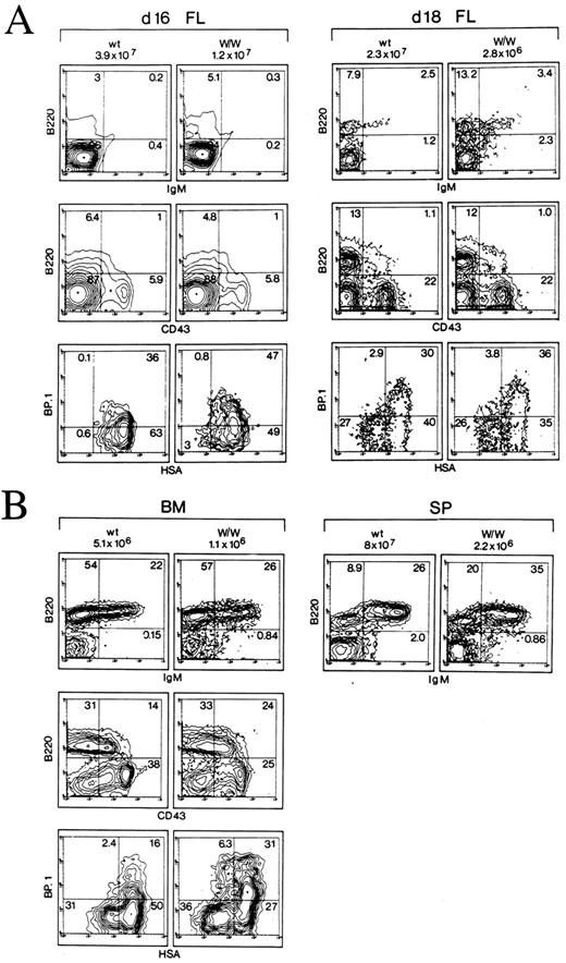
B lymphocyte development in W/W mice.Early stages of B-cell development were analyzed in FL (fetal day 16 and 18, Fig 1A), bone marrow and spleen (day 5 after birth, Fig 1B) comparing wildtype and W/W mice. For the dissection of distinct stages of B-cell differentiation, cell surface expression of B220 (CD45R), CD43, BP-1, heat stable antigen (HSA), and IgM was analyzed according to Hardy et al.37-39 The majority of precursors in the A fraction (B220+CD43+BP-1− HSA−) are not apparently B-cell precursors: these cells do not express c-kit, although both HSC and more differentiated precursors in the B and C fractions express c-kit.22,40 Proliferation of B220+CD43+ early B-cell precursors in fractions B and C can be stimulated by c-kit ligand in vitro. The expression of cytoplasmic μ-chain in early B-cell precursors inhibits c-kit expression and drives their differentiation into B220+CD43−IgM− late precursors (fraction D).22,41 42
Phenotypic analysis of B-cell compartments in FL and day 5 bone marrow and spleen from wildtype and W/W mice. (A) Analysis of B lineage subpopulations present in day 16 and day 18 fetal liver (FL) of wildtype and W/W mutant mice. Total cell numbers are indicated on top of each column. B lineage cells were analyzed by two-color flow cytometry for expression of B220 versus IgM (top row) and by four-color flow cytometry for expression of B220 versus CD43 versus BP-1 versus HSA. Data are presented as expression of B220 versus CD43 of the whole population of nucleated cells (middle row) and BP-1 versus HSA gated on B220+CD43+ cells (bottom row). Numbers given in the individual quadrants indicate percentages. (B) Analysis of B lineage subpopulations present in bone marrow (BM) and spleen (SP) of day 5 wildtype and W/W mutant mice. The data are presented in the same way as for (A).
Phenotypic analysis of B-cell compartments in FL and day 5 bone marrow and spleen from wildtype and W/W mice. (A) Analysis of B lineage subpopulations present in day 16 and day 18 fetal liver (FL) of wildtype and W/W mutant mice. Total cell numbers are indicated on top of each column. B lineage cells were analyzed by two-color flow cytometry for expression of B220 versus IgM (top row) and by four-color flow cytometry for expression of B220 versus CD43 versus BP-1 versus HSA. Data are presented as expression of B220 versus CD43 of the whole population of nucleated cells (middle row) and BP-1 versus HSA gated on B220+CD43+ cells (bottom row). Numbers given in the individual quadrants indicate percentages. (B) Analysis of B lineage subpopulations present in bone marrow (BM) and spleen (SP) of day 5 wildtype and W/W mutant mice. The data are presented in the same way as for (A).
The comparison of fetal livers, bone marrows and spleens from W/W and wildtype mice showed, as expected, severely reduced total cellularities in all examined organs (Fig 1A and B). However, flow cytometric analysis showed that all stages of B-cell development were phenotypically indistinguishable between W/W and wildtype cells: The absence of c-kit expression does not affect the ratios of B220+CD43+ early precursors versus B220+CD43− late precursors (Figs 1A and B, second row, upper right quadrants v upper left quadrants). The B220+ CD43+ pro B cell fraction was further dissected based on the expression pattern of HSA and BP-1. The ratios of fraction B (HSA+BP-1−) versus fraction C (HSA+BP-1+) (Figs 1A and B, third row, lower right quadrants v upper right quadrants) were also indistinguishable between wildtype and W/W hematopoietic tissues. Collectively, the ratios of cell numbers at different stages of B-cell development are very similar between c-kit null mutant (W/W ) and wildtype litter mates.
c-kit independent donor-type B-cell development in the recipient bone marrow.Since W/W mice, in analogy to fetal Sl/Sl mice,14 are expected to contain reduced numbers of HSC, the diminished cellularity in B-cell compartments in W/W mice may be caused at the HSC level. Moreover, in W/W mice, B-cell development occurs in the absence of competition from c-kit–expressing HSC and pro B cells. To examine B-cell development from c-kit–deficient HSC in a more competitive environment, day 13 W/W and wildtype FL cells were separately injected intravenously into RAG-2−/− mice.
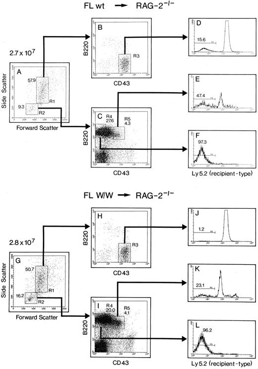
To determine whether c-kit–deficient (donor-type) B-cell precursors can efficiently compete against c-kit+ (host-type) B-cell precursors, the percentages of donor-type cells were measured among the CD43+B220+ subset (gate R5 in Fig 2I). At this stage, B-cell precursors express c-kit43 and are present in RAG-2−/− mice.26 Thus, W/W FL-derived precursors have to compete against host-type cells up to the CD43+B220+ stage in the absence of c-kit–mediated signals. In the recipient reconstituted with W/W FL cells, donor-type cells accounted for 23.1% of the early B precursors in the bone marrow (Fig 2K), indicating that early B precursors from c-kit–deficient mice are indeed capable of competing with a larger number of wildtype precursors. In contrast to the observed B-cell development, the reconstitution of nonlymphoid precursors (gate R3 in Fig 2H) by W/W FL cells was poor and accounted for only 1.2% of the CD43highB220− population (Fig 2J). In contrast, injected wildtype FL cells gave rise to 15.6% of the CD43highB220− population (Fig 2D). These observations are in agreement with previous studies demonstrating that W/WV bone marrow cells can neither make sizable spleen colonies nor reconstitute hematopoiesis in lethally irradiated recipients (for review see Russell15 ).
Analysis of the contribution of transferred wildtype or W/W FL cells to B-cell and myeloid development in the bone marrow of reconstituted RAG-2−/−. A total of 8 × 10⁵ FL cells from day 13 wildtype or W/W mice were injected intravenously into 4Gy irradiated RAG-2−/− mice, and reconstituted mice were analyzed 3 weeks posttransfer. Bone marrow cells from Ly5.2+RAG-2−/− mice reconstituted with Ly5.1+ wildtype FL cells (A through F ) or with Ly5.1+W/W FL cells (G through L) were analyzed by three-color flow cytometry for the expression of Ly5.2, CD43, and B220. Total cell counts are shown on top of (A) and (G). Bone marrow cells are gated, according to forward/side scatter analysis, into nonlymphoid (R1) and lymphoid (R2) gates. Cells in the R1 and R2 gates are further analyzed for the expression of B220 versus CD43 (B, C, H, and I). Percentage of donor-type cells (Ly5.2−) are determined among the nonlymphoid (D and J), B220+CD43+ early B-cell precursor (E and K) and B220+CD43− late B-cell precursor (F and L) populations. Numbers given on the gates indicate percentages.
Analysis of the contribution of transferred wildtype or W/W FL cells to B-cell and myeloid development in the bone marrow of reconstituted RAG-2−/−. A total of 8 × 10⁵ FL cells from day 13 wildtype or W/W mice were injected intravenously into 4Gy irradiated RAG-2−/− mice, and reconstituted mice were analyzed 3 weeks posttransfer. Bone marrow cells from Ly5.2+RAG-2−/− mice reconstituted with Ly5.1+ wildtype FL cells (A through F ) or with Ly5.1+W/W FL cells (G through L) were analyzed by three-color flow cytometry for the expression of Ly5.2, CD43, and B220. Total cell counts are shown on top of (A) and (G). Bone marrow cells are gated, according to forward/side scatter analysis, into nonlymphoid (R1) and lymphoid (R2) gates. Cells in the R1 and R2 gates are further analyzed for the expression of B220 versus CD43 (B, C, H, and I). Percentage of donor-type cells (Ly5.2−) are determined among the nonlymphoid (D and J), B220+CD43+ early B-cell precursor (E and K) and B220+CD43− late B-cell precursor (F and L) populations. Numbers given on the gates indicate percentages.
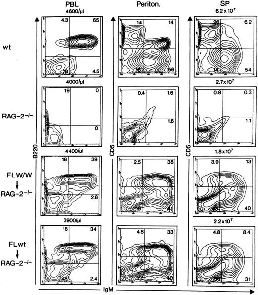
Reconstitution of peripheral B-cell compartments.In our preceding experiments,44 c-kithighB220− cells, which contain HSC,13,45,46 were purified from adult bone marrow and injected into RAG-2−/− mice. The injected cells were found to give rise to surface IgM+ cells in the recipient bone marrow in a synchronized manner at 13 days posttransfer.44 Thus, the initial reconstitution of peripheral B cells by injected HSC can be assessed around 20 days posttransfer. Next, we determined whether the peripheral B-cell compartments were populated equivalently from wildtype or W/W FL cells. To this end, peripheral blood, peritoneal cells, and spleen cells from reconstituted RAG-2−/− mice were analyzed by flow cytometry at 25 days posttransfer (Fig 3). Following transfer of W/W FL cells, both B-1 and B-2 B cell lineages47 were quantitatively reconstituted to levels obtained following injection of wildtype FL cells (compare the third and fourth rows in Fig 3). At 3 weeks posttransfer, the mean percentage (±1 standard error) of B lymphocytes among the nucleated peripheral blood cells in 19 reconstituted mice receiving W/W FL cells was 14.7% ± 11.7%, while that in 21 animals receiving wildtype FL cells was 18.6% ± 14.7%. Thus, there were no significant differences in the reconstitution following transfers of W/W and wildtype FL cells. However, among individual recipients, we observed variable levels of B-lymphocyte reconstitution.
Generation of mature B-1 and B-2 cells in vivo occurs independently of c-kit–mediated signals. A total of 8 × 10⁵ total FL cells from day 13 wildtype or W/W mice were injected intravenously into 4Gy irradiated RAG-2−/− mice. Twenty-five days after cell transfer, the reconstituted mice were analyzed for the presence of IgM+ cells in peripheral blood (PBL), peritoneal cavity (Periton) and spleen (SP). Total cell counts in various tissues are shown on top of each panel. Wildtype C57BL/6 (top row), unreconstituted RAG-2−/− mice (second row), RAG-2−/− mice reconstituted with W/W FL cells (third row) or those reconstituted with wildtype FL cells (fourth row) were analyzed by two-color flow cytometry for expression of B220 versus IgM (PBL), and CD5 versus IgM (Periton and SP). Note that CD5+IgM+ as well as CD5+IgM+ cells appeared in both types of reconstituted mice (third and fourth row).
Generation of mature B-1 and B-2 cells in vivo occurs independently of c-kit–mediated signals. A total of 8 × 10⁵ total FL cells from day 13 wildtype or W/W mice were injected intravenously into 4Gy irradiated RAG-2−/− mice. Twenty-five days after cell transfer, the reconstituted mice were analyzed for the presence of IgM+ cells in peripheral blood (PBL), peritoneal cavity (Periton) and spleen (SP). Total cell counts in various tissues are shown on top of each panel. Wildtype C57BL/6 (top row), unreconstituted RAG-2−/− mice (second row), RAG-2−/− mice reconstituted with W/W FL cells (third row) or those reconstituted with wildtype FL cells (fourth row) were analyzed by two-color flow cytometry for expression of B220 versus IgM (PBL), and CD5 versus IgM (Periton and SP). Note that CD5+IgM+ as well as CD5+IgM+ cells appeared in both types of reconstituted mice (third and fourth row).
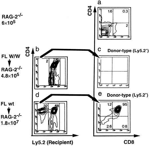
Failure of W/W FL cells to reconstitute donor-type thymocytes in RAG-2−/− recipients.In previous experiments, intrathymically expressed c-kit ligand was shown to drive the expansion of c-kit+ intrathymic precursors.48 To analyze whether donor-type thymocytes can be generated from c-kit–deficient HSC, thymopoiesis was analyzed in RAG-2−/− recipients injected with either wildtype or W/W FL (Fig 4). RAG-2−/− recipients were analyzed at 3 weeks posttransfer.49 Transferred wildtype FL cells yielded 5.5 × 10⁶ donor-type (Ly 5.2−) thymocytes (Fig 4d), which contained >90% CD4+CD8+ double-positive thymocytes, and at this time point, very few mature single-positive thymocytes (Fig 4e). In contrast, no donor-type (Ly5.2−) thymocytes were detectable in the recipient mice that had received W/W FL cells (Fig 4b and c). This finding is in marked contrast with the observed normal B-cell development following transfer of W/W FL.
W/W FL cells are not capable of reconstituting thymocytes in RAG-2−/− recipient mice. Thymocytes from Ly5.2+RAG-2−/− mice (first row), Ly5.2+RAG-2−/− mice reconstituted with day 13 Ly5.1+W/W FL cells (second row) and Ly5.2+RAG-2−/− mice reconstituted with day 13 Ly5.1+ wildtype FL cells (third row) were analyzed by three-color flow cytometry for the presence of Ly5.2, CD4, and CD8. Total cell counts are shown at the left of each panel. Two parameters are shown at a time: Ly5.2 versus CD4 (left column) and CD4 versus CD8 (right column). The contour plots of CD4 versus C8 of the reconstituted mice (c and e) are gated on donor-derived (Ly5.2−) cells only. Numbers given in quadrants and gates indicate percentages.
W/W FL cells are not capable of reconstituting thymocytes in RAG-2−/− recipient mice. Thymocytes from Ly5.2+RAG-2−/− mice (first row), Ly5.2+RAG-2−/− mice reconstituted with day 13 Ly5.1+W/W FL cells (second row) and Ly5.2+RAG-2−/− mice reconstituted with day 13 Ly5.1+ wildtype FL cells (third row) were analyzed by three-color flow cytometry for the presence of Ly5.2, CD4, and CD8. Total cell counts are shown at the left of each panel. Two parameters are shown at a time: Ly5.2 versus CD4 (left column) and CD4 versus CD8 (right column). The contour plots of CD4 versus C8 of the reconstituted mice (c and e) are gated on donor-derived (Ly5.2−) cells only. Numbers given in quadrants and gates indicate percentages.
DISCUSSION
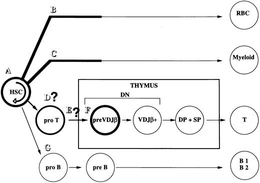
Interactions between c-kit and SCF are known to play important roles in the generation of various hematopoietic lineages. Based on the findings that (1) W/W-derived HSC fail to compete against wildtype HSC following in vivo cell transfers, and (2) most colony-forming units-spleen (CFU-S) are eliminated following administration of antagonistic anti–c-kit antibody, a critical role for SCF/c-kit in the proliferation of HSC had been demonstrated (Fig 5A).13,15,45,46 Also, very rapid and complete elimination of erythroid and myeloid precursors by anti–c-kit antibody indicates that the expansion of committed hematopoietic precursors in steady-state hematopoiesis is strongly dependent on c-kit50 (Fig 5B and C; for review see Galli et al8 and Russell15 ). In T-cell development, SCF–c-kit interactions were previously shown to drive early thymocyte proliferation at early intrathymic stages preceding T-cell receptor (TCR) V(D)Jβ chain rearrangements (Fig 5F ).48 In addition, it is possible that c-kit plays a role in the generation of thymic precursors (Fig 5D) and/or the thymus colonization (Fig 5E).
Hematopoietic pathways affected by interactions of c-kit and SCF. Differentiation pathways and cellular compartments strongly perturbed in the absence of c-kit are depicted in bold print. Fine print denotes pathways unaffected by lack of c-kit. c-kit plays a more important role in the self-renewal of HSC (A), the expansion of early erythroid (B), and myeloid (C) precursors. It is not clear whether the expansion of prethymic precursors (D) and the colonization of these cells at the thymus (E) require c-kit–mediated signals. c-kit plays little role in B-cell development of both B1 and B2 lineages (G).
Hematopoietic pathways affected by interactions of c-kit and SCF. Differentiation pathways and cellular compartments strongly perturbed in the absence of c-kit are depicted in bold print. Fine print denotes pathways unaffected by lack of c-kit. c-kit plays a more important role in the self-renewal of HSC (A), the expansion of early erythroid (B), and myeloid (C) precursors. It is not clear whether the expansion of prethymic precursors (D) and the colonization of these cells at the thymus (E) require c-kit–mediated signals. c-kit plays little role in B-cell development of both B1 and B2 lineages (G).
Here we show that B-cell development can occur normally in the absence of c-kit from the following two experiments: (1) Analysis of fetal and early postnatal W/W and wildtype littermates, and (2) reconstitution of immature and mature B lineage cells in RAG-2−/−c-kit+ recipients by W/W or wildtype FL cells.
The comparison of various stages of B-cell development in W/W and wildtype littermates showed severely reduced hematopoietic cell numbers, but no distortion of B lymphopoiesis. While this analysis shows that B cells can be generated from c-kit–deficient precursors, it does not exclude the possibility that c-kit− B-cell precursors are inferior compared with c-kit+ B-cell precursors in a competitive repopulation assay. To address this point, we compared B lymphocyte development following transfer of 8 × 10⁵ day 13 FL derived from W/W or wildtype mice into 4Gy irradiated RAG-2−/− mice. DHJH joints at the IgH locus are hardly detectable in day 13 fetal liver,51 and B220+ B-cell precursors do not appear until day 15/16 of gestation.39 Thus, day 13 fetal liver is expected to contain few, if any, committed B-cell precursors. Since the content of HSC in day 13 fetal liver from SCF null-mutant mice is very similar to that from wildtype mice,14W/W and wild-type day 13 fetal livers are expected to contain similar percentages of HSC. Upon transfer of W/W or wildtype day 13 FL cells, we could thus follow the initial reconstitution of B-cell development from similar numbers of injected HSC. In addition, the transfer experiment was expected to magnify a defect in the expansion of W/W lymphohematopoietic precursors, because transferred c-kit− precursors need to compete with larger numbers of endogenous c-kit+ precursors.
The data show that comparable reconstitution of both B-1 and B-2 lineages was observed following transfer of W/W and wildtype day 13 FL. In both groups of recipients, similar numbers of donor-type early B220+CD43+ precursors existed 3 weeks posttransfer. Hardy et al39 showed that wildtype B220+CD43+ proB cells injected into severe combined immunodeficient (SCID) mice underwent B-cell development transiently and, at 3 weeks posttransfer, virtually all donor-type cells had already left the recipient bone marrow for the periphery.39 Hence, the presence of donor-type CD43+B220+ early B precursors as late as 3 weeks posttransfer indicates that injected HSC, rather than committed B-cell precursors, were mostly responsible for B-cell development in both groups of recipients. Thus, injected c-kit−HSC may have colonized the recipient bone marrow and differentiate into B cells with similar efficiency as injected wildtype HSC (Fig 5G). This conclusion is in agreement with the previous observation showing that injected W/WV bone marrow cells generated the same numbers of spleen colonies, but with much reduced sizes in lethally-irradiated recipient mice, when compared with injected wildtype bone marrow cells.15
A previous study suggested that W/WV bone marrow cells reconstituted B220+ B-cell precursors in lethally irradiated wildtype littermates at a sevenfold reduced level compared with bone marrow cells from wildtype mice.52 By contrast, the same study showed that WX/WX day 16 FL cells gave rise to B-cell precursors at an only slightly reduced level compared with day 16 FL cells from wildtype mice, and the present study shows no difference in the reconstitution by day 13 W/W and wildtype FL cells. These different results (reconstitution by bone marrow v fetal liver) could be explained by the differences in the content of HSC; the content of HSC in adult W/WV bone marrow is much lower than that in wildtype bone marrow,52 while day 13 W/W and wildtype FL may contain HSC at comparable frequencies.14 Alternatively, since injected FL cells, but not adult bone marrow cells, can give rise to B1 cells,39 HSC of FL might be different from HSC of adult bone marrow with respect to the requirement of c-kit–mediated signals.
Since flk2 (flt3), another receptor tyrosine kinase, exhibits strong similarities in both sequence and expression pattern to c-kit, the two molecules may play similar and overlapping roles in lymphohematopoiesis.53,54 While flk2 null mutant (flk2−/−) and W/WV mice showed a slight decrease in the overall cellularity of bone marrow, W/WVFlk2−/− double-mutant mice exhibited a very significant reduction.18 This finding demonstrates functional redundancy between c-kit and flk2 in hematopoiesis. Given that W/W mice exhibit much more severe anemia than flk2−/− mice, c-kit plays a more important role than flk2 in the self-renewal of HSC (Fig 5A) and the expansion of early myeloid precursors (Fig 5B and C). On the other hand, Flk2−/− mice showed specific deficiencies in B-cell development, which were amplified following transfer of Flk2−/− bone marrow cells into wildtype mice.18 Taking normal B-cell development from transferred W/W HSC into account, flk2 may play a more important role than c-kit in B-cell development (Fig 5G).
ACKNOWLEDGMENT
We thank Drs F. Alt (The Children's Hospital, Boston, MA) and Clarence Reeder (NCI, Frederick, MD) for generously providing RAG-2−/− and B6Ly5.2 congenic mice, respectively, Dr S.-I. Nishikawa (Kyoto University, Kyoto, Japan) for generously providing anti–c-kit MoAb ACK-4. E. Wagner and the Animal Facility staff (Basel) are acknowledged for their care in the maintenance of mouse colonies, M. Dessing and S. Meyer for cell sorter expertise, and H. Stahlberger for artwork. We thank Drs Fritz Melchers, Klaus Karjalainen, Kerry Campbell, Toru Nakano, and Koichi Ikuta for discussions and critical reading of the manuscript.
The Basel Institute for Immunology was founded and is supported by F. Hoffmann La Roche, Basel, Switzerland. The Bayer-chair department is sponsored by Bayer Yakuhin, Japan.
Address reprint requests to Shunichi Takeda, MD, PhD, Bayer-Chair Department of Molecular Immunology, Medical School, Kyoto University, Konoe Yoshida Sakyo-ku, Kyoto 606 Japan.

This feature is available to Subscribers Only
Sign In or Create an Account Close Modal