Abstract
We have previously reported the development of in vivo functional assays for primitive human hematopoietic cells based on their ability to repopulate the bone marrow (BM) of severe combined immunodeficient (SCID) and nonobese diabetic/SCID (NOD/SCID) mice following intravenous transplantation. Accumulated data from gene marking and cell purification experiments indicate that the engrafting cells (defined as SCID-repopulating cells or SRC) are biologically distinct from and more primitive than most cells that can be assayed in vitro. Here we demonstrate through limiting dilution analysis that the NOD/SCID xenotransplant model provides a quantitative assay for SRC. Using this assay, the frequency of SRC in cord blood (CB) was found to be 1 in 9.3 × 10⁵ cells. This was significantly higher than the frequency of 1 SRC in 3.0 × 10⁶ adult BM cells or 1 in 6.0 × 10⁶ mobilized peripheral blood (PB) cells from normal donors. Mice transplanted with limiting numbers of SRC were engrafted with both lymphoid and multilineage myeloid human cells. This functional assay is currently the only available method for quantitative analysis of human hematopoietic cells with repopulating capacity. Both CB and mobilized PB are increasingly being used as alternative sources of hematopoietic stem cells in allogeneic transplantation. Thus, the findings reported here will have important clinical as well as biologic implications.
A QUANTITATIVE ASSAY for human stem cells is essential for the study of the biologic properties of these cells. Such an assay would also allow a more rational approach to the development of clinical treatments involving transplantation, ex vivo stem cell expansion, and gene therapy. To date, quantitative analysis of primitive hematopoietic cells has been limited to in vitro studies using colony assays or long-term cultures (LTC). However, colony assays detect only committed and multipotent progenitors (colony-forming cells, CFC). LTC assays detect more primitive cells (LTC-initiating cells, LTC-IC) capable of giving rise to CFC after at least 5 weeks of culture on competent feeder layers.1,2 However, LTC-IC are functionally heterogeneous: characteristics associated with very immature cells such as quiescence and the capacity to generate progenitors in extended culture are found in only a small subpopulation of LTC-IC.3 In addition, the relationship between LTC-IC and in vivo repopulating human stem cells is not clear.
We have previously reported the development of an in vivo functional assay for primitive human hematopoietic cells based on their ability to repopulate the bone marrow (BM) of severe combined immunodeficient (SCID) and nonobese diabetic/SCID (NOD/SCID) mice after intravenous injection.4-6 Transplantation of human BM or umbilical cord blood (CB) results in the engraftment of primitive cells that proliferate and differentiate to multiple lineages in the murine BM.4,5 We have operationally defined the engrafting human cell as a SCID-repopulating cell (SRC). Kinetic experiments showed that only 0.1% of injected CFC and LTC-IC are detectable in the murine BM 2 days posttransplant, and that there is a large expansion of these cells as well as of primitive CD34+ and CD34+Thy-1+ cells over the next 4 weeks, implying their production from a more primitive cell.7 Recent experiments using retroviral gene transfer showed that while CFC and LTC-IC are easily transduced, these gene-marked cells do not contribute significantly to the repopulation of engrafted mice; a corollary to this finding is that the efficiency of gene transfer into SRC is low.8 Finally, cell purification experiments showed that SRC are exclusively CD34+CD38−,8 9 in contrast to CFC and LTC-IC, which are also found in the CD34+CD38+ fraction. Together, these data indicate that SRC are biologically distinct from and more primitive than most CFC and LTC-IC.
In this report we show that the NOD/SCID xenotransplant model, or SRC assay, provides a quantitative in vivo assay for primitive human hematopoietic cells. We have used this assay to measure and compare the frequency of SRC in human CB, normal adult BM, and mobilized peripheral blood (PB) from normal donors. Our results show that CB is enriched for these primitive hematopoietic cells compared with BM or mobilized PB, an observation that has important implications for clinical transplantation and stem cell expansion strategies.
MATERIALS AND METHODS
Donor samples.CB samples were obtained from umbilical and placental tissues scheduled for discard. BM and granulocyte colony-stimulating factor (G-CSF )–mobilized PB samples from normal adult donors were obtained as leftover cells from harvests for allogeneic transplantation according to procedures approved by the Human Experimentation Committee at the Princess Margaret Hospital, Toronto, Ontario, Canada. CB and BM samples were diluted 1:2 or 1:3 in Iscove's modified Dulbecco's medium (IMDM; GIBCO-BRL, Burlington, Ontario, Canada) containing 10% fetal calf serum (FCS; Cansera, Rexdale, Ontario, Canada) and enriched for mononuclear cells by centrifugation on Ficoll-Paque (Pharmacia, Baie d'Urfé, Quebec, Canada). Mobilized PB was collected by leukapheresis on days 4 and 5 from normal individuals treated with G-CSF 5 to 10 μg/kg subcutaneously on days 1 through 4.
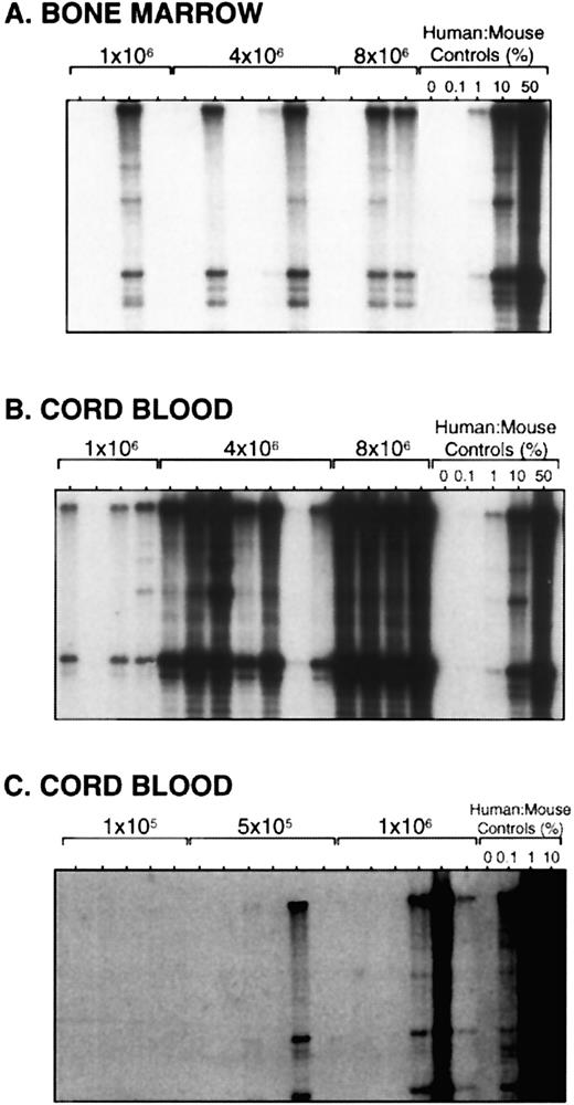
Limiting dilution assays of adult BM and umbilical CB. (A) Southern blot analysis of human cell engraftment in the BM of mice transplanted with 1 to 8 × 10⁶ BM cells. Mice were treated with alternate-day injections of human cytokines and killed 6 weeks posttransplant. Human:mouse DNA controls are given as percent human DNA. (B and C) Southern blot analysis of mice transplanted with 1 × 10⁵ to 8 × 10⁶ CB cells and killed after 6 weeks.
Limiting dilution assays of adult BM and umbilical CB. (A) Southern blot analysis of human cell engraftment in the BM of mice transplanted with 1 to 8 × 10⁶ BM cells. Mice were treated with alternate-day injections of human cytokines and killed 6 weeks posttransplant. Human:mouse DNA controls are given as percent human DNA. (B and C) Southern blot analysis of mice transplanted with 1 × 10⁵ to 8 × 10⁶ CB cells and killed after 6 weeks.
Transplantation of human cells into NOD/SCID mice.CB, BM, or mobilized PB cells were transplanted by tail-vein injection into sublethally irradiated (375 to 400 cGy using a 137Cs γ-irradiator) 8-week-old NOD/LtSz-scid/scid (NOD/SCID) mice according to our standard protocol.4-6 Mice transplanted with BM or PB received alternate-day intraperitoneal injections of human cytokines (huSCF 10 μg, huIL-3 and huGM-CSF 6 μg each; all from Amgen, Thousand Oaks, CA). NOD/SCID mice were bred and maintained in the defined flora animal colony at the Ontario Cancer Institute, Toronto, and the animal experiments were approved by the Animal Care Committee of the Hospital for Sick Children and the Ontario Cancer Institute. Mice were killed 4 to 6 weeks after transplantation, and the BM from 2 femurs, 2 tibiae, and 2 iliac crests was flushed into IMDM plus 10% FCS.
Limiting Dilution Analysis of Human CB, Adult BM, and Mobilized PB
| Cell Source . | Cell Dose . | No. of Negative Mice . | No. of Transplanted Mice . | Percentage of Mice Negative . |
|---|---|---|---|---|
| CB (n = 7) | 1 × 10⁵ | 11 | 11 | 100 |
| 5 × 10⁵ | 8 | 12 | 67 | |
| 1 × 10⁶ | 10 | 37 | 27 | |
| 4 × 10⁶ | 2 | 24 | 8 | |
| 8 × 10⁶ | 0 | 7 | 0 | |
| BM (n = 5) | 5 × 10⁵ | 6 | 9 | 67 |
| 1 × 10⁶ | 18 | 25 | 72 | |
| 2 × 10⁶ | 6 | 9 | 67 | |
| 4 × 10⁶ | 4 | 19 | 21 | |
| 8 × 10⁶ | 1 | 5 | 20 | |
| Mobilized PB (n = 7) | 2.5 × 10⁶ | 3 | 8 | 38 |
| 5 × 10⁶ | 17 | 32 | 53 | |
| 10 × 106 | 2 | 14 | 14 | |
| 20 × 106 | 2 | 16 | 13 |
NOD/SCID mice were transplanted with serial doses of mononuclear cells from human CB (n mice = 91), BM (n = 67), or G-CSF–mobilized PB (n = 70) and the murine BM was analyzed after 4 to 6 weeks by Southern blot to determine the level of human cell engraftment. Mice were scored as negative if no human cells were detectable. The number of experiments from which the pooled data are taken is indicated for each cell source in parentheses.
Analysis of human cell engraftment.High-molecular-weight DNA was isolated from the BM of transplanted mice by phenol/chloroform extraction using standard protocols. The proportion of human cells in the murine BM was quantified by Southern blot analysis using a human chromosome 17-specific α-satellite probe (p17H8)10 as previously described (limit of detection approximately 0.05% human cells).4,5 This technique is more reliable than flow cytometry in detecting very low levels of human cell engraftment. To determine whether human progenitors were present in the BM of engrafted mice, BM cells from transplanted mice were plated in methylcellulose cultures as previously described5,11 under conditions that are selective for the growth of human progenitors and that do not support coexisting mouse progenitors. Colonies were scored at 14 days. The presence of human lymphoid cells in the BM of some mice was assessed by flow cytometry on a FACScan analyzer (Becton Dickinson, San Jose, CA) using human-specific monoclonal antibodies directed against the pan-B-cell marker CD19 (B4; Coulter Immunology, Hialeah, FL) in combination with anti-CD45 (HLe-1) or anti-CD38 (Leu-17) antibodies (both from Becton Dickinson).5 More detailed lineage analysis by flow cytometry was carried out in some mice as described elsewhere.9
Statistical analysis.For purposes of our limiting dilution assays, a transplanted mouse was scored as positive (engrafted) if any human cells were detectable in the murine BM by Southern blot analysis. For each cell source, the data from several limiting dilution experiments were pooled and analyzed by applying Poisson statistics to the single-hit model.12 The major assumptions of this model are that transplantation of only 1 SRC is required to generate a positive response (an engrafted mouse), and that every transplanted SRC will generate a positive response.13 The frequency of SRC in each cell source was calculated using the maximum likelihood estimator.12,13 χ2 provides a measure of the legitimacy of using pooled data and of the validity of applying the single-hit model.12 13
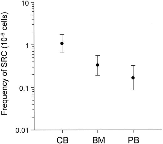
Comparison of the frequency of SRC in CB, BM, and mobilized PB. The frequency of SRC was calculated using Poisson statistics. Bars indicate the 95% confidence limits.
Comparison of the frequency of SRC in CB, BM, and mobilized PB. The frequency of SRC was calculated using Poisson statistics. Bars indicate the 95% confidence limits.
Multilineage Engraftment in the BM of Mice Transplanted With SRC in Limiting Doses
| Mouse . | CD19+ cells (%*) . | BFU-E† . | CFC† . |
|---|---|---|---|
| C15.6 | 0.2 | 1485 | 14,108 |
| C16.2 | 0.1 | 312 | 3,978 |
| C16.7 | 2.4 | 855 | 5,580 |
| C16.8 | 0.2 | 54 | 216 |
| C16.9 | 1.8 | 0 | 1,953 |
| C16.11 | 0.7 | 68 | 1,080 |
| C16.12 | 0.2 | 462 | 1,596 |
| C18.1 | 26.0 | TNTC | TNTC |
| C18.3 | 0.2 | 0 | 2,184 |
| C18.5 | 0.8 | 81 | 324 |
| C18.9 | 7.9 | 23 | 1,685 |
| C19.11 | 2.2 | 2925 | 8,700 |
NOD/SCID mice were transplanted with 1 × 10⁶ mononuclear CB cells and the murine BM was analyzed after 6 weeks by flow cytometry to assess the presence of human CD19+ B-lymphoid cells, and by plating in methylcellulose cultures to assess the presence of human myeloerythroid progenitors.
Abbreviations: BFU-E, burst-forming unit-erythroid; CFC includes granulocyte, macrophage, and granulocyte-macrophage colony-forming cells; TNTC, too numerous to count.
Percentage of total leukocytes in the murine BM.
Number per 6 bones (2 femurs, 2 tibiae, and 2 iliac crests).
RESULTS
Limiting dilution assays of CB, adult BM, and mobilized PB.Groups of sublethally irradiated mice were transplanted with replicate doses of mononuclear cells from CB or normal adult BM over a range of doses which resulted in nonengraftment in a fraction of the mice. Six weeks posttransplant, the murine BM was analyzed by Southern blot, and mice were scored as positive or negative for human cell engraftment. The Southern blot analysis of three representative experiments is shown in Fig 1. Transplantation of 1 × 10⁶ to 8 × 10⁶ BM cells resulted in low levels of engraftment, with positive and negative mice at each dose (Fig 1A). In contrast, transplantation of CB cells over the same dose range resulted in much higher levels of engraftment overall, and all of the mice transplanted with 4 × 10⁶ or 8 × 10⁶ cells were engrafted (Fig 1B). Transplantation of lower numbers of CB cells gave similar results as those seen with the higher doses of BM (Fig 1C).
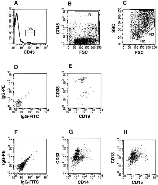
Multilineage engraftment in the BM of a mouse transplanted with CB cells at limiting dilution. BM from a mouse transplanted with 1 × 10⁶ CB cells and killed after 6 weeks was stained with human-specific monoclonal antibodies and analyzed by flow cytometry. (A) Histogram of pan-leukocyte marker CD45 expression demonstrating 3% human cell engraftment in this mouse. (B) Cells for analysis were acquired in a live gate (R1) based on CD45 positivity and medium to high forward scatter. (C) Cells were further gated based on forward and side scatter properties into lymphoid/blast (R2) and myeloid (R3) windows. (D) Isotype control for nonspecific IgG staining of cells in R2. (E) Expression of CD38 and CD19, a pan-B-cell marker, on cells in R2. (F ) Isotype control for cells in R3. (G) Expression of myeloid marker CD33 and monocytic marker CD14 on cells in R3. (H) Expression of myeloid marker CD13 and mature granulocyte marker CD15 on cells in R3.
Multilineage engraftment in the BM of a mouse transplanted with CB cells at limiting dilution. BM from a mouse transplanted with 1 × 10⁶ CB cells and killed after 6 weeks was stained with human-specific monoclonal antibodies and analyzed by flow cytometry. (A) Histogram of pan-leukocyte marker CD45 expression demonstrating 3% human cell engraftment in this mouse. (B) Cells for analysis were acquired in a live gate (R1) based on CD45 positivity and medium to high forward scatter. (C) Cells were further gated based on forward and side scatter properties into lymphoid/blast (R2) and myeloid (R3) windows. (D) Isotype control for nonspecific IgG staining of cells in R2. (E) Expression of CD38 and CD19, a pan-B-cell marker, on cells in R2. (F ) Isotype control for cells in R3. (G) Expression of myeloid marker CD33 and monocytic marker CD14 on cells in R3. (H) Expression of myeloid marker CD13 and mature granulocyte marker CD15 on cells in R3.
To determine whether our model could also be used to assay primitive hematopoietic cells in G-CSF–mobilized PB from normal individuals, we transplanted mononuclear cells from leukapheresis products into NOD/SCID mice using our standard protocol. Flow cytometric analysis and progenitor assays of the BM of engrafted mice demonstrated B-lymphoid and multilineage myeloid engraftment similar to results we obtained with transplantation of human BM or CB (data not shown). Transplantation of mobilized PB in limiting dilution assays resulted in low levels of engraftment in a proportion of transplanted mice after 4 to 6 weeks, as was observed with BM and CB. The data from the limiting dilution assays of CB (n experiments = 7), adult BM (n = 5), and mobilized PB (n = 7) are shown in Table 1. Of the total number of mice in which human cells were detectable by Southern blot, 92% were engrafted at a level of ≥0.1% and 95% had human progenitors in the murine BM.
Frequency of SRC in various hematopoietic tissues.Data from the limiting dilution assays of each cell source were pooled for statistical analysis, according to the method described by Porter and Berry.12 The frequency of SRC was calculated using the maximum likelihood estimator.12,13 The value of χ2 in all cases was not statistically significant (P > .05), demonstrating internal consistency in our assays and allowing pooling of the data. The frequency of SRC in CB was 1 in 9.3 × 10⁵ mononuclear cells (95% confidence limits [CL] 1 in 5.8 × 10⁵ to 1 in 1.5 × 10⁶). This was significantly higher than the calculated frequency of 1 SRC in 3.0 × 10⁶ BM cells (95% CL 1 in 1.8 × 10⁶ to 1 in 5.2 × 10⁶) or 1 in 6.0 × 10⁶ mobilized PB cells (95% CL 1 in 3.1 × 10⁶ to 1 in 1.2 × 10⁷) (Fig 2). As confirmation of the validity of applying the single-hit Poisson model to our assay, the frequency of SRC was also determined by minimum χ2 estimation.13 Calculated frequencies using this second method were similar, and χ2 was again not significant in all cases (P > .05, data not shown).
To assess the differentiative capacity of SRC transplanted in limiting doses, we analyzed additional groups of mice injected with 1 × 10⁶ mononuclear CB cells for evidence of both lymphoid and myeloid differentiation. At this dose, 12 of 30 mice had engraftment detectable by flow cytometry, and by Poisson statistics one half to three quarters of these likely received only 1 SRC. All of the engrafted mice had human CD19+ B-lymphoid cells as well as multiple lineages of human myeloid clonogenic progenitors in their BM (Table 2). Detailed flow cytometric analysis in a number of mice (n = 3) provided independent confirmation that multiple myeloid lineages as well as B cells were present in the BM of engrafted mice (Fig 3).
DISCUSSION
We have shown through limiting dilution analysis that the NOD/SCID xenotransplant model provides an in vivo quantitative assay for primitive human hematopoietic cells. Using this assay we measured the frequency of SRC in various hematopoietic tissues, and showed that CB is enriched for these primitive repopulating cells compared with adult BM or mobilized PB. The higher frequency of SRC in CB compared with adult tissues suggests that CB may be a better source of stem cells for ex vivo manipulations such as retroviral infection or stem cell expansion.
For this analysis we assumed a single-hit Poisson model for the generation of a positive response (an engrafted mouse). The underlying postulate of this statistical model is that only one cell of only one cell type is necessary for a positive response.13 The applicability of the single-hit model to our NOD/SCID assay, as validated by the χ2 test, further supports the hypothesis that the human graft is initiated from a single cell type (the SRC) capable of multilineage differentiation, rather than from many lineage-restricted cells. Importantly, the ability to quantitate SRC allowed us to assess their differentiative capacity more directly by analyzing the human hematopoietic lineages present in the BM of mice transplanted with limiting numbers of SRC. The presence of both lymphoid cells and multiple lineages of myeloid progenitors in all of the engrafted mice, most of which likely received a single SRC, provides strong evidence that this assay detects a primitive cell in the human hematopoietic hierarchy.
Previous studies have reported that the frequency of LTC-IC in normal human BM is approximately 1 in 1 × 10⁴ to 1 in 3 × 10⁴ cells, and that the proportion of LTC-IC in mobilized PB is similar to or higher than that in BM and CB.2,14 The higher frequency of LTC-IC compared with SRC reflects the fact that in vitro LTC assays detect a functionally heterogeneous cell population that includes more mature progenitors in addition to primitive cells. Mobilized PB is enriched for more mature hematopoietic precursors15; however, we have evidence that these cells do not read out in the SRC assay.8 Because the SRC assay quantitatively detects a very primitive hematopoietic cell, it can provide a more clinically relevant measure of the degree of stem cell enrichment achieved by various purification strategies.9
The NOD/SCID xenotransplant model will be an important tool in studies to define the proliferative, differentiative, and self-renewal capacities of primitive human hematopoietic cells. As well, this assay now provides a means to quantify changes in stem cell function in response to a variety of conditions in vivo and in vitro, and to assess how ex vivo manipulations such as retroviral infection protocols or expansion culture techniques affect the maintenance of stem cell activity as measured by repopulating capacity (Bhatia et al, submitted for publication). Both CB and mobilized PB from normal donors are increasingly being used as alternative sources of hematopoietic stem cells in allogeneic transplantation.16-18 Thus, the findings reported here will have important clinical as well as biologic implications.
ACKNOWLEDGMENT
The authors thank N. Jamal and H. Messner for providing bone marrow and mobilized peripheral blood samples, L. McWhirter and S. Lye for providing cord blood specimens, I. McNiece (Amgen, Thousand Oaks, CA) for providing cytokines, and members of the lab for critically reviewing the manuscript.
Supported by grants from the Medical Research Council (MRC) of Canada, the National Cancer Institute of Canada (NCIC) with funds from the Canadian Cancer Society, and AMGEN, with postdoctoral fellowships from the Leukemia Research Fund of Canada and the MRC (J.C.Y.W.), a Research Scientist award from the NCIC (J.E.D.), and an MRC Scientist Award (J.E.D.)
Address reprint requests to John E. Dick, PhD, Department of Genetics, Research Institute, Hospital for Sick Children, 555 University Ave, Toronto, Ontario, Canada M5G 1X8.


This feature is available to Subscribers Only
Sign In or Create an Account Close Modal