Abstract
Quantification of apoptotic cell death in vivo has become an important area of investigation in patients with acute lymphoblastic leukemia (ALL). We have devised a noninvasive analytical method to estimate the percentage of apoptotic lymphoblasts in doxorubicin-treated Jurkat T-cell ALL cultures, using proton nuclear magnetic resonance spectroscopy (1H NMR). We have found that the ratio of the methylene (CH2 ) resonance (at 1.3 ppm) to the methyl (CH3 ) resonance (at 0.9 ppm) signal intensity, as observed by 1H NMR, is directly proportional to the percentage of apoptotic lymphoblasts in vitro. The correlation between the CH2/CH3 signal intensity ratio and the percentage of apoptotic lymphoblasts was optimal 24 to 28 hours after doxorubicin treatment (r2 = .947, N = 27 samples). There was also a direct temporal relationship between an increase in the CH2/CH3 signal intensity ratio and the onset of apoptosis as detected by nuclear morphologic analysis, fluorescein-annexin V flow cytometry, and DNA gel electrophoresis. Thin-layer chromatography confirmed that a dynamic and/or compositional change of the plasma membrane, rather than increases in lipase activity or fatty acid production, appears to account for the increase in the CH2/CH3 signal intensity ratio during apoptosis. 1H NMR may have clinical utility for the early noninvasive assessment of chemotherapeutic efficacy in patients with ALL.
A GROWING NUMBER of clinical investigators have engaged in efforts to quantify apoptosis.1-10 Their ultimate goal is to show the clinical utility of quantifying apoptosis in a number of disease states, including acute lymphoblastic leukemia (ALL). Estimates of the degree of cytoreduction during induction therapy using bone marrow aspirates have provided prognostic information in children with ALL.11,12 Other investigators have also shown a direct relationship between the degree of apoptotic cell death and subsequent tumor growth delay in murine models of leukemia.13 14 However, all methods currently used to quantify apoptosis are invasive and require aspirated or biopsied material.
To date, proton nuclear magnetic resonance spectroscopy (1H NMR) has been the only noninvasive method to quantify the degree of cytoreduction in leukemic patients.15-17 These measurements rely on the ratio of water to lipid proton resonance signal intensities and thus reflect the relative percentages of cellular and fatty bone marrow (percent cellularity) within a defined three-dimensional volume (voxel).18 However, the percentage of cellularity of leukemic marrow does not change significantly until several weeks after the start of therapy and does not directly correlate to apoptotic cell death, which begins to occur within the first several days of treatment.5 6
More recently, 1H NMR spectroscopy has been applied to the study of apoptotic cell death in vitro.19 We have found that the onset of apoptosis is accompanied by a greater than twofold increase in the signal intensity of the membrane lipid methylene (CH2 ) resonance (at 1.3 ppm) as observed by 1H NMR. We now have devised an analytical method based on the magnitude of the CH2 resonance signal intensity to quantify in vitro the fraction of apoptotic cells in Jurkat T-cell cultures treated with the chemotherapeutic agent doxorubicin. We also show that increases in the CH2 resonance signal intensity parallel the surface expression of phosphatidylserine (PS) an early marker of apoptosis,20-23 as determined by fluorescein-annexin V flow cytometry.
Our results show that 1H NMR can detect apoptosis and also quantify apoptotic cell death in vitro. Quantification of apoptosis in vivo by 1H NMR may prove clinically useful in circumstances in which noninvasive methods of early determination of chemotherapeutic efficacy are required.
MATERIALS AND METHODS
Cell culture technique.Jurkat T-cell ALL cultures were grown in RPMI 1640 medium (Applied Scientific, South San Francisco, CA) supplemented with 10% (vol/vol) fetal calf serum and 290 μg/mL L-glutamine, 100 U/mL penicillin, and 100 μg/mL streptomycin. Cells were maintained in the logarithmic growth phase at a concentration of 1 to 5 × 10⁵ cells/mL at 37°C in a 5% CO2 air incubator under sterile conditions.24
DNA fragmentation assay.DNA ladder formation was detected by an assay as previously described.19 Briefly, 5 × 10⁶ cells were harvested and washed with phosphate-buffered saline (PBS). Total DNA was isolated by incubating washed cells in 400 μL lysis buffer containing 50 mmol/L Tris, pH 7.5, 10 mmol/L EDTA, 0.5% sodium dodecyl sulfate (SDS), 100 μg/mL RNase, and 4 μg/mL proteinase K for at least 2 hours at 60°C. After the addition of 75 μL of 8 mol/L KAc, the DNA was extracted with phenol/chloroform, precipitated with ethanol, and suspended in TE (10 mmol/L Tris-HCl, pH 7.5, 1 mmol/L EDTA) with 100 μg/mL of heat-treated RNase. The DNA from 1 × 10⁶ cells was then analyzed in a composite 1% agarose/0.5% Nusieve gel. The fraction of total DNA that underwent DNA fragmentation (DNA ladder formation) was calculated from digitized images using a BioRad Gel Doc System with Molecular Analyst Software (Bio-Rad Laboratories, Richmond, CA).
Cell morphologic analysis.Apoptotic nuclei were identified after staining with Hoechst #33342 (10 μmol/L final concentration) as described by Dive et al.25 Cells were observed with a fluorescence microscope with an excitation wavelength of 365 nm. Cells were considered nonapoptotic when nuclei were morphologically normal with homogeneously distributed blue-stained chromatin. Apoptotic cells showed blue peripherally clumped or fragmented chromatin.
Cell viability was assessed in control and treated cultures before and after 1H NMR spectral analysis using fluorescent microscopy of doubly stained cell samples (with the Hoechst and propidium iodide stains), as previously described.19 There were no changes in the fractions of viable, apoptotic, late stage apoptotic (ie, with irreversible membrane failure) or necrotic cells after 1H NMR spectroscopic analysis of either control or treated cultures (data not shown).
Fluorescein isothiocyanate (FITC) annexin V flow cytometry. Recombinant annexin V was prepared by cytoplasmic expression in Escherichia coli by modifications of a previous vector and method.26 Annexin V is an endogenous human lipocortin that has a high affinity (kd = 10 nmol/L) for PS, which is exposed on the cell surface during apoptotic cell death. Recombinant annexin V was labeled with FITC (Molecular Probes, Eugene, OR), purified to yield a fraction containing a single mole of fluorescein per mole of protein, and quantitated by absorbance at 494 nm, as previously described.27 For flow cytometry, 1 × 10⁶ cells were stained with 100 nmol/L of FITC-Annexin V in deficient RPMI 1640 with 3% fetal calf serum and incubated for 15 minutes on ice, washed twice, and then fixed with 0.5% paraformaldehyde. Fixed cells underwent flow cytometry using a FACStar Plus (Becton Dickinson, Mountain View, CA) within 6 hours of FITC-annexin V labeling.
1H NMR spectral analysis.1H NMR spectroscopy was performed in vitro using methods previously described by Blankenberg et al.19 Briefly, 5 × 10⁷ cells were harvested and washed twice with PBS made with D20 (99.9% purity), suspended in a final volume of 500 μL, and placed immediately on ice until data acquisition. Samples were analyzed on a 400 MHz high resolution Bruker CSI Omega spectrometer (Bruker; Karlsruhe, Germany) at 18°C, pulse-acquire, 90° flip angle, repetition time 10 seconds, 64 or 128 excitations (depending on desired signal to noise), 8k points, 5 kHz bandwidth. A coaxial tube filled with trimethysialoproponic acid (TSP), 0.1% solution in D20 was used as reference (0.0 ppm) for each experiment. The relative areas underneath the CH2 and methyl (CH3 ) resonances (at 1.3 and 0.9 ppm, respectively) were calculated by integration of the proton spectrum using the trough between the CH2 and CH3 resonances as a baseline reference. Changes during spectroscopic analysis were minimal, as confirmed by rescanning cell suspensions after storage on ice for 4 hours (data not shown).
Thin-layer chromatography (TLC) of lipid extracts.Samples containing 5 × 10⁷ cells were added to ice-cold solvent containing 5 mL methanol/chloroform (2:1, vol/vol), 2.5 mL chloroform, and 1 to 3 mL water using methods described by Mitchell et al.28 After vortexing, suspensions were centrifuged (1,000g for 5 minutes at room temperature), and organic (bottom) layers were collected. Aqueous layers were extracted again by adding 2.5 mL of chloroform, centrifuging as described above, and combining organic layers with those collected previously. After solvent removal under a gentle nitrogen stream, extracts were suspended in chloroform/methanol (2:1, vol/vol), and 0.05 vol of each extract was spotted on Silica Gel HLF TLC plates (Analtech, Inc, Newark, DE). DPPC (dipalmitoylphosphatidylcholine), DSPC (distearoylphosphatidylcholine), DOPC (dioleoylphosphatidylcholine), lyso LPC (lauroyl lysophosphatidylcholine), DLPE (dilauroylphosphatidylethanolamine; Sigma Chemical Co, St Louis, MO), and DMPS (dimyristoylphosphatidylserine; Avanti Polar Lipids, Inc, Alabaster, AL) standard solutions were also spotted as indicated. Plates were developed in chloroform/methanol/formic acid (65:30:8, vol/vol), treated with iodine vapor to detect lipid components, and image-scanned (Tamarack Telecom, Inc, Orange, CA; image resolution: 266 ppi).
Spot assignments were confirmed further by fluorescamine (Sigma) detection of primary amine groups under long-wave UV light.29 After development of TLC plates to detect lipid components, the entire area of each plate was sprayed with fluorescamine. After 20 minutes at room temperature, plates were viewed under long-wave UV light. Any spots containing primary amines (ie, PS, PE) using this procedure fluoresce under observation with long-wave UV light.
RESULTS
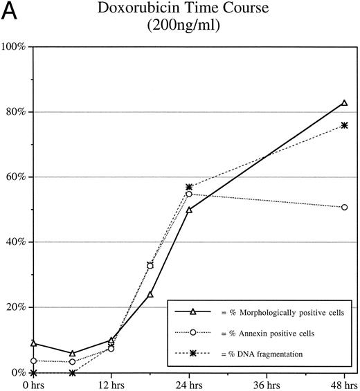
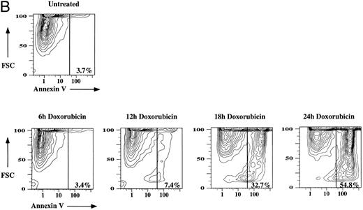
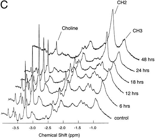
Time course of the spectral changes associated with the exposure of PS (apoptosis).Jurkat T-cell cultures were treated with 200 ng/mL of doxorubicin and harvested at 0, 6, 12, 18, 24, and 48 hours. Cells were then analyzed by FITC-annexin V flow cytometry, DNA gel electrophoresis nuclear morphologic analysis, and 1H NMR. Annexin V positivity, the percentage of DNA fragmentation, the percentage of apoptosis as observed by morphologic analysis, and the CH2/CH3 signal intensity ratio all increased to greater than baseline values at 18 hours (Fig 1A, B, and C). Annexin V positivity (fraction of cells expressing phosphatidylserine on the surface of the plasma membrane) and the CH2/CH3 signal intensity ratio showed a linear correlation (r2 = .883) up to 24 hours, after which annexin V positivity plateaued, whereas the CH2/CH3 signal intensity ratio continued to increase. Annexin V positivity also demonstrated an excellent linear correlation between the percentage of DNA fragmentation (r2 = .996) and nuclear morphologic analysis (r2 = .966) up to 24 hours after doxorubicin treatment. The percentage of DNA fragmentation and the percentage of apoptosis as observed by morphologic analysis correlated to the CH2/CH3 signal intensity ratio (r2 = .886, and r2 = .982, respectively) during the entire 48 hours of doxorubicin treatment.
Time course data of doxorubicin-treated Jurkat cultures. Jurkat cultures were incubated with 200 ng/mL of doxorubicin for 0, 6, 12, 18, 24, and 48 hours. (A) The percentage of apoptotic cells in culture as observed by nuclear morphologic analysis, FITC-annexin V flow cytometry, and DNA gel electrophoresis from 0 to 48 hours after doxorubicin treatment. (B) FITC-annexin V flow cytometric data from 0 to 24 hours after doxorubicin treatment. FCS, forward scatter; FITC-annexin V, fluorescent intensity at 494 nm plotted on a logarithmic scale. (C) 1H NMR spectra obtained (with 64 excitations) at 400 MHz. The spectral resonances of choline protons (-N(CH3 )) at 3.2 ppm, methylene protons (-CH2-) at 1.3 ppm, and methyl protons (-CH3 ) at 0.9 ppm are indicated. The CH2/CH3 signal intensity ratios were 0.28, 0.56, 0.33, 0.89, 2.41, and 4.42 at 0, 6, 12, 18, 24, and 48 hours after doxorubicin treatment, respectively.
Time course data of doxorubicin-treated Jurkat cultures. Jurkat cultures were incubated with 200 ng/mL of doxorubicin for 0, 6, 12, 18, 24, and 48 hours. (A) The percentage of apoptotic cells in culture as observed by nuclear morphologic analysis, FITC-annexin V flow cytometry, and DNA gel electrophoresis from 0 to 48 hours after doxorubicin treatment. (B) FITC-annexin V flow cytometric data from 0 to 24 hours after doxorubicin treatment. FCS, forward scatter; FITC-annexin V, fluorescent intensity at 494 nm plotted on a logarithmic scale. (C) 1H NMR spectra obtained (with 64 excitations) at 400 MHz. The spectral resonances of choline protons (-N(CH3 )) at 3.2 ppm, methylene protons (-CH2-) at 1.3 ppm, and methyl protons (-CH3 ) at 0.9 ppm are indicated. The CH2/CH3 signal intensity ratios were 0.28, 0.56, 0.33, 0.89, 2.41, and 4.42 at 0, 6, 12, 18, 24, and 48 hours after doxorubicin treatment, respectively.
There was also a progressive decrease in the choline [N(CH3 )3]/CH3 signal intensity ratio starting at 18 hours that paralleled the increase in the CH2/CH3 signal intensity ratio and the onset of apoptosis (data not shown). The choline/CH3 signal intensity ratio showed a weak correlation with annexin V positivity over the 24-hour period (r2 = .698) of doxorubicin treatment. The percentage of DNA fragmentation and the percentage of apoptosis, as observed by morphologic analysis, weakly correlated with the choline/CH3 signal intensity ratio (r2 = .76 and r2 = .74, respectively) over the entire 48 hours of doxorubicin treatment.
To more accurately determine the correlation between CH2/CH3 signal intensity ratio and annexin V positivity during the narrow window between induction of apoptosis and the plateauing of the annexin V positivity, additional Jurkat T-cell cultures were treated (200 ng/mL of doxorubicin) and harvested at 0, 18, 20, 22, and 24 hours. Again, annexin V positivity and the fraction of apoptotic cells determined by nuclear morphology showed an excellent linear correlation (r2 = .905, N = 13, data not shown) between 18 and 24 hours of doxorubicin treatment. However, annexin V positivity showed only a weak correlation with the CH2/CH3 signal-intensity ratio (r2 = .642, N = 13) between 18 and 24 hours. Morphologic analysis also showed a weak correlation with the CH2/CH3 signal intensity ratio (r2 = .764, N = 13, data not shown) between 18 and 24 hours.
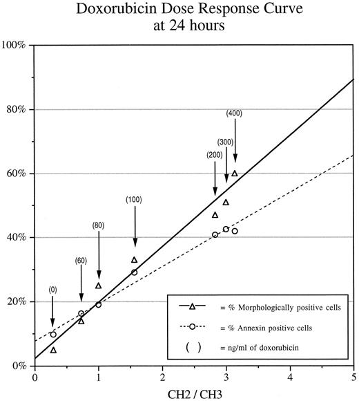
The spectral changes seen in the doxorubicin dose response curves at 24 and 48 hours.Jurkat T-cell cultures underwent harvesting and analysis after treatment with 0, 60, 80, 100, 200, 300, and 400 ng/mL of doxorubicin for 24 hours. There was excellent correlation between annexin V positivity and the CH2/CH3 signal intensity ratio at 24 hours after doxorubicin treatment (Fig 2). There was also excellent correlation between the fraction of apoptotic cells as seen by morphologic analysis and the CH2/CH3 signal intensity ratio at 24 hours after doxorubicin treatment. Additional Jurkat T-cell cultures were treated with 0, 25, 50, 100, 150, 200, and 400 ng/mL of doxorubicin, after which cells were harvested at 48 hours and analyzed. Annexin V positivity remained linearly correlated up to a dose of 150 ng/mL doxorubicin at 48 hours, after which the fraction of annexin-positive cells plateaued (Fig 3). However, the fraction of apoptotic cells as seen morphologically showed an excellent correlation throughout the dose range (25 to 400 ng/mL) with the CH2/CH3 signal intensity ratio. Three data sets of doxorubicin dose-response curves performed at 24 and 48 hours after treatment were combined and analyzed as a composite. There was excellent correlation between the fraction of apoptotic cells as seen morphologically and the CH2/CH3 signal intensity ratio (Fig 4). There was a weak correlation between the fraction of apoptotic cells as seen by morphologic analysis and the decrease in the choline/CH3 signal intensity ratio at 24 and 48 hours (r2 = .304, N = 18 and r2 = .67, N = 9, respectively; data not shown). All other resolvable resonances did not change with apoptotic cell death.
Dose-response data of doxorubicin-treated Jurkat cultures at 24 hours. Jurkat cultures were treated with 0, 60, 80, 100, 200, 300, and 400 ng/mL of doxorubicin for 24 hours. The percentage of apoptotic cells in each culture is plotted against the CH2/CH3 signal intensity ratio determined by the 1H NMR spectra obtained at 400 MHz (64 excitations). The solid line and dashed lines are the best fit lines determined by linear regression analyses corresponding to the percentage of apoptotic cells as measured by morphologic analysis (r2 = .965) and FITC-annexin V flow cytometry (r2 = .985), respectively.
Dose-response data of doxorubicin-treated Jurkat cultures at 24 hours. Jurkat cultures were treated with 0, 60, 80, 100, 200, 300, and 400 ng/mL of doxorubicin for 24 hours. The percentage of apoptotic cells in each culture is plotted against the CH2/CH3 signal intensity ratio determined by the 1H NMR spectra obtained at 400 MHz (64 excitations). The solid line and dashed lines are the best fit lines determined by linear regression analyses corresponding to the percentage of apoptotic cells as measured by morphologic analysis (r2 = .965) and FITC-annexin V flow cytometry (r2 = .985), respectively.
Dose-response data of doxorubicin-treated Jurkat cultures at 48 hours. Jurkat cultures were treated with 0, 25, 50, 100, 150, 200, and 400 ng/mL of doxorubicin for 48 hours. The percentage of apoptotic cells in each culture is plotted against the CH2/CH3 signal intensity ratio determined by the 1H NMR spectra obtained at 400 MHz (64 excitations). The solid line represents the best fit line determined by linear regression analysis corresponding to the percentage of apoptotic cells as measured by morphologic analysis (r2 = .964). The dashed line is the best fit line determined by linear regression analysis corresponding to the fraction of apoptotic cells determined by FITC-annexin V flow cytometry assay up to a dose of 150 ng/mL (r2 = .978).
Dose-response data of doxorubicin-treated Jurkat cultures at 48 hours. Jurkat cultures were treated with 0, 25, 50, 100, 150, 200, and 400 ng/mL of doxorubicin for 48 hours. The percentage of apoptotic cells in each culture is plotted against the CH2/CH3 signal intensity ratio determined by the 1H NMR spectra obtained at 400 MHz (64 excitations). The solid line represents the best fit line determined by linear regression analysis corresponding to the percentage of apoptotic cells as measured by morphologic analysis (r2 = .964). The dashed line is the best fit line determined by linear regression analysis corresponding to the fraction of apoptotic cells determined by FITC-annexin V flow cytometry assay up to a dose of 150 ng/mL (r2 = .978).
Composite of dose-response data from 24 and 48 hours. Data from 24 and 48 hours as shown in Figs 2B and 3B were combined with an additional dose-response experiment in which Jurkat cultures were treated with 0, 90, 110, 120, 140, 160, 180, and 200 ng/mL of doxorubicin for 24 hours. The CH2/CH3 signal intensity ratio was plotted against the percentage of apoptotic cells as seen by nuclear morphologic analysis (r2 = .947, N = 27 samples). The solid line is the best fit line determined by linear regression analysis; dashed lines mark the upper and lower limits of a 95% confidence interval plotted over the entire range of data.
Composite of dose-response data from 24 and 48 hours. Data from 24 and 48 hours as shown in Figs 2B and 3B were combined with an additional dose-response experiment in which Jurkat cultures were treated with 0, 90, 110, 120, 140, 160, 180, and 200 ng/mL of doxorubicin for 24 hours. The CH2/CH3 signal intensity ratio was plotted against the percentage of apoptotic cells as seen by nuclear morphologic analysis (r2 = .947, N = 27 samples). The solid line is the best fit line determined by linear regression analysis; dashed lines mark the upper and lower limits of a 95% confidence interval plotted over the entire range of data.
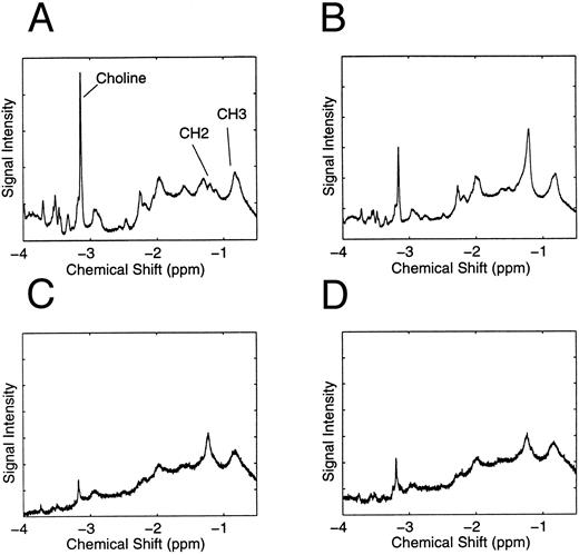
TLC of apoptotic cell cultures treated with doxorubicin, serum starvation, and CH11 IgM anti-Fas antibody.Apoptosis was induced in Jurkat T-cell cultures with doxorubicin (200 ng/mL for 24 hours), serum deprivation (for 72 hours), or CH 11 IgM anti-Fas monoclonal antibody (Immunotech Inc, Westbrook, ME; 200 ng/mL for 48 hours). 1H NMR spectroscopy was performed on control, doxorubicin-treated, serum-deprived, and CH11 IgM anti-Fas monoclonal antibody-treated Jurkat cell cultures (Fig 5A, B, C, and D, respectively). TLC was performed on the lipid extracts of each of the four Jurkat cell cultures (Fig 6). No differences were noted between control and apoptotic cultures with any of the three treatments by TLC despite marked increases in the CH2/CH3 signal intensity ratios above baseline. In particular, there were no increases in the intensity (amount) of lysolipid staining (Rf ≈ 0.10) in apoptotic cultures that would indicate lipase activity with subsequent degradation of diacyl phospholipid. In addition, the results of fluorescamine staining confirmed that the only spots containing primary amines were the ones corresponding to the standards containing PS (eg, Fig 6, lane 1e, DMPS; Rf values [0.55 to 0.68]) and PE (Fig 6, lane 2a and 3a DLPE; Rf values [0.69 to 0.75]) as well as all classes of the lysolipids containing PS and PE from the cell lipid extracts center lanes of TLC #1, #2, and #3 (Rf values; ≈ 0.10). The extracts of each of the four cell cultures demonstrated that the spots corresponding to the lipid fractions containing PS (Rf values; TLC #1 [0.65 to 0.75], TLC #2 [0.70 to 0.88], TLC #3 [0.70 to 0.88]) and phosphatidylcholine (Rf values; TLC #1 [0.26 to 0.45], TLC #2 [0.45 to 0.65], TLC #3 [0.45 to 0.65]) remained unchanged in apoptotic cultures with respect to intensity (amount) and Rf values. These data indicate a lack of significant change in lipid polarity (such as with head group exchange, change in acyl chain length or increases in the degree of unsaturation of acyl chains) with apoptotic cell death.
Spectral changes associated with apoptotic Jurkat cultures with different therapies. The 1H NMR spectra (at 400 MHz) corresponding to the Jurkat cultures before lipid extraction and TLC shown in Fig 6. (A) Control culture. The resonance peaks for choline, CH2 , and CH3 protons are shown. (B) Jurkat cells treated with 200 ng/mL of doxorubicin for 24 hours. (C) Jurkat cells deprived of serum for 3 days. (D) Jurkat cells treated with 200 ng/mL of CH 11 IgM Fas antibody for 48 hours. The CH2/CH3 signal intensity ratio, the percentages of apoptotic cells seen by nuclear morphologic analysis, and FITC-annexin V flow cytometric analysis for each culture were as follows: control (0.29, 5%, and 9.8%, respectively), doxorubicin-treated (2.55, 51%, and 47%, respectively), serum-starved (1.82, 54%, and 46.8%, respectively), and Fas antibody (1.63, 69%, and 71%, respectively).
Spectral changes associated with apoptotic Jurkat cultures with different therapies. The 1H NMR spectra (at 400 MHz) corresponding to the Jurkat cultures before lipid extraction and TLC shown in Fig 6. (A) Control culture. The resonance peaks for choline, CH2 , and CH3 protons are shown. (B) Jurkat cells treated with 200 ng/mL of doxorubicin for 24 hours. (C) Jurkat cells deprived of serum for 3 days. (D) Jurkat cells treated with 200 ng/mL of CH 11 IgM Fas antibody for 48 hours. The CH2/CH3 signal intensity ratio, the percentages of apoptotic cells seen by nuclear morphologic analysis, and FITC-annexin V flow cytometric analysis for each culture were as follows: control (0.29, 5%, and 9.8%, respectively), doxorubicin-treated (2.55, 51%, and 47%, respectively), serum-starved (1.82, 54%, and 46.8%, respectively), and Fas antibody (1.63, 69%, and 71%, respectively).
Thin-layer chromatograms of lipid extracts of apoptotic Jurkat T-cell cultures with different therapies. TLC was performed using the lipid extracts of untreated (control) and treated (apoptotic) Jurkat T-cell cultures. Three separate TLC are shown (ie, TLC #1, #2, and #3) each performed on different dates. Lipid standards (outside lanes) and lipid extracts from cell cultures (inner 3 lanes) were spotted and developed as described. TLC #1 (lanes 1A through 1E): 1A, standard; 1B, doxorubicin-treated cells; 1C untreated (control) cells; 1D, doxorubicin-treated cells; 1E, standard. TLC #2 (lanes 2A through 2E): 2A, standard; 2B, untreated cells; 2C, serum-starved cells; 2D, CH11 IgM anti-Fas antibody-treated cells; 2E, standard. TLC #3 (lanes 3A through 3E): 3A, standard; 3B, untreated cells; 3C, serum-starved cells; 3D, CH11 IgM anti-Fas antibody-treated cells; 3E, standard. Rf scale is the fractional height of spots relative to the total length of TLC plates from origin. Rf = 0.0 origin; Rf = 1.0 solvent front. Doxorubicin treatment (200 ng/mL for 24 hours); serum-starved cells (for 72 hours); CH11 IgM anti-Fas antibody treatment (200 ng/mL for 48 hours). Standards: Lyso LPC = lyso (mono) [12:0] PC; DMPS = di [14:0] PS; DOPC = di [18:1] PC; DLPE = di [12:0] PE; DPPC = di [16:0] PC; DSPC = di [18:0] PC. NB, [n1 :n2], where n1 is the chain length of fatty acyl group and n2 is the number of unsaturations of fatty acyl group. di, mono = number of acyl chains. PC, PS, and PE refer to type of head phospholipid group.
Thin-layer chromatograms of lipid extracts of apoptotic Jurkat T-cell cultures with different therapies. TLC was performed using the lipid extracts of untreated (control) and treated (apoptotic) Jurkat T-cell cultures. Three separate TLC are shown (ie, TLC #1, #2, and #3) each performed on different dates. Lipid standards (outside lanes) and lipid extracts from cell cultures (inner 3 lanes) were spotted and developed as described. TLC #1 (lanes 1A through 1E): 1A, standard; 1B, doxorubicin-treated cells; 1C untreated (control) cells; 1D, doxorubicin-treated cells; 1E, standard. TLC #2 (lanes 2A through 2E): 2A, standard; 2B, untreated cells; 2C, serum-starved cells; 2D, CH11 IgM anti-Fas antibody-treated cells; 2E, standard. TLC #3 (lanes 3A through 3E): 3A, standard; 3B, untreated cells; 3C, serum-starved cells; 3D, CH11 IgM anti-Fas antibody-treated cells; 3E, standard. Rf scale is the fractional height of spots relative to the total length of TLC plates from origin. Rf = 0.0 origin; Rf = 1.0 solvent front. Doxorubicin treatment (200 ng/mL for 24 hours); serum-starved cells (for 72 hours); CH11 IgM anti-Fas antibody treatment (200 ng/mL for 48 hours). Standards: Lyso LPC = lyso (mono) [12:0] PC; DMPS = di [14:0] PS; DOPC = di [18:1] PC; DLPE = di [12:0] PE; DPPC = di [16:0] PC; DSPC = di [18:0] PC. NB, [n1 :n2], where n1 is the chain length of fatty acyl group and n2 is the number of unsaturations of fatty acyl group. di, mono = number of acyl chains. PC, PS, and PE refer to type of head phospholipid group.
DISCUSSION
The signal intensity of CH3 resonance does not change with apoptotic cell death.19 The CH2 resonance signal intensity increases to a maximal value of 5 to 6 times the CH3 resonance signal intensity during apoptosis. The ratio of the integrated areas of CH2 and CH3 resonance peaks, therefore, can be used to estimate the fraction of apoptotic cells in culture. We found that the CH2/CH3 signal-intensity ratio, as measured by 1H NMR, correlated with the fraction of apoptotic cells seen morphologically and by DNA fragmentation throughout the 48-hour time period, whereas annexin V positivity plateaued after 24 hours with doses of 200 ng/mL of doxorubicin or greater.
The plateauing of annexin V positivity after 24 hours may suggest that the concentration of FITC-annexin V (100 nmol/L) used in our assay was insufficient to saturate all annexin V receptors present in the later stages of apoptosis. The incomplete saturation of annexin V receptors may have resulted in a plateauing of annexin V positivity seen by flow cytometry. Because there were no significant changes in the plasma membrane attributable to phospholipid degradation or head group exchange, it is unlikely that there was a loss or degradation of exposed PS (ie, annexin V receptors) in the later (after 24 hours) stages of apoptosis.
The correlation between the CH2/CH3 signal intensity ratio and the fraction of apoptotic cells as seen morphologically was optimized at 24 and 48 hours after treatment with doxorubicin. This implies that there is a time dependence before 24 hours of the magnitude of the increase in the CH2/CH3 signal intensity ratio with doxorubicin-induced apoptotic cell death.
The increase in the CH2/CH3 signal intensity ratio is coincident with increases in annexin V-positive cells, DNA fragmentation, and the number of apoptotic cells seen morphologically. It is unclear what event(s) in the apoptotic cascade is directly associated or responsible for the increase in the CH2 resonance.
Prior work has shown that the prominent lipid spectra in malignant cell lines are due to dynamic changes in microviscosity of the plasma membrane.30,31 Our current study has shown no gross changes in the lipid profiles of cells undergoing apoptosis, as observed by TLC. If activation of phospholipase A2 was responsible for our spectroscopic observations, one would expect to see increased expression of the lysolipid spot (Rf ≈ 1.0) with apoptotic cell death. Furthermore, if phospholipase D (and probably C as well) were being activated, one would also expect to see enhanced fluorescamine staining closer to the origin. In addition, we showed no significant changes in fatty acyl chain length (longer chain lengths have higher Rf values; see TLC #2 and #3, DPPC v DSPC) or degree of unsaturation (unsaturated fatty acids tend to have increased Rf values with respect to saturated fatty acids of the same chain length; see TLC #3 DOPC v DSPC). Therefore, a dynamic change of the plasma membrane that may reflect a change in lipid bilayer composition rather than lipase activity, increases in fatty acid production, or degree of fatty acid unsaturation would be a more plausible explanation for the increase in the methylene resonance occurring with apoptotic cell death.19
Of note, the CH3 resonance at 0.9 ppm did not change with apoptotic cell death. The CH3 resonance is well defined (relatively mobile protons) even in control (untreated) cell cultures and also arises in part from non–lipid-related CH3 protons such as nucleic acids and other compounds.32 33 Therefore, the CH3 resonance is not likely to be greatly affected by dynamic changes of the plasma membrane. Similarly, the N(CH3)3 protons from the phospholipid head groups are also well defined because these head groups are also relatively mobile (on the outer or inner leaflet of the lipid bilayer) and also are not likely to be affected significantly by dynamic changes of the plasma membrane. The gradual decrease in choline/CH3 signal intensity ratio that begins at the onset of apoptosis (and increase in the CH2/CH3 signal intensity ratio) may be due to the loss of choline moieties not related to the lipid bilayer. These small molecule (choline) metabolites may not be replenished as the cell prepares to die. The variability in the rate of loss of choline metabolites (in treated cells) may in part explain the poor correlation between the fraction of apoptotic cells and the choline/CH3 signal intensity ratio.
It is possible that the loss of cytoskeletal architecture is partly responsible for the surface expression of PS seen with apoptosis.34 The loss of cytoskeletal architecture may also allow for increased mobility of the plasma membrane lipids. The increase in extracellular PS exposure during apoptosis also appears to involve a downregulation of the aminophospholipid translocase that translocates PS from the outer face to the inner face of the plasma membrane35 to maintain normal plasma membrane structural asymmetry. There is also an increase in the activity of a phospholipid scramblase during apoptosis35 that allows all phospholipid classes to equilibrate rapidly across the plasma membrane. Both these changes may allow for increased lipid mobility with a subsequent decrease in the microviscosity of the plasma membrane during apoptosis.36 There also may be a time dependence on the degree of cytoskeletal architectural destruction and/or loss of aminophospholipid translocase function that would in part explain the time-dependent changes in the methylene signal before 24 hours with doxorubicin therapy, particularly during the critical 18- to 24-hour window after treatment in which the CH2 signal increases to greater than baseline values.
Our study provides the actual analytical methods with which to estimate the percentage of apoptotic leukemic cells in vivo. Efforts at studying tumor-bearing animals and patients with leukemia are ongoing.
ACKNOWLEDGMENT
The authors thank Dr Linda Brunauer (Stanford University, Stanford, CA) for helpful discussion about the TLC studies, particularly in regard to the composition of the TLC solvent system; and Dr Wray H. Huestis (Stanford University) for helpful discussion and the use of reagents and facilities for TLC and lipid extractions.
Supported by the Office of Technology Licensing at Stanford University, CA. P.D.K. was supported by National Institutes of Health Grant No. CA 42509, awarded to Leonard A. Herzenberg (Professor of Genetics, Stanford University, Stanford, CA). C.B. was graciously supported by the Alberta Heritage Foundation for Medical Research.
Address reprint requests to Francis G. Blankenberg, MD, Lucile Suter Packard Children's Hospital, 725 Welch Rd, Palo Alto, CA 94304.



![Fig. 6. Thin-layer chromatograms of lipid extracts of apoptotic Jurkat T-cell cultures with different therapies. TLC was performed using the lipid extracts of untreated (control) and treated (apoptotic) Jurkat T-cell cultures. Three separate TLC are shown (ie, TLC #1, #2, and #3) each performed on different dates. Lipid standards (outside lanes) and lipid extracts from cell cultures (inner 3 lanes) were spotted and developed as described. TLC #1 (lanes 1A through 1E): 1A, standard; 1B, doxorubicin-treated cells; 1C untreated (control) cells; 1D, doxorubicin-treated cells; 1E, standard. TLC #2 (lanes 2A through 2E): 2A, standard; 2B, untreated cells; 2C, serum-starved cells; 2D, CH11 IgM anti-Fas antibody-treated cells; 2E, standard. TLC #3 (lanes 3A through 3E): 3A, standard; 3B, untreated cells; 3C, serum-starved cells; 3D, CH11 IgM anti-Fas antibody-treated cells; 3E, standard. Rf scale is the fractional height of spots relative to the total length of TLC plates from origin. Rf = 0.0 origin; Rf = 1.0 solvent front. Doxorubicin treatment (200 ng/mL for 24 hours); serum-starved cells (for 72 hours); CH11 IgM anti-Fas antibody treatment (200 ng/mL for 48 hours). Standards: Lyso LPC = lyso (mono) [12:0] PC; DMPS = di [14:0] PS; DOPC = di [18:1] PC; DLPE = di [12:0] PE; DPPC = di [16:0] PC; DSPC = di [18:0] PC. NB, [n1 :n2], where n1 is the chain length of fatty acyl group and n2 is the number of unsaturations of fatty acyl group. di, mono = number of acyl chains. PC, PS, and PE refer to type of head phospholipid group.](https://ash.silverchair-cdn.com/ash/content_public/journal/blood/89/10/10.1182_blood.v89.10.3778/3/m_bl_0029f6.jpeg?Expires=1766424341&Signature=hLRPZm3khAGImPiDzseXA8ik7Hxg8l7AOSVwYh3wlgsxtOnS06ohEknSmaKKIKoNlW5Nr9UtH4NdlltvHxY0DILYZU90Itzvp07-5LZJ9nr9FBbbJePoyovJeaNR4-Ui-uHSc8q1o96baygtGTX5qo7mbxD~-J3prRumDzHYxH5NHhJzGWxPZ5ZHYMLVwZnL0xA7HyZwUA3RIbikMdigRxZMsOWtfxaXCF1wzyQ8q1559KQxl5QYhZsfyJE0mjg8hNxzSoYMYHQJpxqFIh8x~zhWaBZqxVrcruYO5EjCBKib8jbzvBCP6QYKZUI6bIo06WiNLN6J2FHr-6TL3iu9mg__&Key-Pair-Id=APKAIE5G5CRDK6RD3PGA)
This feature is available to Subscribers Only
Sign In or Create an Account Close Modal