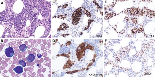
A 59-year-old man was diagnosed in 2018 with mantle cell lymphoma (MCL), leukemic presentation with TP53 mutation after presenting with leukocytosis and splenomegaly without lymphadenopathy. He achieved complete remission with ibrutinib, rituximab, and hyper-CVAD. In 2022, he relapsed with fluorodeoxyglucose-avid lymphadenopathy and right tonsillar enlargement. Biopsy of the tonsil confirmed pleomorphic MCL. After chimeric antigen receptor T-cell therapy, the patient achieved a second remission. Fourteen months later, he developed lymphocytosis. A restaging bone marrow biopsy showed large, pleomorphic lymphoma cells in an intrasinusoidal pattern (panel A: core biopsy, original magnification ×400; panel B: aspirate smear, original magnification ×1000), with immunohistochemical staining positive for PAX5, cyclin D1, SOX11, and P53 (panels C-F: original magnification ×400). Flow cytometry detected a CD5+CD10−CD200− κ-monoclonal B-cell population. Cytogenetic study identified a complex karyotype with IGH::CCND1, and next-generation sequencing identified mutations in TP53, NOTCH2, and CCND1, consistent with clonal evolution and disease progression. The patient was refractory to multiple lines of salvage chemoimmunotherapy and died 3 months later.
Intrasinusoidal involvement by B-cell lymphomas is characteristic of intravascular large B-cell lymphoma and splenic marginal zone lymphoma, but it is rare in MCL. This case underscores the importance of recognizing this rare, unusual intrasinusoidal pattern of involvement by pleomorphic MCL that may mimic intravascular large B-cell lymphoma, thus avoiding a diagnostic pitfall.
For additional images, visit the ASH Image Bank, a reference and teaching tool that is continually updated with new atlas and case study images. For more information, visit https://imagebank.hematology.org.


This feature is available to Subscribers Only
Sign In or Create an Account Close Modal