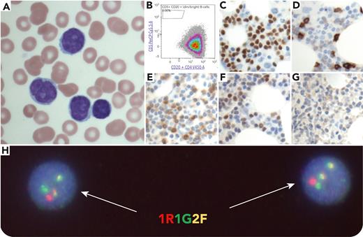
A 76-year-old woman presented with lymphocytosis without lymphadenopathy or splenomegaly. Peripheral blood flow cytometry was initially interpreted to be suggestive of marginal zone lymphoma. Over 2 years, the patient developed worsening fatigue, progressive anemia (96 g/L), leukocytosis (149.7 × 10⁹/L), and mild thrombocytopenia (133 × 109/L). A bone marrow biopsy was performed. The peripheral blood showed medium-sized lymphocytes with ovoid/indented/cleaved nuclei, slightly dispersed chromatin, distinct nucleoli, and scant cytoplasm (panel A, May-Grünwald stain, original magnification ×1000). Marrow flow cytometry was nonspecific, showing κ-restricted B cells negative for CD5 (panel B) and CD10 and dimly positive for CD23. Immunohistochemistry (IHC) showed PAX5-positive B cells (panel C, original magnification ×400) with a subset expression of CD5 (weak; panel D, original magnification ×400; CD5-strong cells are T cells), CD23 (panel E, original magnification ×400), and, notably, cyclin D1 (weak; panel F, original magnification 400×); SOX11 was negative (panel G, original magnification ×400). BRAF p.V600E and MYD88 p.L265P mutations were not detected. Fluorescence in situ hybridization (FISH) identified the IGH::CCND1 rearrangement in 69% of nuclei (panel H; 2 fused signals), confirming a diagnosis of leukemic nonnodal mantle cell lymphoma (LNNMCL).
LNNMCL is characterized by blood, marrow, and sometimes splenic involvement, with absent or minimal lymphadenopathy. Diminished CD5 expression is unusual for LNNMCL but has been reported. This phenotype can be confused with those of other small B-cell leukemias/lymphomas. Here, the mantle cell lymphoma diagnosis was suggested by weak cyclin D1 IHC expression and confirmed via FISH.
For additional images, visit the ASH Image Bank, a reference and teaching tool that is continually updated with new atlas and case study images. For more information, visit https://imagebank.hematology.org.


This feature is available to Subscribers Only
Sign In or Create an Account Close Modal