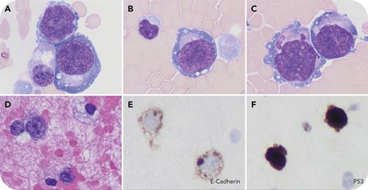
A 64-year-old man with hypoplastic TP53-mutated myelodysplastic syndrome (MDS), confirmed by a bone marrow (BM) procedure 2 weeks earlier, was admitted for stem cell transplant evaluation. Computed tomography scan detected recurrent bilateral pleural effusions, which were negative for carcinoma and acute myeloid leukemia (limited measurable residual disease panel by flow cytometry). The thoracentesis fluid cytospins revealed scattered large atypical cells displaying immature erythroblast morphology, characterized by finely reticulated chromatin, several distinct nucleoli, a deep basophilic agranular cytoplasm, and occasional cytoplasmic vacuoles (panels A-C: Wright-Giemsa stain, 100× lens objective). The cell block showed discohesive large cells positive for E-cadherin and p53 (panel D: hematoxylin and eosin stain; panels E-F: E-cadherin and p53 immunostains, 40× lens objective), but negative for CD45, myeloperoxidase, and CD34, suggesting an extramedullary erythroid leukemia. This prompted an immediate BM procedure, which revealed 46% pronormoblasts and 36% normoblasts on touch preparation, with the majority of marrow cellularity showing strong nuclear p53 expression. Flow cytometric immunophenotyping demonstrated blasts with the phenotype of CD4−/CD5(partial+)/CD7(partial+)/CD13−/CD14−/CD15−/CD19−/CD25−/CD33−/CD34(subset+)/CD36+/CD38(partial+)/CD45(decreased+)/CD54+/CD56−/CD64−/CD71+/CD117(partial+)/CD123−/HLA-DR−. The next-generation sequencing leukemia panel on BM aspirates identified dual TP53 mutations (c.989T>G p.L330R, variant allelic frequency: 26%; and c.949C>T p.Q317∗, variant allelic frequency: 15%), confirming a diagnosis of acute erythroid leukemia (AEL).
This case illustrates that the presence of immature erythroblasts in pleural effusion may precede the diagnosis of AEL in BM. It also emphasizes the need for thorough examination of body fluids in MDS patients and highlights the importance of a comprehensive workup in diagnosing AEL.
For additional images, visit the ASH Image Bank, a reference and teaching tool that is continually updated with new atlas and case study images. For more information, visit https://imagebank.hematology.org.


This feature is available to Subscribers Only
Sign In or Create an Account Close Modal