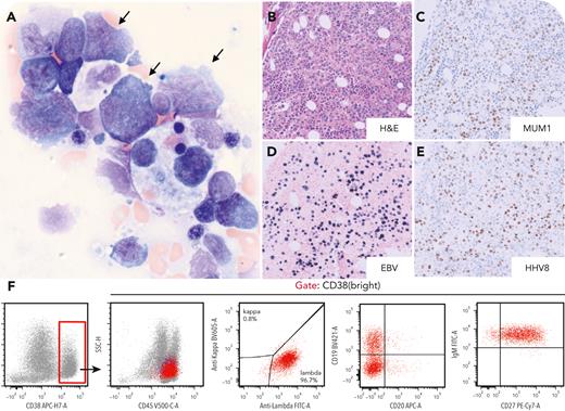
A 64-year-old man with history of liver transplant presented with pancytopenia and Epstein-Barr virus (EBV) viremia. Positron emission tomography–computed tomography showed hypermetabolism in the spleen, retroperitoneal lymph nodes, and bone marrow. Bone marrow revealed large neoplastic cells with coarse chromatin, nucleoli, eccentric nuclei, and blue cytoplasm in a background of dyserythropoiesis and rare hemophagocytosis (panel A: Wright-Giemsa stain, 60× lens objective, total magnification ×600). Neoplastic cells expressed multiple myeloma oncogene 1 (MUM1), human herpesvirus 8 (HHV-8), and EBV (panel B: hematoxylin-eosin [H&E] stain; panels C-E: immunostain; 20× lens objective, total magnification ×200). Flow cytometry showed neoplastic cells expressing CD38, CD45, lambda light chain, CD19 (partial), immunoglobulin M, and CD27 (variable), and not expressing CD20, CD22, CD24, CD138, or B-cell maturation antigen (panel F, and data not shown). B-cell clonality and immunoglobulin gene heavy-chain variable (IGHV) somatic hypermutation by next-generation sequencing showed a dominant IGHV1-2∗02/IGHJ4∗02 gene rearrangement (60.5% of the total rearranged IGH V-J sequence reads), which was unmutated. No pathogenetic mutations were identified, but karyotyping demonstrated hypertetraploidy (XXYY,+Y,+2,−6,−9,+10,−14,−15). Retroperitoneal lymph node biopsy showed Kaposi sarcoma.
This is a case of HHV-8+ diffuse large B-cell lymphoma (DLBCL) with EBV coinfection and is distinguished from extracavitary primary effusion lymphoma (ecPEL) based on lack of somatic hypermutation. The neoplastic cells of HHV-8+ DLBCL correspond to naive, immunoglobulin M–producing B cells without immunoglobulin somatic hypermutation, whereas the neoplastic cells of ecPEL are terminally differentiated B cells with immunoglobulin somatic hypermutation. Although ecPEL typically demonstrates HHV-8/EBV coinfection, rare cases of HHV-8+ DLBCL may also show EBV coinfection.
For additional images, visit the ASH Image Bank, a reference and teaching tool that is continually updated with new atlas and case study images. For more information, visit https://imagebank.hematology.org.


This feature is available to Subscribers Only
Sign In or Create an Account Close Modal