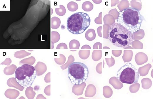
A full-term baby girl with a long history of respiratory insufficiency, increased levels of alkaline phosphatase, and oral tolerance failure was admitted with acute on chronic hypoxic respiratory failure. Upon physical examination, the patient demonstrated periorbital edema, central hypotonia, metatarsus adductus deformity of the left foot (L; panel A), and peripheral spasticity. Her white blood cell count was 19.2 × 10⁹/L, with lymphocytosis; hemoglobin 8.7 g/dL; and platelets 431 × 109/L. The blood smear demonstrated frequent lymphocytes with prominent vacuolization, suggesting a metabolic disease (panels B-F: Wright-Giemsa stain, 100× lens objective). Other white cells showed unremarkable morphology. Urine liquid chromatography–tandem mass spectrometry revealed elevated keratan sulfate (5.25 g/mol creatinine, reference range ≤1.61 g/mol creatinine), consistent with a deficiency of β-galactosidase enzyme activity. Subsequent clinical exome-sequence analysis revealed 2 autosomal-recessive mutations in intron 2 c.245+1 G>A and exon 2 c.202 C>T (p.Arg68Trp) of the GLB1 gene, confirming the diagnosis of GM1 gangliosidosis (type 1, infantile form).
GM1 gangliosidosis is a lysosomal-storage disease associated with progressive neurodegeneration caused by mutations in the GLB1 gene that encodes the enzyme β-galactosidase. This leads to the accumulation of β-linked galactose-containing glycoconjugates, including GM1 ganglioside, in neuronal tissue. Prominent vacuolization within lymphocytes may be attributed to the accumulation of metabolic byproducts. The lymphocyte morphology may be a helpful diagnostic clue in patients without an established history of metabolic disorders.
For additional images, visit the ASH Image Bank, a reference and teaching tool that is continually updated with new atlas and case study images. For more information, visit https://imagebank.hematology.org.


This feature is available to Subscribers Only
Sign In or Create an Account Close Modal