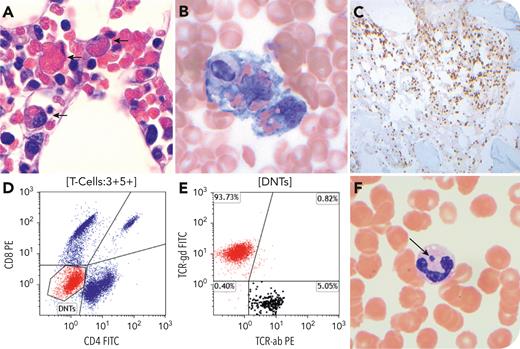
A 67-year-old man with no known relevant medical history presented with altered mental status, fever (39.4°C), and splenomegaly. A complete blood count revealed leukopenia (1400 white blood cells per μL), thrombocytopenia (10 000 platelets per μL), and normal hemoglobin (13.1 g/dL). Other studies included hypertriglyceridemia (258 mg/dL), hypofibrinogenemia (141 mg/dL), elevated liver enzymes (aspartate transaminase 158 U/L, alanine aminotransferase 58 U/L), elevated ferritin (27 238 ng/mL), and soluble interleukin-2R/CD25 (8534 pg/mL). Bone marrow biopsy (panel A; hematoxylin and eosin stain, 100× objective) and aspirate (panel B; Wright-Giemsa stain, 100× objective) revealed hemophagocytic forms and significant T-cell infiltrate (panel C; CD3 immunostain, 10× objective). Flow cytometry identified expansion of γ/δ double-negative T cells (CD3+/CD5+/CD4−/CD8−/T-cell receptor-γδ+), representing 10% of total (panels D and E). Cytogenetic analysis was normal and there was no T-cell receptor gene rearrangement. Peripheral blood smear revealed rare neutrophils with intracytoplasmic morulae (panel F; Wright-Giemsa stain, 100× objective) and serum polymerase chain reaction was positive for Anaplasma phagocytophilum. The patient was diagnosed with hemophagocytic lymphohistiocytosis (HLH) secondary to anaplasmosis. He was treated with doxycycline and recovered with complete symptom resolution. Given his satisfactory evolution, steroids were not administered.
Both infections and T-cell lymphoma can cause secondary HLH. This patient had anaplasmosis leading to expansion of reactive double-negative γ/δ T cells mimicking T-cell lymphoma. Cases with clonal expansion of γ/δ T cells resolving after antibiotic treatment have been documented. Although immunosuppression is used to treat HLH, it is not necessary and could be counterproductive in some cases of infection-associated HLH, highlighting the importance of a thorough diagnostic work-up.
For additional images, visit the ASH Image Bank, a reference and teaching tool that is continually updated with new atlas and case study images. For more information, visit https://imagebank.hematology.org.


This feature is available to Subscribers Only
Sign In or Create an Account Close Modal