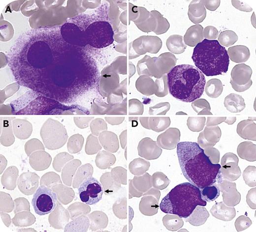
A 51-year-old man presented with unexplained pancytopenia (hemoglobin 114 g/L, platelets 65 × 109/L, neutrophils 1.7 × 10⁹/L) alongside left elbow tendinitis and microcrystalline arthritis in the left wrist. Relevant laboratory findings included an elevated C-reactive protein (36 mg/L), mean corpuscular volume (102 femtoliters), and reticulocyte count (548 × 109/L). No paraprotein was detected. The patient had good performance status without fever, skin involvement, pulmonary infiltrate, vasculitis, ear or nose chondritis, or venous thromboembolism. Bone marrow was normocellular with increased erythroid lineage (61%) and decreased granulocytic lineage (28%). Significant dysmegakaryopoiesis with megakaryocytes with separated nuclei (panel A) and dyserythropoiesis with nuclear budding and basophilic stipplings (panel B) were observed (all images, May-Grünwald-Giemsa stain, 100× lens objective). Reactive changes such as toxic granules were present in granulocytes (panel C), but no significant vacuolization was observed in bone marrow precursors (<10%). Blast cells (panel D) were increased (5%), defining myelodysplastic syndrome with excess blasts (myelodysplastic syndrome with excess blasts per International Consensus Classification, myelodysplastic neoplasm with increased blasts–1 per the World Health Organization). Karyotype was normal. Next-generation sequencing revealed S56F UBA1 mutation (VAF 69%), indicative of VEXAS (vacuoles, E1 enzyme, X-linked, autoinflammatory, somatic) syndrome, with no other mutations.
S56F UBA1 is a recently described somatic mutation in VEXAS syndrome, characterized by increased erythropoiesis, pronounced cytopenia and dysplastic changes, mild inflammatory phenotype, and minimal bone marrow vacuoles. These hematological features contrast with classical VEXAS syndrome, indicating a subtype likely predisposed to manifest as MDS.
For additional images, visit the ASH Image Bank, a reference and teaching tool that is continually updated with new atlas and case study images. For more information, visit https://imagebank.hematology.org.


This feature is available to Subscribers Only
Sign In or Create an Account Close Modal